
GC102 Fever After Chemotherapy Infections In Immunocompromised Hosts
Febrile neutropenia and opportunistic infections occurring in immunocompromised patients following chemotherapy, requiring urgent evaluation and empiric broad-spectrum antimicrobial therapy due to impaired host defenses.
Fever After Chemotherapy: Infections in Immunocompromised Hosts
This lecture centres on a single critical clinical scenario: a patient who develops fever during the neutropenic nadir following chemotherapy. From this anchor case, the lecture systematically expands outward to cover the entire landscape of infections in immunocompromised hosts — not just neutropenia, but every major immune defect (cellular, humoral, barrier, device-related, splenectomy, biologics) and the specific pathogen spectrum each defect predicts.
Why this matters: Febrile neutropenia is a medical emergency. Untreated, it progresses to septic shock and death within hours because neutropenic patients cannot mount an adequate inflammatory response. You must start empirical broad-spectrum antibiotics before culture results. The lecture teaches you to: (1) recognize the immune defect, (2) predict the pathogen, (3) investigate efficiently, (4) treat empirically, and (5) prevent infections proactively.
Core Definitions
Lecture-era wording: oral temperature > 37.6°C more than once within a day; or single oral temperature > 38.3°C; or sustained ≥38.0°C for > 1 hour [1]
The low 37.6°C twice-daily threshold is not a universal modern definition. Common oncology protocols use a single oral temperature ≥38.3°C or ≥38.0°C sustained for 1 hour, while NICE defines neutropenic sepsis as neutrophils ≤0.5 × 10⁹/L plus temperature > 38°C or other clinically significant sepsis features. Follow the local oncology pathway; do not wait for a rigid fever threshold when the patient is unwell [11,12]. Fever is driven mainly through IL-1, IL-6 and TNF signalling with hypothalamic prostaglandin E₂—not the slide's inclusion of IL-4 as a principal pyrogen.
Fever may be blunted by steroids, chemotherapy, and NSAIDs [1]
This is a crucial clinical trap: a patient on dexamethasone for brain metastases or on high-dose steroids post-transplant can have raging sepsis with only a low-grade temperature or even be normothermic. Never be reassured by the absence of high fever in an immunocompromised patient.
Absolute Neutrophil Count (ANC) thresholds: [1]
- ANC < 1.5 × 10⁹/L → Neutropenia
- ANC < 0.5 × 10⁹/L → Severe neutropenia
- ANC < 0.1 × 10⁹/L → Profound neutropenia
Neutropenic fever is defined as ANC ≤ 0.5 × 10⁹/L (or ANC ≤ 1.0 expected to decline to ≤ 0.5 within 48h) PLUS single oral temperature ≥ 38.3°C OR sustained ≥ 38.0°C for > 1 hour [1][2]
Why ANC 0.5? This is the threshold below which the risk of clinically significant infection rises sharply, with especially high risk at ANC < 0.1 × 10⁹/L. The relationship is inversely proportional — the lower the ANC and the longer the duration of neutropenia, the higher the infection risk.
Lecture pattern: post-chemotherapy nadir often 7–14 days; duration may be 14–28 days. The slide states that 100% developed fever if neutropenic for ≥3 weeks. [1]
Treat the nadir and “100%” figures as regimen- and cohort-dependent historical teaching, not a prediction for every patient. The robust principle is that deeper and longer neutropenia increases infection risk.
High Yield – Past Paper Direct Hit
2024 4th Summative MCQ Q84 asked: "For fever after chemotherapy, the risk of infection or bacteraemia is MOST RELATED to…?" Answer: The degree and duration of neutropenia [4]. Although mucositis and Hickman catheters contribute, the lecture explicitly frames severity and duration of neutropenia as the primary determinant.
M/25, AML-M2. Induction chemotherapy (Ara-C + daunorubicin). Day 7: 37.4°C, nausea, WBC 1.3, ANC 0.7. Day 10: sudden rigor, 39.2°C, sore throat/mouth, retrosternal pain on swallowing, watery diarrhoea 4×/day. P/E: diffuse oral erythema & ulcerations. Hb 7.2, WBC 0.1, Plt 30. [1]
Unpacking the case:
- By Day 10, the total WBC is 0.1 × 10⁹/L, making profound neutropenia very likely at the expected nadir; formally calculate the ANC from the differential rather than equating total WBC with ANC
- Rigor + 39.2°C = significant bacteraemia until proven otherwise
- Oral erythema/ulceration = chemotherapy-induced mucositis → the broken mucosal barrier allows translocation of oral/GI flora into the bloodstream
- Retrosternal pain on swallowing = likely oesophageal mucositis ± Candida oesophagitis or HSV oesophagitis
- Watery diarrhoea = GI mucosal damage ± C. difficile ± viral enteritis
- Hb 7.2, Plt 30 = pancytopenia from marrow suppression after chemotherapy. Thrombocytopenia primarily changes bleeding/procedural risk; it is not the defining driver of infection risk here
This case perfectly illustrates the convergence of multiple risk factors: neutropenia + mucositis + indwelling catheter (likely Hickman) + thrombocytopenia.
Reading the Early Visual Sequence (Slides 4–7)
- Neutrophil kinetics (Slide 4): maturation requires a marrow reservoir and several days, whereas circulating neutrophils have a short half-life. Cytoreductive chemotherapy therefore produces a delayed nadir and recovery cannot be instantaneous. The count also does not capture qualitative defects in adhesion, chemotaxis, phagocytosis or oxidative killing.
- Oral photograph (Slide 5): confluent erythema and irregular ulcers demonstrate mucosal-barrier injury rather than a pathogen-specific appearance. Mucositis creates pain, poor intake and a portal for oral flora; swab/biopsy is guided by suspected superinfection, not appearance alone.
- Time-course diagram (Slide 6): mucositis rises as granulocytes fall; fever and bacteraemia occur during the deepest nadir, while catheter infection and pulmonary infiltrates can emerge later. It is a conceptual overlay, not a fixed timetable.
- Catheter photograph (Slide 7): focal erythema at/around a line site may be the only external clue. Inspect and palpate the exit site and tunnel, but remember that absence of redness does not exclude catheter-related bloodstream infection.
An individual with alterations in innate & adaptive immunity as a result of underlying diseases & their therapy, which predispose the host to infections and/or neoplasia [1]
The Virulence–Immunity Spectrum
Outcome of infection determined by interplay between microbial virulence and host immunocompetence [1]
Slide 9 presents this as a microbe–host–therapy triangle: the syndrome reflects the organism and inoculum, the patient's specific immune deficit, and how antimicrobials or immunosuppression have altered both. Slide 11's graph makes the same point from another angle—the weaker the host defence, the lower the microbial virulence needed to produce disease. Slide 12 then moves from intact skin/mucosa through phagocytes and complement to lymphocyte-mediated adaptive immunity; locating the failed layer is more useful than simply labelling someone “immunocompromised.”
| Organism | Virulence | Causes Disease in Normal Host? | Significance in Immunocompromised |
|---|---|---|---|
| Rabies virus, Bacillus anthracis | Very high | Yes — always | Still pathogenic, same or worse |
| Dimorphic fungi (Histoplasma, etc.) | Moderate | Sometimes (geography-dependent) | Much more likely to cause disseminated disease |
| CMV, Candida albicans, Bacillus cereus | Low | Rarely (self-limiting or commensal) | Can cause life-threatening invasive disease |
Key principle: In the immunocompromised host, organisms that would normally be harmless commensals or environmental contaminants become lethal pathogens. This is why you must think of a completely different pathogen list.
Classification [1]:
- HIV vs Non-HIV
- Transplantation vs Non-transplantation
- BMT vs Solid organ (heart/lungs, liver, kidneys)
- Malignancy vs Non-malignancy
- Haematological vs Solid tumour
- Autoimmune disease
- Chronic diseases
- Splenectomy
- Congenital immunodeficiency
Why this classification matters: Different categories have different dominant immune defects. A patient with CLL has humoral immune dysfunction. A patient post-allogeneic HSCT has neutropenia early, then cellular immune dysfunction late. An HIV patient with CD4 < 50 has severe T-cell depletion. Each predicts a different pathogen profile.
| # | Mechanism | Examples | Pathogen Implications |
|---|---|---|---|
| 1 | Granulocytopenia (Neutropenia) | Chemo, AML, aplastic anaemia, BMT | Bacteria (GNR, GPC), Candida, Aspergillus |
| 2 | Cellular immune dysfunction (T lymphocytes) | HIV/AIDS, post-transplant IS, lymphoma | Intracellular bacteria, fungi, viruses, protozoa, helminths |
| 3 | Humoral immune dysfunction (B lymphocytes/Antibody) | CLL, myeloma, post-splenectomy, CVID | Encapsulated bacteria |
| 4 | Skin/mucosal barrier damage | Burns, chemo/RT mucositis | Skin and gut flora translocation |
| 5 | Medical/surgical procedures, indwelling devices, antimicrobial therapy | Hickman catheter, urinary catheter, broad-spectrum antibiotics | Catheter-related BSI, resistant organisms, C. difficile |
| 6 | Major organ dysfunction | Tumour obstruction, neurological dysfunction (loss of gag reflex → aspiration), splenectomy, cirrhosis, uraemia, heart failure, COPD, gastric hypochlorhydria | Site-specific infections |
| 7 | Miscellaneous | Thrombocytopenia, complement deficiency, iron overload, malnutrition ( < 75% IBW), autoantibodies vs cytokines (e.g. anti-IFN-γ) | Variable |
| 8 | Biologics | MnAb vs TNF, IL-2, IL-6R, IL-12, IL-23; JAK/STAT kinase inhibitors | TB reactivation (anti-TNF), opportunistic infections |
Why Biologics Matter – Exam Trend
The lecture specifically highlights monoclonal antibodies against cytokines/receptors (e.g. anti-TNF, anti-IL-2, anti-IL-6R, anti-IL-12, anti-IL-23) and JAK/STAT kinase inhibitors as modern causes of immunosuppression [1]. Anti-TNF agents have a major TB/opportunistic-infection warning, while JAK inhibitors disrupt multiple cytokine pathways and increase the risk of herpes zoster and other serious/opportunistic infections [29,32].
Spectrum of Pathogens by Specific Immune Defect
This is the conceptual backbone of the entire lecture: know the defect → predict the pathogen.
1. Neutropenia
Pathogens in neutropenia [1]:
| Category | Organisms | Source |
|---|---|---|
| Gram-negative rods (gut) | Escherichia coli, Klebsiella pneumoniae, Pseudomonas aeruginosa | GI translocation through damaged mucosa |
| Gram-positive cocci (skin) | Staphylococcus epidermidis, Staphylococcus aureus, Viridans streptococci (S. mitis), Enterococcus spp. | Skin flora, catheter, oral mucosa |
| Yeasts | Candida spp., Trichosporon spp. | GI/GU mucosal translocation |
| Moulds | Aspergillus, Mucoraceae, Fusarium spp. | Air spores (inhalation) |
The lecture's temporal progression is a useful risk pattern:
- Bacteria dominate early, especially with mucosal injury or a line
- Yeasts (Candida) become more important with persistent neutropenia, mucosal damage and antibacterial exposure
- Moulds (Aspergillus and Mucorales) become more important with prolonged profound neutropenia or additional host risks
The severity and duration of neutropenia determine the pathogen spectrum [1]
This is not a deterministic clock: prophylaxis, prior colonisation, local ecology, corticosteroids, transplant type and the exact duration/depth of neutropenia can shift the sequence substantially.
Slides 10 and 16 use unlabelled microscopy to reinforce the breadth of possible organisms. They are not safe standalone identification tests: interpret morphology only with stain, specimen, host context and confirmatory culture or molecular/antigen testing.
Chemotherapy-induced mucositis causes damage to mucosa throughout the alimentary system. The endogenous bacterial/fungal flora seeds the bloodstream and causes the majority of neutropenic fever cases. [2]
Slides 19–20 should be read together. The body-map contrasts normally sterile internal sites with densely colonised surfaces (especially dental plaque, saliva and the large bowel). The next diagram shows three linked processes:
- Chemotherapy injures mucosa from the oral cavity through the bowel.
- Antibiotics select the flora, suppressing susceptible commensals and allowing resistant potential pathogens to dominate.
- Barrier breach permits translocation, while a skin catheter provides a second direct route to systemic infection.
The numerical microbial loads on Slide 19 are illustrative historical values; the important point is the steep gradient between colonised surfaces and normally sterile tissue. The original note's “CPJ/GEJ/ICJ” labels are not shown in this deck and should not be memorised as a required model.
Bacterial biofilm on intravascular catheters leads to persistence [1]
Slide 21 pairs a mildly inflamed catheter exit site with scanning microscopy of adherent organisms. Biofilm can protect microbes from host defence and antimicrobials, explaining persistence or relapse. Catheter removal is organism-, complication- and stability-dependent; it is not an automatic first step for every initial fever episode.
In 968 episodes of fever and neutropenia (Ann Intern Med 1994) [1]:
- Microbiologically defined infection: 44% (35% bacteraemia, 9% non-bacteraemic)
- Clinically defined infection: 17%
- Unexplained fever: 39%
Slide 46 is a 1994 cohort, so its proportions are historical rather than current incidence estimates. Its durable message is that many episodes lack microbiological proof; negative cultures do not make early empiric therapy optional.
Pathogens in cellular immune dysfunction [1]:
| Category | Organisms | Key Features |
|---|---|---|
| Bacteria | Listeria, Salmonella, Legionella, Mycobacteria (TB & NTM), Nocardia | Usually intracellular pathogens of the RES |
| Fungi | Cryptococcus neoformans, Dimorphic fungi, Pneumocystis jirovecii, Microsporidia | PCP is the hallmark AIDS-defining illness |
| Viruses | VZV, CMV, HSV, EBV, HHV-6, Respiratory viruses, HPV | Reactivation of latent herpesviruses |
| Protozoa | Toxoplasma, Cryptosporidium | Toxoplasma brain abscess in AIDS |
| Helminths | Strongyloides stercoralis | Hyperinfection syndrome |
Why intracellular pathogens? T-helper cells (CD4+) are essential for activating macrophages to kill intracellular organisms. Without functional CD4+ T cells, organisms that hide inside macrophages (Listeria, Salmonella, Mycobacteria) or inside other cells (CMV, Toxoplasma) replicate unchecked.
How to read Slide 24: the helper T cell is the central coordinator. Its cytokines support cytotoxic T cells and NK-cell antiviral killing and activate macrophages against intracellular organisms. Losing helper-T-cell function therefore weakens several effector arms simultaneously, explaining why the pathogen list spans bacteria, fungi, viruses, protozoa and Strongyloides rather than one narrow class.
Slides 22, 23, 26 and 27 are largely unlabelled radiographic, clinical or microscopy images: a chest radiograph with pulmonary opacity, an intracellular yeast-like organism in a person with advanced HIV, a widespread vesicular/haemorrhagic eruption compatible with disseminated herpesvirus infection, and encapsulated budding yeast-like forms. They teach pattern recognition, but organism identity cannot be confirmed from an unlabelled image alone; use clinical context plus imaging distribution, microscopy characteristics, antigen/PCR and culture.
Classic clinical context: HIV with CD4 < 200 → PCP; CD4 < 100 → Toxoplasma brain abscess, Cryptosporidium; CD4 < 50 → CMV retinitis, disseminated MAC, disseminated Talaromyces (Penicillium) marneffei [1][5].
These are overlapping risk zones rather than diagnostic cut-offs. Tuberculosis can occur at any CD4 count; PCP risk rises substantially below 200 cells/mm³; toxoplasmosis, cryptococcosis and talaromycosis are mainly diseases of advanced HIV (often below 100); and CMV retinitis and disseminated MAC classically cluster below 50 in people without effective antiretroviral therapy [22–26].
Pathogens in humoral immune dysfunction and post-splenectomy [1]:
| Category | Organisms | Mechanism |
|---|---|---|
| Encapsulated bacteria | Streptococcus pneumoniae, Haemophilus influenzae, Neisseria meningitidis, Capnocytophaga canimorsus | Need antibody/complement-mediated opsonisation and splenic clearance |
| Protozoa | Babesia microti | Intra-erythrocytic parasite filtered by spleen |
| Others | Salmonella, Neisseria, Plasmodium | Also require splenic clearance |
Why encapsulated bacteria? Their polysaccharide capsules resist phagocytosis unless opsonised by specific antibody and complement. Slide 28 shows antigen presentation, helper-T-cell support, B-cell proliferation and antibody secretion; a defect at any of those steps can weaken opsonisation. Slide 29 then zooms into the splenic marginal zone, where macrophages and marginal-zone B cells sample blood-borne antigen. Loss of splenic filtration and rapid antibody responses permits overwhelming post-splenectomy infection (OPSI), which can progress to shock and purpura fulminans within hours.
High Yield – Past Paper: 2021 SAQ Q12c
The scenario of a patient 4 years post-BMT who did not return for routine vaccination, presenting with sudden high fever, hypotension, purpuric rash, and death within 24 hours (with Howell-Jolly bodies on blood film indicating functional asplenia) asks for 3 organisms: S. pneumoniae, H. influenzae, N. meningitidis (encapsulated bacteria causing OPSI) [6].
Temporal Relationship of Infections Post-HCT
This is one of the most commonly tested frameworks from this lecture.
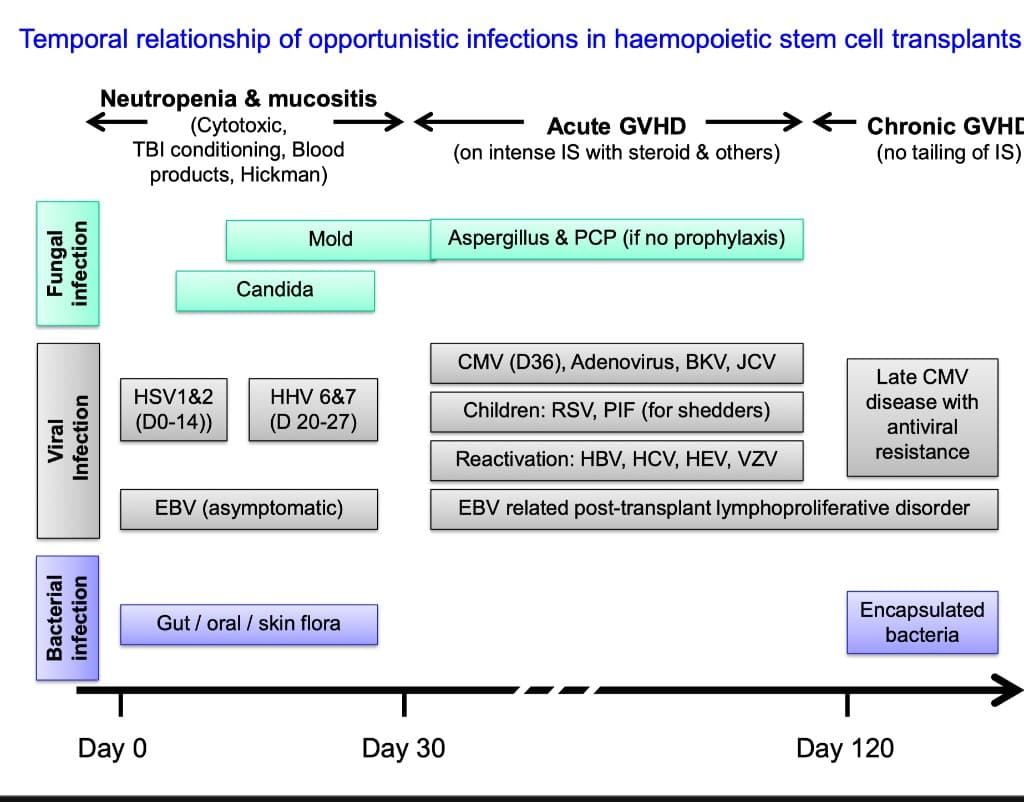
Lecture timeline of opportunistic infections after haematopoietic cell transplant (HCT) [1]:
How to use this timeline
Read Slide 51 as overlapping immune-defect bands, not a calendar that diagnoses the organism. Pre-engraftment neutropenia/mucositis favours bacterial and invasive fungal infection; acute or chronic GVHD and its treatment prolong cellular immune dysfunction; later functional hyposplenism favours encapsulated bacteria. Donor/recipient serostatus, graft type, conditioning, GVHD, immune recovery, prophylaxis and molecular surveillance can shift every boundary.
- Dominant defect: Neutropenia + mucosal barrier damage (from conditioning chemotherapy ± TBI)
- Risk factors: Cytotoxic conditioning, TBI, blood products, Hickman catheter
- Pathogens:
- Bacteria (gut/oral/skin flora): E. coli, Klebsiella, Pseudomonas, Staph, Strep
- HSV-1 & HSV-2 reactivation (Day 0–14) — from orolabial/genital latent infection
- HHV-6 & HHV-7 (Day 20–27)
- Candida → Moulds (Aspergillus) as duration increases
- Dominant defect: Cellular immune dysfunction from acute GVHD and its treatment (intense immunosuppression with steroids and other agents)
- Pathogens:
- CMV (labelled “D36” on this historical slide), adenovirus, BK virus, JC virus
- EBV-related PTLD (post-transplant lymphoproliferative disorder)
- Aspergillus + PCP (if prophylaxis not taken)
- Reactivation of HBV, HCV, HEV, VZV
- Children: RSV, parainfluenza (for shedders)
- Dominant defect: Chronic GVHD (if immunosuppression not tapered) → functional asplenia + ongoing cellular immune dysfunction
- Pathogens:
- Encapsulated bacteria (S. pneumoniae, H. influenzae) — especially if no revaccination
- Late CMV disease with antiviral resistance
- VZV reactivation
The CMV band is especially lecture-era. Modern CMV-seropositive allogeneic-HCT recipients may receive letermovir prophylaxis, which significantly reduces clinically significant CMV infection; PCR surveillance and pre-emptive treatment strategies also reshape timing. Therefore, “Day 36” is not a contemporary diagnostic rule [19,20].
High Yield – Past Paper: 2021 SAQ Q12
Part (a): Day 10 post-conditioning, ANC < 0.1 — name common blood culture organisms → GNR (E. coli, Klebsiella, Pseudomonas) + GPC (S. epidermidis, Viridans streptococci) [6]
Part (b): Day 28 post-engraftment, fever + impaired LFT + diarrhoea + CXR infiltrates, didn't take prophylaxis → CMV (hepatitis + colitis + pneumonitis), PCP (CXR infiltrates), Aspergillus (lung infiltrates) [6]
Part (c): 4 years post-transplant, fulminant sepsis with purpura, Howell-Jolly bodies → S. pneumoniae, H. influenzae, N. meningitidis (OPSI) [6]
2022 4th Summative MCQ Q19-21 [7]:
- Q19: HIV, CD4 60, disseminated skin papules, bilateral infiltrates, hepatosplenomegaly → Talaromyces marneffei (dimorphic fungus, endemic in SE Asia, CD4 < 100)
- Q20: AML, severe prolonged neutropenia, on meropenem, breakthrough fever + worsening oral mucositis, blood culture positive → Streptococcus mitis (Viridans streptococcus — breakthrough bacteraemia during meropenem, associated with severe mucositis)
- Q21: Post-liver transplant, on prednisolone + tacrolimus + MMF, low-grade fever + diarrhoea + abdominal pain, endoscopic biopsy shows inclusion bodies → CMV (CMV colitis with characteristic "owl's eye" inclusion bodies)
Exam Trap – S. mitis vs E. coli
When a neutropenic patient on broad-spectrum antibiotics develops breakthrough fever with severe oral mucositis, viridans streptococci—especially S. mitis—are an important clue because they colonise the oral cavity and can cross a damaged mucosal barrier [7]. Do not convert that exam association into a class-wide claim that carbapenems lack viridans activity: meropenem is an accepted febrile-neutropenia option, although individual isolates can be resistant and cultures still guide definitive therapy [12,28].
Special features of infection in immunocompromised hosts [1]:
| Feature | Explanation |
|---|---|
| 1. Spectrum predicted by specific defect | Know the defect → know the pathogen |
| 2. Unusual pathogens | (a) Environmental organisms or normal commensals become pathogenic; (b) Reactivation of latent organisms (old TB foci, herpesviruses) |
| 3. Rapid clinical deterioration | Without adequate immune response, infection spreads unchecked → fulminant sepsis |
| 4. Subtle clinical presentation | Minimal inflammation → no pus, no consolidation on CXR, no meningism despite meningitis → fever may be the ONLY sign |
| 5. Multiple organisms | Co-infection is common (e.g. bacterial + fungal, CMV + PCP) |
| 6. Virus-associated cancers | Prolonged immunosuppression → oncogenic virus-driven neoplasia: EBV-related PTLD, HHV-8 Kaposi's sarcoma, HPV-related anogenital cancers |
Why subtle presentation? Pus formation and many radiographic/CSF inflammatory findings depend on recruited neutrophils. Profound neutropenia can therefore blunt abscess formation, pulmonary consolidation or CSF pleocytosis—but does not make these findings impossible. Maintain a high index of suspicion despite minimal signs.
Slides 34–35 make the examination-and-sampling point visually: a dark, subtle perianal lesion can be clinically important in neutropenia, and tissue microscopy may reveal organisms despite little surface inflammation. Neither unlabelled image is organism-specific. Inspect hidden sites gently, avoid traumatic rectal instrumentation, and obtain imaging/tissue or aspirate when clinically safe rather than relying on visible pus.
Clinical Approach to Neutropenic Fever
Full Hx & P/E (daily): [1]
- Skin (including catheter exit sites, bone marrow biopsy sites)
- Oral cavity (mucositis, oral candidiasis, HSV ulcers)
- Lungs (subtle crackles, even normal examination doesn't exclude pneumonia)
- Abdomen (tenderness — may indicate typhlitis/neutropenic enterocolitis)
- Perianal area (abscess — DO NOT do digital rectal examination in neutropenic patients as this can cause bacteraemia)
- Hickman catheter exit site (erythema, discharge, tunnel tenderness)
- Bone marrow biopsy site
- Over nasal sinuses (sinusitis — especially Aspergillus)
Approach to investigation of neutropenic fever [1]:
| Investigation | Indication | What You're Looking For |
|---|---|---|
| CBP, LFT, RFT | All patients | Baseline; trends in counts; organ dysfunction |
| Blood cultures | Obtain adequate-volume sets before antibiotics without delaying treatment; with a central line, sample line lumen(s) and a peripheral vein when feasible | Identify organism; paired simultaneous samples permit differential time to positivity |
| CXR | Only when clinically indicated by respiratory findings | May show infiltrate but a normal film does not exclude infection |
| CT chest | Clinical suspicion of invasive pulmonary aspergillosis, even if CXR is normal | Nodules, consolidation, halo or other patterns; none is organism-specific |
| CT abdomen | Abdominal pain, tenderness, diarrhoea | Typhlitis (caecal wall thickening), hepatosplenic candidiasis |
| Stool for C. difficile | New clinically significant diarrhoea; test unformed stool according to the local algorithm | NAAT/GDH plus toxin testing as locally validated; culture/cytotoxicity is no longer the routine bedside wording |
| Urine, skin swab, sputum | If symptoms or clinical/radiological lesions present | Culture + sensitivity |
| BAL (bronchoalveolar lavage) | Pulmonary infiltrates not responding to empirical therapy | PCP, Aspergillus, CMV, other fungi |
| Serum/BAL galactomannan | Selected high-risk groups (haematological malignancy/HCT) or suspected invasive aspergillosis | Supports diagnosis in context; routine serum screening performs poorly during mould-active prophylaxis |
| 1,3 β-D-glucan | Selected high-risk patients | A nonspecific fungal marker; does not identify Aspergillus and is usually absent in Mucorales/cryptococcosis |
Why Blood Cultures From 2 Ports?
Sampling separate catheter lumens can improve detection, but differential time to positivity requires a simultaneously collected central and peripheral pair. If the central sample grows the same organism ≥2 hours before the peripheral sample, a catheter source is supported. Do not delay immediate empiric antibiotics in an unstable patient merely to complete cultures [13].
Current diagnostic guidance supports clinically indicated imaging rather than routine CXR, CT whenever invasive pulmonary aspergillosis is suspected, and context-dependent use of galactomannan/β-D-glucan [11,14]. For C. difficile, test an appropriate unformed stool sample and interpret NAAT with toxin/clinical context to avoid treating colonisation [15].
6-Step Management Framework (RECOGNITION to MONITORING)
The lecture presents a 6-step management framework; historical treatment examples are qualified below [1]:
| Step | Action | Details |
|---|---|---|
| 1. RECOGNITION | Identify the specific immune defect | Neutropenia? T-cell dysfunction? Humoral? Barrier? |
| 2. SUSPICION | Maintain high suspicion for infection from minimal symptoms | Low-grade fever, mental dullness may be the ONLY signs |
| 3. TAKING | Collect appropriate clinical specimens | Blood cultures, BAL, etc. for direct microscopy, antigen detection, culture |
| 4. COOPERATION | Collaborate with clinical microbiologist | Request unusual pathogens (PCP, fungi, Nocardia) — don't just request "routine culture" |
| 5. THERAPY | (a) Early aggressive empirical antimicrobial therapy BEFORE definitive culture results; (b) historical handout: “buffy coat”/granulocyte transfusion; (c) specialist option: ex-vivo expanded virus-specific T lymphocytes | Suspected neutropenic sepsis is a medical emergency; the latter interventions are not routine |
| 6. MONITORING | Monitor for efficacy and side effects | Drug assays (e.g. vancomycin levels, voriconazole levels) |
The handout specifically mentions intravenous imipenem [2]. That is an example, not a universal prescription. Give the local first-line antipseudomonal β-lactam immediately, adjusted for allergy, renal function, prior colonisation/infection and local resistance. NICE uses piperacillin–tazobactam monotherapy when no patient-specific or microbiological contraindication exists; other major guidelines include cefepime or a carbapenem for appropriate patients. Carbapenem exposure should be justified rather than automatic [11,12].
Why antipseudomonal coverage? Pseudomonas aeruginosa bacteraemia in a neutropenic patient carries extremely high mortality if not covered from the start. Even though GPC (S. epidermidis) are now the most commonly isolated organisms, Pseudomonas remains the most dangerous, so empirical regimens must cover it.
Do not add vancomycin routinely: a central line or mucositis alone does not automatically require empiric glycopeptide therapy. Add anti-MRSA/Gram-positive coverage for a specific indication such as haemodynamic instability, pneumonia, convincing catheter/skin-soft-tissue infection, a Gram-positive blood culture awaiting identification, or relevant resistant-organism history/local protocol; stop it if that indication is not confirmed [11,12].
When to consider antifungals: persistent fever after about 4–7 days matters mainly in patients expected to have prolonged high-risk neutropenia, or earlier when imaging/clinical findings suggest invasive fungal disease. Empiric mould-active therapy or a CT/biomarker-driven pre-emptive strategy may be used; routine empiric antifungal therapy is not recommended for an otherwise stable patient whose neutropenia is expected to last < 10 days [14].
Historical handout option: “buffy coat”/granulocyte transfusion for refractory infection with persistent neutropenia [2]
Adoptive transfer of ex-vivo expanded virus-specific T lymphocytes: for drug-resistant CMV, disseminated adenovirus, EBV-related PTLD [1][2]
Paradoxical deterioration: Immune Reconstitution Inflammatory Syndrome (IRIS) — can occur with recovery of CD4 count after antiretroviral therapy; or Myeloid Reconstitution Syndrome — can occur with ANC recovery after chemotherapy [2]
Modern granulocyte transfusion is a specialised rescue intervention, not routine “replacement.” The RING randomised trial did not show a significant overall benefit on its primary composite outcome, although inadequate granulocyte dosing limited interpretation [21]. Virus-specific T-cell products are likewise specialised and may be centre-specific or investigational.
Why immune-reconstitution inflammation? When cellular or myeloid immunity recovers, inflammation against a known or previously subclinical infection can paradoxically worsen. It is a diagnosis of exclusion: new infection, resistance, inadequate source control, drug toxicity and treatment failure still need active reassessment.
| Strategy | Details | Why |
|---|---|---|
| 1. Avoid unnecessary invasive procedures or antimicrobials | Don't disturb the ecological balance of microbial flora | Broad-spectrum antibiotics → C. difficile, resistant organisms, fungal overgrowth |
| 2. Assess and manage genuine infection foci before immunosuppression | Dental, skin, sinus, urinary or anorectal symptoms require assessment and source control where indicated | Reduces avoidable portals and untreated infection |
| 3a. Protective environment + food safety | HEPA-filtered positive-pressure rooms are used for selected allogeneic HCT recipients; use meticulous hand hygiene and safe food handling | Reduces environmental mould exposure and food-borne infection |
| 3b. Prophylactic antibiotics | Fluoroquinolone prophylaxis may be used for selected patients expected to have profound, protracted neutropenia | Benefit must be balanced against toxicity and local resistance |
| 3c. Prophylactic antivirals | Examples include acyclovir for HSV/VZV risk, letermovir for eligible CMV-seropositive allogeneic-HCT recipients, and entecavir or tenofovir for appropriate HBV-reactivation risk | Agent and duration depend on serostatus, transplant and immunosuppressive regimen |
| 3d. Prophylactic antifungals | Fluconazole is yeast-active; posaconazole/voriconazole are mould-active in selected settings; co-trimoxazole prevents PCP | Match prophylaxis to the predicted defect rather than prescribing every agent |
| 3e. Prophylactic antiparasitic | Septrin (Toxoplasma gondii) | Septrin does double duty: PCP + Toxoplasma |
| 3f. Active immunisation | Post-transplant revaccination schedule | Immune memory lost after HSCT; revaccination prevents late infections |
| 3g. Selected immune replacement | IVIG for defined antibody deficiency/recurrent serious infection—not every immunocompromised patient | Replaces deficient antibodies when there is a genuine indication |
| 3h. Planned acceleration of immune recovery | G-CSF when it is part of the cancer regimen or indicated by risk-based prophylaxis guidance | It is not routine adjunctive treatment for every episode of neutropenic sepsis |
| 3i. Adoptive T-cell transfer | Ex-vivo expanded virus-specific T lymphocytes in specialist transplant programmes | May provide virus-specific immunity in selected refractory/high-risk cases |
Current-practice corrections to the handout
The handout is valuable historical teaching, but several items are not universal modern practice. Do not screen for or treat asymptomatic bacteriuria automatically before chemotherapy: evidence in high-risk neutropenia is insufficient, so follow the oncology/infectious-diseases pathway [16]. A restrictive “neutropenic diet” has not shown superiority to a regular diet with rigorous food safety [18]. Antibacterial, antiviral and antifungal prophylaxis is reserved for defined risk groups [27]; letermovir has substantially changed CMV prevention after allogeneic HCT [19,20].
Reading the Environmental-Prevention Visuals (Slides 47–50)
The room photographs and airflow diagram depict a protective environment, not ordinary contact isolation. For allogeneic HCT recipients, the core engineering controls are HEPA-filtered supply air, positive pressure relative to the corridor, a well-sealed room and sufficient air exchanges. These measures reduce airborne fungal exposure; they do not replace hand hygiene, device care or transmission-based precautions when another infection requires them [17].
Septrin Does Triple Duty
Co-trimoxazole (Septrin) is used for prophylaxis against: (1) Pneumocystis jirovecii, (2) Toxoplasma gondii, and (3) has some activity against Nocardia. It is one of the most important prophylactic agents in immunocompromised patients [1].
Slides 37–39 are serial chest radiographs, not CT images. The Day 10 film has no convincing focal consolidation; by Day 13 an evolving focal opacity is apparent. This sequence teaches that neutropenia can blunt early radiographic inflammation and that a later abnormality may emerge. A chest radiograph cannot identify Aspergillus, and the deck does not show a CT halo sign. A halo sign may occur on CT in angioinvasive fungal disease, but it is not organism-specific [14].
Slides 40–42 then show fungal colonies, microscopy and a culture labelled Aspergillus fumigatus. Colony and conidiophore morphology support laboratory identification, but invasive pulmonary aspergillosis is diagnosed by integrating host risk, CT, serum/BAL galactomannan where appropriate, respiratory sampling/culture and sometimes histopathology. Current guidance recommends chest CT whenever invasive pulmonary aspergillosis is suspected, even if the radiograph is normal. If clinical suspicion is strong, begin mould-active treatment while completing the diagnostic evaluation; voriconazole is a common primary option, while isavuconazole or liposomal amphotericin B may be preferable according to interactions, prior prophylaxis, resistance and host factors [14].
Low CD4 lymphocyte count < 50/mm³ in HIV [1]
The slide uses CD4 count to illustrate increasing opportunistic-infection risk. A safer current reading is:
| Approximate risk zone | Examples and caveats |
|---|---|
| Any count | TB can occur throughout HIV disease; CD4 count modifies phenotype and dissemination risk rather than excluding TB |
| Usually < 200 cells/mm³ | PCP risk rises substantially; prophylaxis decisions also consider viral load, ART and prior disease [22] |
| Often < 100 cells/mm³ | Cerebral toxoplasmosis, cryptococcosis, talaromycosis in endemic settings and chronic protozoal enteritis become more likely [23,25] |
| Classically < 50 cells/mm³ without effective ART | CMV retinitis and disseminated MAC [24,26] |
| No single reliable threshold | PML may occur with advanced immunosuppression but should not be assigned to one rigid CD4 cut-off |
Monoclonal antibodies (MnAbs) against interconnecting cytokine/chemokine & receptors [1]
Slides 31–32 map drug targets across antigen-presenting cells, T cells and B cells, then show how many cytokines converge on JAK–STAT signalling. The visual message is that infection risk follows the specific blocked immune function; inhibiting a shared signalling node can affect several defence pathways at once. Risk is agent-, dose-, combination- and patient-specific rather than identical across an entire class.
- Anti-TNF agents are strongly associated with reactivation/dissemination of TB and other serious opportunistic infections [29].
- IL-6 blockade can suppress CRP and other inflammatory signals, so infection may appear deceptively quiet [30].
- Anti-CD20 therapy produces B-cell depletion and can cause hypogammaglobulinaemia; HBV reactivation is a key preventable risk, while PML is rare [31].
- JAK inhibitors affect multiple cytokine signals and are associated with herpes zoster and other serious/opportunistic infections [32].
Before treatment, use the specific product label and specialty protocol for infection screening, vaccination and prophylaxis; do not extrapolate one agent's warning to every biologic.
| Related Lecture | Connection |
|---|---|
| GC 048 (Fever) | General fever definitions and mechanisms; this lecture applies them to the immunocompromised setting |
| GC 060 (High WCC) | AML diagnosis, chemotherapy complications, neutropenic fever as a haematological emergency |
| GC 096 (Why do I always get sick) | Primary immunodeficiencies — congenital counterpart to acquired immunodeficiency |
| GC 100 (Defence against microbes) | Innate and adaptive immunity basics that underpin understanding of each immune defect |
| GC 101 (Diagnosis of infections) | How to investigate infections (cultures, serology, molecular); applies directly to specimens in febrile neutropenia |
| GC 106 (Practical issues in antibiotic use) | Empirical vs targeted therapy, de-escalation, drug levels |
| GC 144 (Recurrent infections in children) | Primary immunodeficiencies — overlap with phagocytic defects (CGD), complement deficiency |
High Yield Summary
- Treat suspected neutropenic sepsis as an emergency. Obtain cultures promptly and start an appropriate antipseudomonal β-lactam without waiting for results; use the local fever/ANC pathway because definitions vary [11,12].
- Depth and duration of neutropenia drive risk. Mucositis and catheters add portals of entry, while steroids may blunt fever and local inflammation.
- Defect predicts pathogen: neutropenia/barrier loss → bacterial and invasive fungal infection; T-cell dysfunction → intracellular/opportunistic organisms; antibody loss or hyposplenism → encapsulated bacteria.
- Timelines are risk maps, not clocks. HCT phase, GVHD, prophylaxis, immune recovery and prior colonisation shift the expected organisms.
- Investigate by syndrome. Pair central and peripheral cultures when feasible, use CT for suspected invasive pulmonary aspergillosis even with a normal radiograph, and interpret fungal biomarkers in context [13–15].
- Prevention is individualised. Use hand hygiene, device care, safe food handling and risk-specific prophylaxis/vaccination; do not apply every handout intervention to every patient [16–20,27].
Active Recall - Fever After Chemotherapy & Infections in Immunocompromised Hosts
[1] Lecture slides: GC 102. Fever after chemotherapy infections in immunocompromised hosts.pdf [2] Lecture slides: GC 102. Fever after chemotherapy infections in immunocompromised hosts [Handout].pdf [3] Senior notes: MBBS Final MB (Medicine) (Felix PY Lai).pdf (p1329-1332) [4] Past papers: 2024 Fourth Summative MCQ.pdf (Q84) [5] Senior notes: Block A - HIV positive_ HIV related diseases, accidental needle prick injury.pdf [6] Past papers: 2021 Fourth Summative SAQ.pdf (Q12) [7] Past papers: 2022 Fourth Summative MCQ.pdf (Q19-21) [8] Senior notes: Adrian Lui Pediatrics Notes.pdf (p423) [9] Senior notes: Ryan Ho Haemtology.pdf (p70) [11] NICE. Neutropenic sepsis: prevention and management in people with cancer (CG151)—recommendations. [12] Taplitz RA, et al. Outpatient management of fever and neutropenia in adults treated for malignancy: ASCO and IDSA clinical practice guideline update. J Clin Oncol. 2018. [13] SCCM/IDSA. Guidelines for evaluating new fever in adult patients in the ICU. [14] IDSA. Clinical practice guideline for the diagnosis and management of aspergillosis. [15] CDC. C. diff: facts for clinicians—diagnosis and testing. [16] IDSA. Clinical practice guideline for the management of asymptomatic bacteriuria. [17] CDC. Protective environment recommendations for allogeneic haematopoietic stem-cell transplant recipients. [18] National Cancer Institute. Nutrition in Cancer Care (PDQ)—neutropenic diets and food safety. [19] Marty FM, et al. Letermovir prophylaxis for cytomegalovirus in haematopoietic-cell transplantation. N Engl J Med. 2017. [20] FDA. PREVYMIS (letermovir) prescribing information. [21] Price TH, et al. RING: efficacy of transfusion with granulocytes from G-CSF/dexamethasone-treated donors in neutropenic patients with infection. Blood. 2015. [22] NIH. Pneumocystis pneumonia: adult and adolescent opportunistic-infection guideline. [23] NIH. Toxoplasma gondii encephalitis: adult and adolescent opportunistic-infection guideline. [24] NIH. Cytomegalovirus disease: adult and adolescent opportunistic-infection guideline. [25] NIH. Talaromycosis: adult and adolescent opportunistic-infection guideline. [26] NIH. Disseminated Mycobacterium avium complex: adult and adolescent opportunistic-infection guideline. [27] Taplitz RA, et al. Antimicrobial prophylaxis for adult patients with cancer-related immunosuppression: ASCO and IDSA guideline update. J Clin Oncol. 2018. [28] Chua CC, et al. Risk factors for viridans-group streptococcal bacteraemia in neutropenic and non-neutropenic patients: a single-centre case-case-control study. Open Forum Infect Dis. 2017. [29] FDA. HUMIRA (adalimumab) prescribing information. [30] FDA. ACTEMRA (tocilizumab) prescribing information. [31] FDA. RITUXAN (rituximab) prescribing information. [32] FDA. XELJANZ/XELJANZ XR (tofacitinib) prescribing information.
GC101 Diagnosis Of Infections
Diagnosis of infections is the systematic process of identifying causative pathogens through clinical assessment, microbiological techniques (culture, microscopy, serology, molecular methods), and laboratory markers to guide appropriate antimicrobial therapy.
GC103 Fever After Travelling
Fever occurring in a patient who has recently traveled, particularly to tropical or endemic regions, requiring systematic evaluation for infections such as malaria, dengue, typhoid, and other travel-related diseases.