Upper Gi Bleed
Upper gastrointestinal bleeding is hemorrhage originating from a source proximal to the ligament of Treitz, commonly caused by peptic ulcers, esophageal varices, or Mallory-Weiss tears.
Upper GI Bleed (UGIB)
Upper gastrointestinal bleeding (UGIB) refers to haemorrhage originating from the gastrointestinal tract proximal to the ligament of Treitz (the duodenojejunal flexure) [1][2][3].
- The ligament of Treitz ("Treitz" = a Czech pathologist who described it) is the suspensory ligament of the duodenum — a fibromuscular band connecting the duodenojejunal junction to the right crus of the diaphragm. It serves as the anatomical landmark dividing upper from lower GI bleeding.
- Therefore, UGIB encompasses bleeding from the oesophagus, stomach, and duodenum (up to the 4th part/DJ flexure).
Key terminology to define upfront:
| Term | Definition | Mechanism / Clinical Implication |
|---|---|---|
| Haematemesis | Vomiting of blood (frank red or coffee-ground) | Source must be proximal to ligament of Treitz. Frank blood = brisk/active bleeding; coffee-ground = blood exposed to gastric acid (acid converts haemoglobin → haematin, which is brown/black) |
| Melena | Black, tarry, offensive-smelling stool | Blood degraded by gut bacteria + digestive enzymes during transit. Requires only ~50–100 mL of blood in the upper GIT. Anatomical extent: nasopharynx to proximal colon |
| Haematochezia | Passage of fresh red blood per rectum | Usually lower GI in origin, but can occur with massive UGIB (rapid transit > 1000 mL overwhelms the degradation capacity) |
| Coffee-ground vomitus | Dark, granular emesis | Small-volume / slow-rate bleed that has been partially digested by gastric acid |
| Occult GI bleeding | Bleeding not visible to the patient | Detected only by faecal occult blood test (FOBT) or iron-deficiency anaemia |
Don't Confuse
Melena can also be mimicked by iron supplementation and bismuth (bismuth + sulfur in the gut → bismuth sulfide = black). Always ask about drug history. However, these do NOT produce the characteristic tarry consistency and offensive smell of true melena [2].
GI bleeding of obscure origin [3][4]:
- Defined as bleeding of unknown origin persisting or recurring after negative upper and lower endoscopy ("top and tail")
- Obscure overt: visible bleeding symptoms (haematemesis, melena, haematochezia) but source unidentified
- Obscure occult: iron-deficiency anaemia + FOBT positive, but no visible bleeding and source unidentified
- Most eventually found in the small bowel (between ligament of Treitz and ileocaecal valve)
2. Epidemiology
- UGIB is a common medical emergency with an annual incidence of approximately 50–150 per 100,000 population in Western countries
- In Hong Kong, UGIB remains a significant cause of hospital admissions, particularly among the elderly population given the high prevalence of H. pylori infection and widespread NSAID/aspirin use in an ageing population
- Overall in-hospital mortality: ~5–10% (higher in variceal bleeding: 15–25%)
- Mortality has decreased over the past decades due to advances in endoscopic haemostasis, PPI therapy, and ICU care
- Leading cause of death of peptic ulcer is haemorrhage [5]
- H. pylori prevalence: ~50% in the Hong Kong adult population (declining in younger cohorts due to improved sanitation, but still significant)
- High use of aspirin/antiplatelets in the ageing population for cardiovascular prophylaxis
- Liver disease and hepatitis B virus (HBV) — Hong Kong has a historically high HBV prevalence (~8% carrier rate), leading to cirrhosis and portal hypertension/variceal bleeding
- Alcohol-related liver disease is increasing
- NSAID availability (including over-the-counter) contributes to peptic ulcer bleeding
3. Risk Factors
These can be divided into risk factors for developing UGIB and risk factors for poor outcome/recurrent bleeding once UGIB has occurred.
| Category | Risk Factors | Mechanism |
|---|---|---|
| Drugs | NSAIDs (including aspirin), anticoagulants, antiplatelets, corticosteroids | NSAIDs inhibit COX-1 → ↓ prostaglandins → loss of mucosal protection. Anticoagulants impair clot formation at bleeding sites. Steroids augment NSAID ulcerogenicity |
| H. pylori infection | Gram-negative microaerophilic bacterium | Colonises gastric epithelium → chronic inflammation → disrupts mucous layer → mucosal injury |
| Stress | ICU patients, burns (Curling's ulcer), head injury (Cushing's ulcer), major surgery | Mucosal hypoperfusion → ischaemia → stress ulceration |
| Excess gastric acid | Zollinger-Ellison syndrome (gastrinoma) | Hypergastrinaemia → massive acid hypersecretion → ulceration |
| Liver disease | Portal hypertension → varices, coagulopathy | ↑ Portal pressure → portosystemic collaterals (varices). Also ↓ synthetic function → ↓ clotting factors |
| Alcohol | Chronic alcoholism | Direct mucosal irritant + liver disease + Mallory-Weiss tears from retching |
| Smoking | Impaired mucosal healing, ↑ acid secretion | |
| Previous GI bleed | Prior ulcer or variceal bleed | History of bleeding is the strongest predictor of future bleeding |
| Age | Elderly ( > 60 years) | Reduced mucosal defence, polypharmacy, more comorbidities |
Risk factors for recurrent bleeding:
- Shock on presentation
- Hb < 8.0 g/dL on presentation
- Transfusion requirement
- Age > 60 yrs
- Comorbidity
- Coagulopathy
- Patient already hospitalised (i.e. in-patient bleed — worse prognosis than community-onset)
- Large ulcer
- Ulcer on posterior D1 (because of proximity to gastroduodenal artery)
- Ulcer on higher posterior lesser curve (because of proximity to left gastric artery)
Why Posterior D1 Ulcers Are Dangerous
The gastroduodenal artery (GDA) runs directly behind the first part of the duodenum. A posterior D1 ulcer can erode into the GDA, causing catastrophic arterial haemorrhage that is often difficult to control endoscopically. Similarly, ulcers on the posterior lesser curvature can erode into the left gastric artery. These are the two most feared locations for bleeding peptic ulcers [1][5][6].
4. Anatomy and Function
Understanding the anatomy is essential for knowing why certain lesions bleed, where they bleed from, and what arteries are at risk.
The upper GI tract for the purposes of UGIB extends from the oesophagus → stomach → duodenum (to the ligament of Treitz at the DJ flexure).
- ~25 cm muscular tube from pharynx (C6) to stomach (T11)
- No serosa — this makes the oesophagus susceptible to perforation (Boerhaave syndrome) and means that oesophageal cancers spread early [3]
- The gastro-oesophageal junction (GOJ/GEJ) is the site of:
- Oesophageal varices (submucosal veins draining into both portal and systemic systems)
- Mallory-Weiss tears (mucosal lacerations at the GEJ)
- Blood supply: inferior thyroid arteries (upper), bronchial arteries (mid), left gastric artery and inferior phrenic artery (lower)
- Venous drainage: The lower oesophageal veins drain into the left gastric (coronary) vein (portal system) — this is precisely why oesophageal varices form here in portal hypertension
Arterial supply [2]:
- Greater curvature:
- Short gastric arteries (from splenic artery)
- Left gastro-omental (gastroepiploic) artery (from splenic artery)
- Right gastro-omental (gastroepiploic) artery (from gastroduodenal artery)
- Lesser curvature:
- Left gastric artery (from coeliac trunk — the largest artery supplying the stomach)
- Right gastric artery (from common hepatic artery → proper hepatic artery)
Nerve supply [2]:
- Sympathetic: Greater splanchnic nerve (T5–T9)
- Parasympathetic (vagus nerve):
- Anterior vagal nerve: Stomach + Pylorus + Liver
- Posterior vagal nerve: Stomach + Foregut and midgut down to splenic flexure
Vagotomy in PUD Surgery
Understanding vagal anatomy explains surgical options for PUD. Truncal vagotomy divides both vagal trunks → reduces acid secretion but also denervates the pylorus (causing gastroparesis), so a drainage procedure (pyloroplasty or gastrojejunostomy) is always added. Highly selective vagotomy preserves the nerve of Latarjet (which innervates the antrum/pylorus for gastric motility) and only denervates parietal cell–containing areas — no drainage needed, but technically difficult [5].
Gastric mucosal defence — the balance between aggressive and protective factors:
| Aggressive Factors | Protective Factors |
|---|---|
| Gastric acid (HCl) | Mucus-bicarbonate barrier |
| Pepsin | Mucosal blood flow |
| H. pylori | Prostaglandins (PGE₂, PGI₂) |
| NSAIDs | Epithelial cell renewal |
| Bile reflux | Tight junctions |
| Alcohol, smoking | Trefoil peptides |
The mucosa is a dynamic battleground. Peptic ulceration occurs when aggressive factors overwhelm protective factors.
- Four parts (D1–D4); the ligament of Treitz marks the junction of D4 and jejunum
- D1 (duodenal bulb/cap) is the most common site for duodenal ulcers [2]
- The gastroduodenal artery (GDA) — a branch of the common hepatic artery — runs posterior to D1. This is why posterior D1 ulcers cause severe arterial haemorrhage
- The ampulla of Vater opens into D2 — bleeding from the ampulla (haemobilia) can present as UGIB
5. Etiology (with focus on Hong Kong)
The causes of UGIB can be remembered by anatomical location (oesophagus → stomach → duodenum) or by frequency.
5.1 Common Causes — In Descending Order of Frequency
Per lecture slides [1]:
Causes of upper GI bleeding in descending order of frequency:
- Duodenal or gastric ulcer
- Gastritis
- Esophageal or gastric varices
- Mallory-Weiss syndrome
- Benign or malignant gastric tumour
Definition: A defect in the gastrointestinal mucosa that extends through the muscularis mucosae (distinguishing an ulcer from an erosion, which is superficial) [2].
Locations [2]:
- Duodenum (75%): D1 (duodenal bulb) is the most common site
- Unusual for non-NSAID ulcers to occur outside D1 — if they do, suspect Zollinger-Ellison syndrome or Crohn's disease
- Stomach (20%): Lesser curvature and antrum most common
- Oesophagus, Meckel's diverticulum, stomal ulcers (rare)
Four major risk factors [2][3]:
- H. pylori infection
- NSAIDs
- Stress
- Excess gastric acid
Pathophysiology of each risk factor:
a) H. pylori [2]:
- Microaerophilic Gram-negative coccobacillus (micro = small, aerophilic = oxygen-loving, but only in small amounts)
- Strong urease activity → hydrolyses urea → ammonia → neutralises gastric acid around the bacterium, creating a protective alkaline cloud that allows survival
- Spiral shape + flagella + mucolytic enzymes → enable the organism to burrow through the mucus layer to reach the gastric surface epithelium
- Once established, it causes chronic active gastritis via:
- Direct epithelial injury (cytotoxins: CagA, VacA)
- Inflammatory cascade (IL-8 recruitment of neutrophils)
- Disruption of the mucous barrier → mucosa exposed to acid → ulceration
The pattern of gastritis determines the clinical outcome [2]:
- Antral-predominant gastritis → ↑ gastrin release from G cells → ↑ acid secretion → duodenal ulcer (does NOT predispose to gastric cancer)
- Pangastritis (antrum + body) → eventual atrophy of parietal cells → ↓ acid secretion (hypochlorhydria) → compensatory ↑ gastrin → gastric ulcer + ↑ risk of gastric cancer
b) NSAIDs (including aspirin) [2]:
- Gastric and duodenal mucosa use COX-1 for prostaglandin synthesis
- Prostaglandins (PGE₂) protect the mucosa by:
- Stimulating mucin production
- Stimulating bicarbonate secretion
- Maintaining mucosal blood flow
- Inhibiting gastric acid secretion
- NSAIDs non-selectively inhibit COX-1 and COX-2 → ↓ prostaglandins → loss of mucosal protection → mucosal barrier breakdown → back-diffusion of H⁺ ions → injury
- COX-2 selective inhibitors (celecoxib) preserve COX-1 and therefore have less GI toxicity — but NOT zero risk
Risk factors for NSAID-related ulcers [2]:
- Advanced age ( > 75 years)
- Prior ulcer history or complications
- High dose / long duration / relatively toxic NSAID
- Concurrent corticosteroids or anticoagulants
c) Stress ulcers [2]:
- Occur in critically ill ICU patients — burns (Curling's ulcer), head injury (Cushing's ulcer), sepsis, multi-organ failure
- Mechanism: splanchnic hypoperfusion → mucosal ischaemia → intramural acidosis → epithelial cell death → ulceration
- Cushing's ulcers are unique: head injury → ↑ vagal tone → ↑ acid secretion AND mucosal ischaemia
d) Excess gastric acid — Zollinger-Ellison syndrome [2]:
- Gastrinoma (usually in the gastrinoma triangle — duodenum/pancreas) secretes gastrin autonomously
- ↑↑ Gastric acid → overwhelms mucosal defence → ulceration in unusual locations (2nd part duodenum, jejunum)
- Suspect when: recurrent ulcers despite adequate treatment, unusual ulcer locations, no NSAID/H. pylori, diarrhoea (acid inactivates pancreatic enzymes)
- Diagnosis: ↑ fasting serum gastrin + high gastric acid output
Modified Johnson Classification of Gastric Ulcers [2][5]:
| Type | Location | Acid Secretion |
|---|---|---|
| Type I (most common, 58%) | Along lesser curvature near angular incisure | Normal / ↓ |
| Type II (22%) | Body of stomach (GU) + Duodenum (DU) simultaneously | ↑↑ |
| Type III (20%) | Prepyloric | ↑↑ |
| Type IV (rare) | High on lesser curvature near OGJ | ↓↓ |
| Type V | Any location — medication-induced | Normal |
Clinical Pearl — Johnson Types II and III
Types II and III are associated with acid hypersecretion — similar pathophysiology to duodenal ulcers. Types I and IV are associated with normal or low acid secretion — the problem is impaired mucosal defence, and there is a higher malignant potential. Type V is drug-induced and can occur anywhere [2].
- Inflammation-associated mucosal injury that is typically self-limited [2]
- Causes: NSAIDs/aspirin, alcohol, stress
- Bleeding is usually from superficial erosions (does not extend through muscularis mucosae)
- Diagnosis is made exclusively by endoscopic evaluation of the mucosa
- Gastritis is the second most common cause of UGIB [1]
- A complication of portal hypertension, most commonly due to liver cirrhosis [2][3]
- In Hong Kong, HBV-related cirrhosis is historically the most important cause, though alcohol-related and NAFLD-related cirrhosis are increasing
Pathophysiology [2]:
- Portal hypertension → opening and dilatation of pre-existing portosystemic collateral channels → varices
- Varices decompress the portal circulation by shunting blood back to the systemic venous circulation
- Most common location: distal oesophagus and proximal stomach (around the GEJ)
- Formed when the left gastric (coronary) vein (portal system) collateralises with oesophageal veins (systemic/azygos system)
- Hepatic venous pressure gradient (HVPG) = portal vein pressure – IVC pressure
- Normal: 1–5 mmHg
- Varices develop at HVPG ≥ 10 mmHg
- Bleeding risk increases at HVPG ≥ 12 mmHg
- Unlikely to bleed if HVPG < 12 mmHg [2]
- Laplace's Law: Wall tension (T) ∝ Pressure (P) × Radius (r) / Wall thickness (w)
- Larger varices have greater radius → greater wall tension → higher risk of rupture → larger the varices, the more likely they will bleed [2]
Endoscopic Grading [2]:
- F1: Small straight varices — fully compress with air insufflation
- F2: Enlarged tortuous varices occupying < 1/3 of lumen — fail to compress with air
- F3: Large coil-shaped varices occupying > 1/3 of lumen — fail to compress with air
- Red signs (indicate ↑ bleeding risk):
- Red wale marks = longitudinal red streaks on varices (resemble red corduroy wales)
- Cherry red spots = discrete red cherry-coloured spots overlying varices
Clinical presentation [2]:
- Haematemesis (often large volume, bright red)
- Melena
- Shock
- ± Coma (hepatic encephalopathy — ammonia from blood in the gut + impaired hepatic clearance)
Definition: Longitudinal mucosal (superficial) tears of distal oesophagus at the gastro-oesophageal junction [2][3].
Pathogenesis:
- Forceful retching/vomiting → sudden ↑ intra-abdominal pressure → mechanical tearing of the mucosa at the GEJ
- The lacerations involve the mucosa and submucosa but NOT the full thickness (that would be Boerhaave syndrome)
- Tears expose submucosal arteries → bleeding
Risk factors [3]:
- Chronic alcoholism (most common association — alcohol causes nausea/retching)
- Bulimia
- Severe retching from any cause (chemotherapy, gastroparesis)
- Sudden increase in intra-abdominal pressure (straining, coughing, seizures)
Key distinction from Boerhaave syndrome [3]:
| Feature | Mallory-Weiss | Boerhaave |
|---|---|---|
| Depth | Mucosal/submucosal tear | Full-thickness perforation |
| Location | GEJ | Left posterolateral distal oesophagus |
| Presentation | Haematemesis | Chest pain, surgical emphysema, sepsis |
| Prognosis | Usually self-limited (80–90%) | Surgical emergency, high mortality |
- Benign or malignant gastric tumour [1]
- Oesophageal, gastric, or duodenal cancers are uncommon causes of UGIB
- Usually presents with constitutional symptoms (weight loss, anorexia, early satiety) + chronic occult bleeding → iron-deficiency anaemia rather than massive haematemesis
- In Hong Kong, gastric cancer remains relevant — risk factors include H. pylori, smoking, salt-preserved foods, family history
- Gastrointestinal stromal tumour (GIST) can also bleed
5.2 Less Common Causes
Per lecture slides [1]:
Less common causes of UGIB:
- Esophagitis, esophageal tumour
- Stomal ulcer
- Aortoduodenal fistula
- Haemobilia, haemosuccus pancreaticus
- Vascular malformation, angiodysplasia
- Dieulafoy's lesion
- Duodenal or jejunal diverticulum, jejunal ulcer
- Usually in context of GERD (gastro-oesophageal reflux disease) — acid reflux → mucosal injury
- Other causes: infections (HSV, CMV, Candida — especially in immunocompromised), medications (tetracycline, bisphosphonates — pill oesophagitis), irradiation
- Risk factors: obesity, GERD, immunosuppression
- Portal hypertension → mucosal vascular ectasia in the stomach
- Mucosa is friable → bleeds when ectatic vessels rupture
- More commonly presents as chronic occult bleeding with anaemia rather than acute haemorrhage
- Endoscopic appearance: mosaic-like (snakeskin) pattern of mucosa
- Dilated aberrant submucosal vessel that erodes the overlying epithelium in the absence of a primary ulcer
- Usually located in the proximal stomach along the lesser curvature or near the GEJ (some sources say gastric fundus is MC site) [2][5]
- Can cause massive arterial bleeding from a very small mucosal defect (< 3 mm)
- Diagnosis is endoscopic — a visible vessel protruding through normal-appearing mucosa
- Management: endoscopic therapy (sclerosant injection, endoscopic clips, thermocoagulation)
- Known as "watermelon stomach" because endoscopic appearance shows longitudinal rows of flat, reddish stripes radiating from the pylorus into the antrum
- Distinct from portal hypertensive gastropathy (GAVE occurs in the antrum specifically, PHG is more diffuse/fundal)
- Often responds to argon plasma coagulation (APC)
- Medical emergency with high mortality rate
- Direct communication between the aorta and the GI tract
- Almost always secondary to placement of a prosthetic abdominal aortic vascular graft — should be suspected in any patient with prior AAA repair presenting with GI bleeding
- 3rd or 4th portion of the duodenum is the most common site (because the graft lies directly adjacent)
- Classic presentation: "herald bleed" (initial small bleed) → massive exsanguination
- Diagnosis: CT aortogram [3]
- Bleeding from the hepatobiliary tract (haemo = blood, bilia = bile)
- Should be considered in patients with history of hepatic or biliary instrumentation (percutaneous liver biopsy, transjugular liver biopsy, ERCP, hepatic trauma)
- Classic triad (Quincke's triad): upper GI bleeding + biliary colic + jaundice
- Bleeding from the pancreatic duct (haemo = blood, succus = juice, pancreaticus = pancreas)
- Most often in patients with chronic pancreatitis, pancreatic pseudocysts, or pancreatic tumours
- Mechanism: pseudocyst or inflammation erodes into a peripancreatic artery (often splenic artery) → blood drains via pancreatic duct → ampulla of Vater → duodenum
- Dilated tortuous submucosal vessels (arteriovenous malformations)
- In the upper GI tract, often originates from stomach and duodenum [2]
- More common in elderly, associated with aortic stenosis (Heyde syndrome — acquired type 2A von Willebrand disease) and ESRD [4]
- Diagnosis: endoscopy shows cherry red spots; angiography shows "mother-in-law phenomenon" (early filling, delayed emptying) [4]
- Management: argon plasma coagulation (APC), endoscopic electrocoagulation, or angiographic embolisation [4]
- Erosions or ulcers occurring in the sac of a hiatal hernia
- Mechanism: mechanical trauma of the gastric mucosa at the diaphragmatic hiatus during respiration/sliding
- Occurs at surgical anastomotic sites (e.g., after Billroth I/II, Roux-en-Y)
- Usually due to acid exposure of susceptible jejunal mucosa
Per lecture slide [4], causes include:
Vascular diseases: Angiodysplasia, Angiomas, Dieulafoy lesion, Watermelon stomach, Varices, Haemosuccus pancreaticus, Haemobilia
Ulcerative diseases: Peptic ulcer, Reflux disease, Cameron ulcers, Crohn's disease
Neoplasms: Polyps, Lipoma, Lymphoma, Carcinoid, GIST, Primary small bowel carcinoma, Metastatic cancer, Melanoma
Genetic disorders: Osler-Weber-Rendu syndrome, Blue rubber bleb naevus syndrome, Gardner's syndrome, Hermansky-Pudlak syndrome, Klippel-Trenaunay-Weber syndrome, Neurofibromatosis type I and II, Ehlers-Danlos syndrome
Others: Medications/NSAID, CMV infection, Tuberculosis infection, Meckel's diverticulum, Diverticulosis
6. Classification
UGIB can be classified in multiple ways:
- Variceal vs. Non-variceal — this is the most clinically important distinction because management differs fundamentally
| Feature | Variceal UGIB | Non-Variceal UGIB |
|---|---|---|
| Cause | Portal hypertension (cirrhosis) | PUD, gastritis, Mallory-Weiss, etc. |
| Typical patient | Chronic liver disease, stigmata of cirrhosis | NSAID use, H. pylori, elderly |
| Severity | Often massive, high mortality | Variable — can be massive (GDA erosion) |
| Initial pharmacotherapy | Terlipressin (splanchnic vasoconstrictor) + IV antibiotics | IV PPI (esomeprazole 80 mg bolus → 8 mg/h) |
| Endoscopic Rx | Band ligation (oesophageal) / Histoacryl glue (gastric) | Adrenaline injection + thermal/clips |
| Rescue | Sengstaken-Blakemore tube / TIPSS | Surgery / IR embolisation |
- Mild: stable haemodynamics, Hb > 10, no ongoing bleeding
- Moderate: tachycardia, Hb 7–10, some postural symptoms
- Severe/Massive: shock (tachycardia, hypotension), Hb < 7, ongoing haematemesis/haematochezia, requiring urgent transfusion and intervention
This is the standard classification used during OGD to describe the ulcer base and predict rebleeding risk:
| Forrest Class | Description | Rebleeding Risk (%) | Endoscopic Therapy? |
|---|---|---|---|
| Ia | Spurting arterial haemorrhage | 90% | Yes |
| Ib | Oozing haemorrhage | 50% | Yes |
| IIa | Non-bleeding visible vessel | 50% | Yes |
| IIb | Adherent clot | 30% | Consider (irrigate clot) |
| IIc | Flat pigmented spot | 7% | No |
| III | Clean-base ulcer | 3% | No |
High Yield — Forrest Classification
Forrest Ia, Ib, and IIa are considered high-risk stigmata and require endoscopic haemostasis. Forrest IIb (adherent clot) is controversial — current practice is to attempt to dislodge the clot to assess the underlying ulcer base. Forrest IIc and III are low-risk and do NOT need endoscopic therapy — just PPI and observation.
7. Clinical Features
7.1 Symptoms
The symptoms of UGIB reflect the volume and rate of blood loss, the underlying cause, and the patient's baseline physiology.
| Symptom | Pathophysiological Basis |
|---|---|
| Haematemesis (frank blood) | Active, moderate-to-severe bleeding proximal to ligament of Treitz. Blood has not been in contact with gastric acid long enough to be degraded |
| Coffee-ground vomitus | Slower or more limited bleeding. Blood in the stomach is exposed to HCl → haemoglobin oxidised to haematin (brown/black pigment) → granular appearance |
| Melena (black tarry stool) | Blood (≥50 mL) transits through the GI tract → bacterial degradation + enzymatic digestion → produces haematin and other pigments → characteristic black colour and offensive smell. Loose consistency because blood in the GI tract causes intestinal hurry (is an osmotic and irritant stimulus) [2] |
| Haematochezia (fresh red blood PR) | Usually lower GI, but can occur with massive UGIB — rapid transit ( > 1000 mL) means blood passes through so quickly that there is insufficient time for degradation |
These reflect the volume and rate of blood loss:
| Symptom | Mechanism |
|---|---|
| Dizziness / lightheadedness | ↓ Cerebral perfusion due to hypovolaemia |
| Syncope / pre-syncope | Severe ↓ cerebral blood flow. Postural hypotension initially, then frank syncope |
| Palpitations | Compensatory ↑ heart rate (sympathetic activation) to maintain cardiac output despite ↓ preload |
| Thirst | Hypovolaemia → ADH release + angiotensin II → thirst stimulus |
| Dyspnoea / exertional breathlessness | ↓ Haemoglobin → ↓ oxygen-carrying capacity; also ↑ cardiac output with sympathetic drive |
| Confusion / altered consciousness | Severe ↓ cerebral perfusion → global cerebral hypoxia |
| Oliguria | ↓ Renal perfusion → ↓ GFR → ↓ urine output |
Severity assessment [3]: syncope, postural hypotension, palpitation indicate significant volume loss.
Why Anaemic Symptoms Are UNCOMMON in Acute UGIB
In acute haemorrhage, both plasma and red cells are lost simultaneously — the haematocrit/haemoglobin may be normal initially because the blood is "diluted" proportionally. Classic anaemic symptoms (fatigue, pallor) develop later as the body auto-resuscitates (shifts interstitial fluid into the vascular compartment) and the remaining blood becomes diluted. This is why initial Hb is an unreliable marker of severity in acute UGIB — it may take 24–72 hours to equilibrate [3].
| Symptom | Suggests | Mechanism |
|---|---|---|
| Preceding forceful vomiting then haematemesis | Mallory-Weiss syndrome | Retching → ↑ intra-abdominal pressure → mucosal tear at GEJ [3] |
| Epigastric pain associated with meals | PUD | Gastric ulcer: pain ↑ with eating (food → acid). Duodenal ulcer: pain 2–5h after meals / at night (relieved by food/antacids) [3] |
| Jaundice, easy bruising, distended abdomen, peripheral oedema | Liver failure / cirrhosis (variceal bleed) | Jaundice = ↓ bilirubin conjugation. Bruising = ↓ clotting factors. Ascites = portal hypertension + hypoalbuminaemia. Oedema = hypoalbuminaemia [3] |
| Constitutional symptoms (weight loss, anorexia, night sweats) | Malignancy | Tumour metabolism, cytokine release (TNF-α, IL-6) |
| Dysphagia | CA oesophagus | Progressive luminal obstruction by tumour [3] |
| Persistent epigastric pain | CA stomach | Transmural invasion, peritoneal irritation [3] |
| Heartburn / acid brash | Oesophagitis / GERD | Acid reflux → mucosal inflammation |
| Epigastric pain radiating to back | Pancreatic pathology (haemosuccus pancreaticus) | Retroperitoneal inflammation/erosion into splenic artery |
| Recent liver biopsy / ERCP | Haemobilia | Iatrogenic vascular injury in hepatobiliary tract |
| History of AAA repair | Aorto-enteric fistula | Graft erodes into duodenum (usually D3/D4) [2][3] |
7.2 Signs
| Sign | Pathophysiological Basis |
|---|---|
| Pallor (conjunctivae, palms, nail beds) | ↓ Haemoglobin → ↓ red colour in vascular beds |
| Tachycardia ( > 100 bpm) | Sympathetic compensatory response to ↓ circulating volume (baroreceptor reflex) |
| Hypotension (SBP < 100 mmHg) | Severe intravascular volume depletion exceeding compensatory capacity |
| Postural hypotension (SBP drop ≥ 20 mmHg on standing) | Moderate hypovolaemia — on standing, gravitational pooling in legs + ↓ preload → insufficient cardiac output |
| ↓ Capillary refill time ( > 2 seconds) | Peripheral vasoconstriction (sympathetic response to maintain central perfusion) |
| Cold, clammy extremities | Sympathetic vasoconstriction — shunts blood away from periphery to vital organs |
| Altered mental status | ↓ Cerebral perfusion (Class III–IV shock) |
| Tachycardia as a feature of ongoing bleeding [2] | Persistent sympathetic drive due to continued haemorrhage |
| Dry mucous membranes | Dehydration / hypovolaemia |
This is a must-know framework for assessing blood loss [3]:
| Parameter | Class I | Class II | Class III | Class IV |
|---|---|---|---|---|
| Blood loss (mL) | < 750 | 750–1500 | 1500–2000 | > 2000 |
| Blood loss (%) | < 15% | 15–30% | 30–40% | > 40% |
| Heart rate | < 100 | 100–120 | 120–140 | > 140 |
| Blood pressure | Normal | Normal | ↓ | ↓↓ |
| Pulse pressure | Normal | ↓ | ↓ | ↓↓ |
| Respiratory rate | 14–20 | 20–30 | 30–40 | > 35 |
| Urine output (mL/h) | > 30 | 20–30 | 5–15 | Negligible |
| CNS | Slightly anxious | Anxious | Confused | Lethargic |
ATLS Classification
This is marked as important in the senior notes [3]. Notice that blood pressure does NOT drop until Class III ( > 30% blood loss) — this means a normotensive patient can still have lost up to 1500 mL of blood. Tachycardia is an earlier sign. Don't be falsely reassured by a normal BP.
If you see these signs, think portal hypertension → variceal bleed:
| Sign | Mechanism |
|---|---|
| Jaundice | ↓ Hepatic bilirubin conjugation/excretion |
| Spider naevi (in SVC distribution) | Oestrogen excess (↓ hepatic metabolism) → arteriolar vasodilation |
| Palmar erythema | Hyperdynamic circulation + ↑ oestrogen |
| Gynaecomastia | ↑ Oestrogen (↓ hepatic metabolism) |
| Caput medusae | Umbilical vein recanalisation due to portal hypertension → visible periumbilical venous distension |
| Ascites | Portal hypertension (↑ hydrostatic pressure in splanchnic capillaries) + hypoalbuminaemia (↓ oncotic pressure) + renal sodium/water retention (RAAS activation) |
| Splenomegaly | Portal congestion → splenic venous hypertension |
| Asterixis (liver flap) | Hepatic encephalopathy — ↑ ammonia → impaired neurotransmission |
| Easy bruising / petechiae | ↓ Clotting factor synthesis + thrombocytopenia (hypersplenism) |
| Peripheral oedema | Hypoalbuminaemia → ↓ plasma oncotic pressure → fluid shifts to interstitium |
| Dupuytren's contracture | Associated with alcoholic liver disease (mechanism unclear, possibly fibroblast proliferation) |
| Fetor hepaticus | Volatile dimethyl sulfide in expired air (↓ hepatic clearance) |
| Finding | Significance |
|---|---|
| Epigastric tenderness | PUD, gastritis |
| Hepatomegaly (or small shrunken liver in cirrhosis) | Liver disease |
| Splenomegaly | Portal hypertension |
| Ascites (shifting dullness, fluid thrill) | Portal hypertension + hypoalbuminaemia |
| Surgical scars | Prior AAA repair (→ aorto-enteric fistula), prior gastric surgery (→ stomal ulcer) |
| Abdominal mass | Malignancy |
Always perform a PR examination in suspected GI bleed:
- Melena — black, tarry, offensive stool confirms upper GI source
- Fresh blood — consider massive UGIB or lower GI source
- Rectal mass — malignancy
Features of ongoing bleeding:
- Haematemesis
- Haematochezia
- Fresh PR bleeding
- Fresh blood aspirated from NG tube
- Tachycardia
Important questions to ask [3]:
-
Vomitus: Amount, character
- Haematemesis (fast rate of bleeding)
- Coffee-ground (small amount / slow rate, blood altered by gastric acid)
- Distinguish from haemoptysis (coughing up blood from lungs)
-
Stool: Character
- Melena: fresh (jet-black tarry stool) vs. stale (black-grey dull, mixed with normal stool)
- Haematochezia: fresh PR bleeding — profuse & fast
-
Severity: For deciding urgency of OGD
- Volume loss: syncope, postural hypotension, palpitation
- Rate of bleeding: massive haematemesis + fresh PR bleed
- Anaemic symptoms: UNCOMMON acutely (both plasma & blood cells are lost) [3]
-
Associated symptoms: As above in the aetiological table
-
Risk factors: Drug history (antiplatelet/anticoagulant, NSAID, steroid), H. pylori, liver disease [3]
8. Pre-Endoscopic Assessment
| Investigation | Purpose |
|---|---|
| CBC | Baseline Hb (may be normal initially in acute bleed), platelet count |
| Clotting (PT/INR, APTT) | Coagulopathy assessment (liver disease, anticoagulant use) |
| Cross-match | Prepare for blood transfusion |
| LRFT (Liver + Renal Function Tests) | Liver disease (↑ bilirubin, ↓ albumin, ↑ INR). Renal function + electrolytes |
| Elevated urea:creatinine ratio ( > 100:1) | Hb digestion in gut → amino acids absorbed → urea production + reduced renal perfusion [3] — this is a clue to UGIB even before endoscopy |
| VBG (Venous Blood Gas) | Acidosis — lactic acidosis from tissue hypoperfusion [3] |
| CXR | Pneumoperitoneum (perforated peptic ulcer) + left-sided pleural effusion (Boerhaave's perforation) [3] |
| CT aortogram | If Hx of aortic graft → aorto-enteric fistula [3] |
| Group & save | For potential transfusion |
| ECG | Exclude myocardial ischaemia from anaemia/hypotension in elderly |
Urea:Creatinine Ratio in UGIB
A disproportionately elevated urea relative to creatinine ( > 100:1 in SI units or > 36:1 in conventional units) strongly suggests upper rather than lower GI bleeding. Why? Haemoglobin is broken down to amino acids in the gut, absorbed, and converted to urea in the liver. Additionally, hypovolaemia causes pre-renal uraemia (↓ renal perfusion → ↑ urea reabsorption in proximal tubule). Creatinine is less affected by GI bleeding [3].
| Parameters | Glasgow-Blatchford Score (GBS) | Rockall Score |
|---|---|---|
| Timing | Pre-endoscopy | Pre- and post-endoscopy |
| Components | Clinical + Lab (endoscopic results NOT required) | Clinical (Age, BP, Comorbidities) + Endoscopy (Diagnosis, Evidence of bleeding) |
| Purpose | Identifies patients who need intervention (transfusion, endoscopy, surgery) | Predicts mortality and rebleeding risk |
| Low-risk | GBS = 0 → consider outpatient management | Low Rockall → early discharge |
GBS vs Rockall — When To Use Which
- GBS is done at the bedside BEFORE endoscopy — it helps decide if a patient needs admission/urgent endoscopy. A GBS of 0 identifies very low-risk patients who can potentially be managed as outpatients.
- Rockall incorporates endoscopic findings and is done AFTER OGD — it helps predict rebleeding and mortality, guiding disposition decisions [3].
Pre-endoscopic PPI: IV esomeprazole 80 mg stat → 8 mg/h infusion until OGD
- Rationale: Raising gastric pH promotes clot stability (platelet aggregation and fibrin clot formation are optimal at neutral pH; pepsin is active at low pH and lyses clots)
- Per HO handbook [3]: only if early endoscopy cannot be arranged
- It does NOT replace endoscopy but may downstage the lesion (e.g., convert a Forrest IIa to IIc)
High Yield Summary
Definition: UGIB = bleeding proximal to the ligament of Treitz (oesophagus, stomach, duodenum)
Key Presentations: Haematemesis (frank/coffee-ground), melena, haematochezia (if massive)
Most Common Causes (in order): Duodenal/gastric ulcer > Gastritis > Oesophageal/gastric varices > Mallory-Weiss > Gastric tumour
Critical Classification: Variceal vs Non-variceal — entirely different management pathways
PUD Risk Factors: H. pylori, NSAIDs, stress, excess acid (Zollinger-Ellison)
Dangerous Ulcer Locations: Posterior D1 (GDA) and posterior lesser curve (left gastric artery)
Variceal Bleeding: HVPG ≥ 12 mmHg → bleeding risk; Laplace's law → larger varices = higher risk
Forrest Classification: Ia/Ib/IIa = high risk → need endoscopic therapy; IIc/III = low risk → PPI only
ATLS Shock: BP doesn't drop until Class III ( > 30% loss) — tachycardia is the earlier sign
Risk Factors for Recurrent Bleeding: Shock on presentation, Hb < 8, age > 60, coagulopathy, posterior D1 ulcer, large ulcer, prior hospitalisation
Pre-endoscopy Workup: CBC, clotting, cross-match, LRFT, VBG, urea:Cr ratio ( > 100:1 = UGIB), CXR (pneumoperitoneum), CT aortogram if prior aortic graft
GBS = pre-endoscopy (need for intervention); Rockall = post-endoscopy (mortality prediction)
Pre-endoscopic PPI: Esomeprazole 80 mg IV bolus → 8 mg/h infusion (if early OGD unavailable)
Active Recall - Upper GI Bleed (Pre-Diagnosis)
[1] Lecture slides: GC 198. Profuse vomiting of fresh blood and in shock severe upper GI bleeding.pdf (p7–8, p10, p26–27) [2] Senior notes: felixlai.md (Upper GI bleeding; Peptic ulcer disease sections) [3] Senior notes: maxim.md (3.3 UGIB; 3.6 Benign diseases of stomach) [4] Lecture slides: GC 186. Lower and diffuse abdominal painfresh blood in stool.pdf (p43 — GI bleeding of obscure origin) [5] Senior notes: maxim.md (3.6 Benign diseases of stomach — PUD complication hemorrhage; Johnson classification) [6] Lecture slides: GC 198. Profuse vomiting of fresh blood and in shock severe upper GI bleeding.pdf (p26–27)
Differential Diagnosis of Upper GI Bleed
The differential diagnosis of UGIB is best approached systematically by anatomical site (oesophagus → stomach → duodenum → other) and then ranked by frequency, because the likelihood of each diagnosis directly guides your pre-endoscopy clinical reasoning and resource allocation.
The golden rule: you cannot definitively diagnose the source of UGIB without endoscopy (OGD). The DDx below is your working list before the scope goes in — history, examination, and risk factors help you weight the probabilities.
Presentation of upper GI bleed [1]:
- Haematemesis
- Coffee ground vomiting (methaemoglobin)
- Melaena (haematin) (also named tarry stool)
- Fresh PR bleed (also named haematochezia)
- Occult bleed (symptoms of anaemia)
Any of these presentations should prompt you to run through the full UGIB differential. The character of the presentation gives you a clue about the rate of bleeding, not the source — you still need OGD.
Now let's go through each in detail, grouping by common vs. less common as per the lecture slides.
Common Causes — In Descending Order of Frequency
Per lecture slides [1]:
Causes of upper GI bleeding in descending order of frequency:
- Duodenal or gastric ulcer
- Gastritis
- Esophageal or gastric varices
- Mallory-Weiss syndrome
- Benign or malignant gastric tumour
(The slide marks varices, aortoduodenal fistula, stomal ulcer, and Dieulafoy's lesion as "= more likely severe") [1]
Why is it number one? Because the two most prevalent risk factors — H. pylori and NSAIDs — are extremely common in the general population, especially in Hong Kong's ageing demographic with high NSAID/aspirin use and historically high H. pylori prevalence [2][3].
| Feature | Detail |
|---|---|
| Pathophysiology | Mucosal defect extending through the muscularis mucosae. Bleeding occurs when the ulcer erodes into a submucosal or deeper artery |
| Key risk factors | H. pylori, NSAIDs, stress, excess gastric acid [2] |
| Typical presentation | Epigastric pain related to meals (gastric: ↑ with food; duodenal: 2–5h post-meal, relieved by food). Haematemesis/melena when bleeding |
| Why it bleeds severely | DU: usually posterior wall → GDA involvement → coffee-ground vomiting or melena [5]. GU on lesser curvature → left gastric artery erosion |
| Key clues on history | NSAID/aspirin use, known H. pylori, prior PUD, epigastric pain [3][7] |
| Endoscopic finding | Ulcer crater ± stigmata of recent haemorrhage (Forrest classification) |
Why Duodenal Ulcers on the Posterior Wall Are Dangerous
The posterior wall of D1 is immediately anterior to the gastroduodenal artery (GDA). An ulcer here can erode directly into the GDA — a medium-sized artery that branches from the common hepatic artery. This produces catastrophic arterial haemorrhage. By contrast, anterior D1 ulcers tend to perforate (into the peritoneal cavity) rather than bleed, because there is no major artery anteriorly — just the peritoneum [5].
| Feature | Detail |
|---|---|
| Pathophysiology | Inflammation-associated superficial mucosal injury — erosions do NOT extend through the muscularis mucosae (unlike ulcers). Bleeding is from damaged superficial capillaries/venules |
| Causes | Drug-induced (aspirin/NSAIDs), alcohol-induced, stress-induced [2] |
| Typical presentation | Often mild, self-limited oozing. Coffee-ground vomitus or occult bleeding more common than frank haematemesis |
| Key point | Diagnosis is made exclusively by endoscopic evaluation of the mucosa — you cannot diagnose gastritis on clinical grounds alone [2] |
| Why it's common | Same risk factor pool as PUD (NSAIDs, alcohol, H. pylori), but the injury is more superficial |
| Feature | Detail |
|---|---|
| Pathophysiology | Portal hypertension → opening and dilatation of pre-existing portosystemic collateral channels → varices. Left gastric (coronary) vein collateralises with oesophageal veins at the GEJ [2][7] |
| Why they bleed | Laplace's law: T ∝ P × r / w. Thin-walled, large-radius vessels under high portal pressure → enormous wall tension → rupture |
| Risk threshold | HVPG ≥ 12 mmHg [2] |
| Key clues | Jaundice, easy bruising, distended abdomen, peripheral oedema (signs of liver failure/cirrhosis) [3]. In Hong Kong, HBV carrier status is a critical background |
| Severity | More likely severe [1] — variceal bleeds are often massive, with mortality 15–25% per episode |
| Other variceal sites | Ectopic varices: rectum, umbilicus (caput medusae), intestinal varices [2] |
Differential diagnosis of bleeding in a cirrhotic patient — this is a key exam scenario [7]:
When a cirrhotic patient presents with UGIB, do NOT assume it's varices. The DDx includes:
- Variceal bleeding (oesophageal and gastric) — most common
- Portal hypertensive gastropathy — friable ectatic mucosal vessels
- Generalised bleeding tendency — pancytopenia from hypersplenism + ↓ coagulation factor production
- Peptic ulcer disease — cirrhotic patients can still have PUD
- Mallory-Weiss syndrome — alcoholics retch frequently
| Feature | Detail |
|---|---|
| Definition | Longitudinal mucosal lacerations (intramural dissections) in distal oesophagus and proximal stomach (GEJ) [2][6] |
| Pathophysiology | Forceful retching/vomiting → sudden ↑ intra-abdominal pressure → mechanical shearing of mucosa at the GEJ → lacerations expose submucosal arteries → haemorrhage [2] |
| Key clue | Preceding forceful vomiting THEN haematemesis — the temporal sequence is diagnostic [3] |
| Risk factors | Chronic alcoholism, chemotherapy, sudden increase in abdominal pressure [6] |
| Natural history | 90% of bleeding stops spontaneously [6]. Active bleeding: OGD for adrenaline + clipping |
| Severity | Usually self-limited; occasionally severe |
| Distinguish from Boerhaave | Mallory-Weiss = partial-thickness mucosal tear (bleeds). Boerhaave = full-thickness rupture (perforates — presents with mediastinitis, surgical emphysema, Mackler's triad) [6] |
| Feature | Detail |
|---|---|
| Includes | Gastric adenocarcinoma, GIST (gastrointestinal stromal tumour), lymphoma, carcinoid, benign polyps |
| Why it bleeds | Tumour neovascularisation produces fragile vessels; surface ulceration of tumour exposes these to gastric acid and mechanical trauma |
| Key clues | Constitutional symptoms (weight loss, anorexia, night sweats), dysphagia (CA oesophagus), persistent epigastric pain (CA stomach) [3] |
| Hong Kong relevance | Gastric cancer still common — risk factors include H. pylori (especially pangastritis pattern), smoking, salt-preserved foods |
| Typical bleeding pattern | Usually chronic occult bleeding → iron-deficiency anaemia rather than acute massive haemorrhage, though large tumours can erode into major vessels |
Less Common Causes
Per lecture slides [1]:
Less common causes of UGIB:
- Esophagitis, esophageal tumour
- Stomal ulcer
- Aortoduodenal fistula
- Haemobilia, haemosuccus pancreaticus
- Vascular malformation, angiodysplasia
- Dieulafoy's lesion
- Duodenal or jejunal diverticulum, jejunal ulcer
| Feature | Detail |
|---|---|
| Pathophysiology | Acid reflux (GERD) → chronic mucosal injury → erosive oesophagitis → if severe, ulceration → bleeds from exposed submucosal vessels |
| Risk factors | GERD, obesity, infections (HSV, Candida), medications (NSAIDs, tetracycline, bisphosphonates), irradiation for malignancy [2] |
| Key clue | History of heartburn, acid brash, dysphagia; immunocompromised status (for infectious causes) |
| Pill oesophagitis | Medication lodges in the oesophagus → direct chemical injury. Bisphosphonates are notorious — hence the instruction to take with a full glass of water and remain upright |
- Occurs at surgical anastomotic sites (e.g., after Billroth I/II, Roux-en-Y, gastrojejunostomy)
- Why? The jejunal mucosa lacks the protective mechanisms of gastric/duodenal mucosa (less bicarbonate secretion, no mucus layer adapted for acid) → acid from the gastric remnant causes ulceration at the stoma
- Key clue: history of prior gastric surgery
| Feature | Detail |
|---|---|
| Definition | Direct communication between the aorta and GI tract (usually at the 3rd or 4th part of the duodenum) |
| Pathophysiology | Secondary to placement of a prosthetic abdominal aortic vascular graft — the graft erodes through the duodenal wall over months–years. The duodenum is draped over the aorta at D3/D4, so the graft presses directly against it [2] |
| Key clue | History of AAA repair — aorto-enteric fistula until proven otherwise in this context [8] |
| Classic presentation | Classic triad: UGIB, fever, abdominal pain [8]. "Herald bleed" (initial self-limited bleed) → then massive exsanguination |
| Investigation | OGD (up to D4), contrast-enhanced CT abdomen [8] |
| Severity | Medical emergency with high mortality rate [2]. More likely severe [1] |
- "Haemo" = blood, "bilia" = bile → bleeding from the hepatobiliary tract that drains through the ampulla of Vater into the duodenum
- Causes: hepatic/biliary instrumentation (percutaneous liver biopsy, transjugular liver biopsy, ERCP), hepatic trauma, cholangiocarcinoma [3]
- Quincke's triad: upper GI bleeding + biliary colic + jaundice (blood in the biliary tree causes colicky pain and can obstruct bile flow)
- Why it presents as UGIB: blood flows from the damaged hepatic/biliary vessel → bile ducts → ampulla of Vater → D2 → haematemesis/melena
- "Haemo" = blood, "succus" = juice, "pancreaticus" = pancreas → bleeding from the pancreatic duct
- Mechanism: chronic pancreatitis / pseudocyst / pancreatic tumour → erosion into a peripancreatic artery (most commonly the splenic artery) → blood tracks through the main pancreatic duct → ampulla of Vater → D2
- Rare but important; suspect in patients with known chronic pancreatitis and intermittent GI bleeding
| Feature | Detail |
|---|---|
| Definition | Dilated tortuous submucosal vessels (AVM) |
| Upper GI location | Often stomach and duodenum [2] |
| Associations | Ageing, aortic stenosis (Heyde syndrome — shear stress through stenotic valve destroys vWF multimers → acquired type 2A vWD), ESRD, hereditary haemorrhagic telangiectasia (Osler-Weber-Rendu) |
| Endoscopic appearance | Cherry red spots |
| Key clue | Chronic occult bleeding with anaemia in an elderly patient; association with aortic stenosis or CKD |
| Feature | Detail |
|---|---|
| Definition | Dilated aberrant submucosal vessel that erodes the overlying epithelium in the absence of a primary ulcer [2] |
| Location | Proximal stomach along the lesser curvature or near the GEJ [2] |
| Why it bleeds | A congenitally large-calibre submucosal artery (1–3 mm) sits just beneath a tiny mucosal defect ( < 3 mm). Despite the tiny surface lesion, the vessel is arterial-calibre → can cause massive, intermittent bleeding |
| Diagnosis | Endoscopic — visible vessel protruding through normal-appearing mucosa (no surrounding ulcer) |
| Severity | More likely severe [1] — arterial bleeding from a large vessel |
| Management | Endoscopic therapy (sclerosant, clips, thermocoagulation) [5] |
Dieulafoy's vs. Peptic Ulcer — Key Distinction
In a peptic ulcer, the visible vessel sits within an ulcer crater surrounded by inflamed/fibrotic tissue. In Dieulafoy's lesion, the mucosa looks entirely normal except for a tiny erosion with a pumping vessel — there is no ulcer. This is why Dieulafoy's can be missed on endoscopy if the lesion is not actively bleeding at the time of examination.
- Endoscopic appearance: longitudinal rows of flat, reddish stripes radiating from the pylorus into the antrum — resembling the stripes on a watermelon
- Pathophysiology is distinct from portal hypertensive gastropathy (though both can coexist in cirrhotic patients)
- GAVE is localised to the antrum; PHG is more diffuse/fundal
- Usually presents with chronic occult bleeding and iron-deficiency anaemia
- Treatment: argon plasma coagulation (APC)
- Erosions or ulcers at the neck of a hiatal hernia where the stomach is constricted at the diaphragmatic hiatus
- Mechanism: mechanical trauma from respiratory excursion + possible ischaemia at the constriction point
- Usually presents as chronic occult bleeding → IDA rather than acute haemorrhage
- Diverticula (outpouchings) of the duodenal/jejunal wall can harbour ulceration or erosion → bleeding
- Periampullary diverticula are relatively common in elderly
- Usually an incidental finding but can cause significant bleeding
Per senior notes [3], this is a clean way to organise the DDx:
| Site | Differential Diagnoses |
|---|---|
| Oesophagus | Oesophagitis, oesophageal varices, Mallory-Weiss syndrome, malignancy [3] |
| Stomach | Gastritis, PUD (MC), Dieulafoy's lesion, portal hypertensive gastropathy, gastric varices, malignancy [3] |
| Duodenum | Duodenitis, ulcer, malignancy, haemobilia (cholangiocarcinoma) [3] |
| Extrinsic | Aortic-enteric fistula (after aortic repair with grafts) [3] |
| Any site | AV malformation [3] |
When standard OGD and colonoscopy ("top and tail") are negative, consider the rarer causes. Per lecture slides [4]:
Vascular diseases: Angiodysplasia, Angiomas, Dieulafoy lesion, Watermelon stomach, Varices, Haemosuccus pancreaticus, Haemobilia
Ulcerative diseases: Peptic ulcer, Reflux disease, Cameron ulcers, Crohn's disease
Neoplasms: Polyps, Lipoma, Lymphoma, Carcinoid, GIST, Primary small bowel carcinoma, Metastatic cancer, Melanoma
Genetic disorders: Osler-Weber-Rendu syndrome, Blue rubber bleb naevus syndrome, Gardner's syndrome, Hermansky-Pudlak syndrome, Klippel-Trenaunay-Weber syndrome, Neurofibromatosis type I and II, Ehlers-Danlos syndrome
Others: Medications/NSAID, CMV infection, Tuberculosis infection, Meckel's diverticulum, Diverticulosis
Most obscure GI bleeding is eventually localised to the small bowel — investigated with capsule endoscopy or double-balloon enteroscopy [3].
The DDx list above is long, but at the bedside you narrow it efficiently using history, risk factors, and clinical signs. Here is a structured clinical reasoning approach:
| Clinical Clue | Points Toward | Why |
|---|---|---|
| NSAID/aspirin use | PUD, gastritis | COX-1 inhibition → ↓ prostaglandins → mucosal breakdown [2] |
| Known H. pylori | PUD, gastritis | Chronic mucosal inflammation [2] |
| Chronic liver disease / stigmata of cirrhosis | Varices, PHG, coagulopathy | Portal hypertension → collaterals; ↓ clotting factors [3][7] |
| Preceding forceful vomiting | Mallory-Weiss syndrome | Mechanical tear at GEJ from ↑ intra-abdominal pressure [2][3] |
| Epigastric pain with meals | PUD | Acid-mediated mucosal injury [3] |
| Weight loss, anorexia, dysphagia | Malignancy | Tumour consuming energy (cachexia) or obstructing lumen [3] |
| History of AAA repair with graft | Aorto-enteric fistula | Graft erosion into duodenum [8] |
| Recent liver biopsy / ERCP | Haemobilia | Iatrogenic vascular injury [2] |
| Chronic pancreatitis / pseudocyst | Haemosuccus pancreaticus | Pseudocyst erodes into splenic artery [2] |
| Prior gastric surgery | Stomal ulcer | Acid exposure on unprotected jejunal mucosa [1] |
| Elderly + aortic stenosis + anaemia | Angiodysplasia | Heyde syndrome — acquired vWD [2] |
| Elderly + CKD | Angiodysplasia | Uraemic platelet dysfunction + vascular fragility |
| Anticoagulant / antiplatelet use | Coagulopathy-exacerbated bleed from any source | Impaired haemostasis amplifies bleeding from any lesion [3] |
| Alcoholism | Varices, gastritis, Mallory-Weiss, PUD | Multiple mechanisms: liver damage, direct mucosal irritation, retching [3] |
The lecture slides highlight that certain diagnoses are "more likely severe" [1]:
Conditions marked as more likely to cause severe bleeding:
- Oesophageal or gastric varices
- Aortoduodenal fistula
- Stomal ulcer
- Dieulafoy's lesion
Why these four?
- Varices: thin-walled venous channels under very high pressure → torrential bleeding
- Aorto-enteric fistula: direct communication with the aorta → exsanguination
- Stomal ulcer: anastomotic sites are highly vascular from surgical neo-vascularisation
- Dieulafoy's lesion: congenitally large-calibre artery → arterial-pressure bleeding despite a tiny mucosal defect
Peptic ulcers can also cause severe bleeding (especially posterior D1 eroding into GDA), but they exist on a spectrum — many bleeds are mild and self-limited.
Before finalising a UGIB DDx, always exclude:
| Mimic | How to Distinguish |
|---|---|
| Haemoptysis (coughing up blood from lungs) | Blood is bright red, frothy, coughed not vomited, associated with respiratory symptoms. Ask: "Did you vomit it or cough it up?" [3] |
| Epistaxis (swallowed nasal blood) | History of nosebleed, posterior nasal bleeding can be swallowed → haematemesis or melena |
| Oropharyngeal bleeding | Dental/oral mucosal source |
| Iron / bismuth ingestion | Black stools but NOT tarry, NOT offensive. Stool guaiac test negative. Always check drug history [2] |
High Yield Summary — Differential Diagnosis of UGIB
Most common causes (in order): PUD (MC) > Gastritis > Varices > Mallory-Weiss > Gastric tumour
Anatomical site approach: Oesophagus (varices, oesophagitis, Mallory-Weiss, CA) → Stomach (PUD, gastritis, Dieulafoy, PHG, GAVE, CA) → Duodenum (DU, duodenitis, aorto-enteric fistula, haemobilia) → Any site (angiodysplasia)
"More likely severe" causes: Varices, aorto-enteric fistula, stomal ulcer, Dieulafoy's lesion
Cirrhotic patient bleeding DDx: Varices (commonest) + PHG + coagulopathy + PUD + Mallory-Weiss
Aorto-enteric fistula: suspect in any patient with prior AAA graft + GI bleed → CT aortogram + OGD to D4
Obscure GI bleeding: negative top-and-tail → think small bowel (angiodysplasia, Meckel's, GIST, Crohn's, melanoma metastasis) → capsule endoscopy / double-balloon enteroscopy
Always exclude pseudo-UGIB: haemoptysis, swallowed epistaxis, iron/bismuth ingestion
Active Recall - Differential Diagnosis of UGIB
References
[1] Lecture slides: GC 198. Profuse vomiting of fresh blood and in shock severe upper GI bleeding.pdf (p2, p7–10) [2] Senior notes: felixlai.md (Upper GI bleeding — Differential diagnosis; Peptic ulcer disease sections) [3] Senior notes: maxim.md (3.3 UGIB — Differential diagnosis, Important questions) [4] Lecture slides: GC 186. Lower and diffuse abdominal painfresh blood in stool.pdf (p43 — GI bleeding of obscure origin) [5] Senior notes: maxim.md (3.6 Benign diseases of stomach — PUD complication hemorrhage, Dieulafoy's lesion) [6] Senior notes: maxim.md (Mallory-Weiss syndrome and Boerhaave's syndrome table) [7] Senior notes: felixlai.md (Portal hypertensive gastropathy — Differential diagnosis of bleeding in cirrhotic patients; Variceal hemorrhage) [8] Senior notes: maxim.md (Approach to UGIB with background of AAA repair)
Diagnosis of Upper GI Bleed — Criteria, Algorithm & Investigations
UGIB is not a single disease — it is a presentation. There is no single "diagnostic criterion" in the way we diagnose, say, rheumatoid arthritis. Instead, the diagnostic process has three interlocking goals:
- Confirm that bleeding is from the upper GI tract (clinical features + biochemistry)
- Identify the specific source/aetiology (almost always requires endoscopy)
- Risk-stratify to determine urgency of intervention and predict outcomes (scoring systems)
The definitive diagnostic tool is oesophago-gastro-duodenoscopy (OGD) — everything else is either pre-endoscopic triage or a fallback when OGD is inconclusive [2][3][9].
There is no formal validated "diagnostic criteria" checklist for UGIB, but the diagnosis is established by a combination of:
| Component | What Confirms UGIB | Why |
|---|---|---|
| Haematemesis | Blood vomited must originate proximal to the ligament of Treitz | You cannot vomit blood from a colonic source — the ileocaecal valve and pylorus prevent retrograde flow of distal blood. Haematemesis = upper GI source by definition [2] |
| Coffee-ground vomitus | Acid-altered blood (haemoglobin → methaemoglobin by HCl) [1] | Confirms blood has been in contact with gastric acid → stomach or proximal duodenum |
| Melena | Black tarry stool from degraded blood | Requires ≥50 mL of blood and adequate transit time for bacterial/enzymatic degradation. While most melena is upper GI, it can occasionally come from the proximal colon or even nasopharynx (swallowed blood) [2] |
| Elevated urea:creatinine ratio ( > 100:1) | Disproportionate uraemia from Hb digestion | Blood proteins digested → amino acids absorbed → urea production in liver ↑. Also hypovolaemia → pre-renal azotaemia. This ratio helps distinguish UGIB from LGIB [3] |
| NG aspirate | Fresh blood or coffee-ground material in NG aspirate | Confirms active or recent upper GI bleeding; however, a negative aspirate does NOT exclude UGIB — a duodenal source may not reflux into the stomach if the pylorus is competent [2] |
| OGD findings | Identification of a bleeding source in the oesophagus, stomach, or duodenum | Gold standard and definitive diagnostic modality [1][2][3][9] |
Negative NG Aspirate Does NOT Rule Out UGIB
Up to 15% of patients with confirmed duodenal bleeding have a clear or bile-stained NG aspirate. Why? Because a competent pylorus can prevent duodenal blood from refluxing back into the stomach. Therefore, if clinical suspicion is high (melena, haemodynamic instability, elevated urea:Cr), proceed to OGD regardless of NG aspirate findings [2].
Risk Stratification Scores
Risk stratification scores serve as semi-diagnostic criteria — they don't tell you what is bleeding, but they tell you how urgently you need to find out and what the likely outcome will be.
| Parameter | Score |
|---|---|
| Blood urea (mmol/L): 6.5–7.9 = 2; 8.0–9.9 = 3; 10.0–24.9 = 4; ≥25.0 = 6 | 0–6 |
| Hb (g/dL) — Men: 12.0–12.9 = 1; 10.0–11.9 = 3; < 10.0 = 6 | 0–6 |
| Hb (g/dL) — Women: 10.0–11.9 = 1; < 10.0 = 6 | 0–6 |
| Systolic BP: 100–109 = 1; 90–99 = 2; < 90 = 3 | 0–3 |
| Other markers: pulse ≥ 100 = 1; melena = 1; syncope = 2; hepatic disease = 2; cardiac failure = 2 | 0–2 each |
| Interpretation | |
|---|---|
| GBS = 0 | Manage as outpatient — very low risk, can be safely discharged [3] |
| GBS > 0 | Manage as inpatient [3] |
| GBS > 6 | > 50% risk of requiring intervention (transfusion, endoscopy, surgery) [3] |
Why GBS is useful: It uses only clinical and laboratory parameters — no endoscopy needed. This makes it the ideal triage tool at the emergency department. A GBS of 0 identifies patients who almost certainly do NOT need urgent intervention and can be scoped electively as an outpatient [3].
| Component | 0 | 1 | 2 | 3 |
|---|---|---|---|---|
| Age | < 60 | 60–79 | ≥ 80 | — |
| Shock | No shock (HR < 100, SBP ≥ 100) | Tachycardia (HR ≥ 100, SBP ≥ 100) | Hypotension (SBP < 100) | — |
| Comorbidity | None | — | Cardiac failure, IHD, any major comorbidity | Renal failure, liver failure, disseminated malignancy |
| Diagnosis (post-OGD) | Mallory-Weiss, no lesion found | All other diagnoses | Upper GI malignancy | — |
| Evidence of bleeding (post-OGD) | None or dark spot | — | Blood in upper GIT, adherent clot, visible vessel, spurting | — |
| Interpretation | |
|---|---|
| Score < 3 | Good prognosis [3] |
| Score > 8 | High mortality [3] |
| Role | Better at predicting rebleeding and mortality within 30 days [3] |
Why two scores? GBS answers "Does this patient need admission and urgent OGD?" Rockall answers "Having done the OGD, what is the prognosis?" They complement each other in the clinical pathway [3].
GBS vs Rockall — Exam Favourite
GBS = pre-endoscopy, lab-based, predicts need for intervention. Rockall = includes endoscopy findings (Age, BP, Comorbidities + Diagnosis + Evidence of bleeding [3]), predicts rebleeding and mortality. Know both, but GBS is the one you use in A&E before the scope.
The following algorithm represents the standard clinical approach from presentation to definitive diagnosis. The key decision points are: Is the patient in shock? → Resuscitate first → Then endoscope.
Per lecture slides [1]:
General guideline for ulcer bleeding:
- Bleeding stopped (80%) → History, examination, investigation
- Ongoing bleeding (20%) → Shock → Resuscitation → then endoscopy; No shock → Rapid assessment & endoscopy
Investigation Modalities — Detailed Breakdown
These are done immediately in the emergency department to assess severity, guide resuscitation, and provide pre-endoscopy risk stratification.
| Investigation | Key Findings & Interpretation | Why You Order It |
|---|---|---|
| CBC | Hb may be normal initially (whole blood lost proportionally); Hb falls as haemodilution occurs during resuscitation. Thrombocytopenia if massive blood loss or hypersplenism (cirrhosis). Thrombocytosis if reactive [2] | Baseline Hb for trend monitoring; platelet count for coagulopathy assessment |
| Clotting profile (PT/INR, APTT) | Prolonged in liver disease (↓ factor synthesis), anticoagulant use, DIC from massive haemorrhage | Obtain baseline before performing endoscopy [2]. Guides need for FFP/vitamin K/reversal agents |
| Type and cross-match | — | Indicated for haemodynamically unstable patients [2]. Prepare blood products for transfusion |
| LRFT | LFT: ↑ bilirubin, ↓ albumin, ↑ INR → liver disease (variceal source). RFT: ↑ Urea:Creatinine ratio ( > 100:1) → Hb digestion + pre-renal azotaemia = strong pointer to UGIB [2][3] | Identifies underlying liver/renal disease and supports UGIB diagnosis |
| VBG / ABG | Metabolic acidosis (↑ lactate) → tissue hypoperfusion from hypovolaemic shock [3] | Assesses adequacy of tissue perfusion; lactate is a marker of shock severity |
| Group and save | — | For potential transfusion in less acute presentations |
| ECG | ST changes, arrhythmias | Elderly patients can develop demand ischaemia from anaemia/hypotension → rule out ACS |
| Nasogastric lavage | Fresh blood → active UGIB. Coffee grounds → recent UGIB. Clear/bile-stained → does not exclude UGIB (see above) | Indicated if source of bleeding is unclear; also used to clean the stomach prior to endoscopy [2] |
Interpreting Hb in Acute UGIB
Hb will often be at normal baseline as the patient is losing whole blood [2]. In acute haemorrhage, both plasma and red cells are lost proportionally, so the concentration (Hb) doesn't change initially. Hb will decline as blood is diluted by fluid during resuscitation [2] — this is haemodilution. Therefore, a "normal" Hb does NOT mean the patient hasn't lost significant blood. Always correlate with haemodynamic status.
| Investigation | When to Order | Key Findings |
|---|---|---|
| CXR (erect) | Routinely in acute presentations | Pneumoperitoneum (free gas under diaphragm) → perforated peptic ulcer, NOT a bleeding ulcer — changes management entirely (surgical emergency). Left-sided pleural effusion → Boerhaave's syndrome [3] |
| CT aortogram | If history of aortic graft | Aorto-enteric fistula — perigraft fluid, gas, contrast extravasation into duodenum [3][8] |
| CT angiogram (abdomen + pelvis) | Haemodynamically unstable + suspected active bleeding, or if OGD inconclusive | Contrast extravasation at arterial phase localises active bleeding site. Sensitivity 85.2%, specificity 92.1%. Detects bleeding at 0.3–0.5 mL/min [4][10] |
CXR Before OGD — Don't Skip It
An erect CXR is quick and can be life-saving. If you see pneumoperitoneum, the patient has a perforated (not just bleeding) ulcer and needs surgery, NOT endoscopy. Endoscopy in a perforation risks catastrophic peritoneal contamination. The CXR takes 2 minutes and changes your entire management [3].
3. Oesophago-Gastro-Duodenoscopy (OGD) — The Gold Standard
Role of upper endoscopy [1]:
- Verify bleeding source
- Stratify risk of rebleeding
- Therapy — definitive, temporizing
OGD is unique because it simultaneously diagnoses, risk-stratifies, AND treats — no other single investigation does all three.
| Timing | Indication |
|---|---|
| Emergency OGD | Unstable haemodynamics despite resuscitation, massive haematemesis, suspected variceal bleeding [3] |
| Urgent OGD (within 24h) | All other UGIB patients admitted to hospital (after initial stabilisation) |
| 2nd-look OGD | If gastric ulcer (6–8 weeks) or large/complicated DU — to confirm healing and exclude malignancy [3] |
- Suspected intestinal perforation — insufflation of air would worsen peritoneal contamination
- Unstable cardiac or pulmonary status — sedation risk outweighs diagnostic benefit until stabilised
This is the key endoscopic classification used to describe peptic ulcer bleeding and predict rebleeding risk. This is important and must know [3].
| Forrest Class | Appearance | Prevalence | Risk of Rebleeding (if untreated) | Management |
|---|---|---|---|---|
| Ia | Spurting haemorrhage ("acute spurter") | 10% | ~100% | Endoscopic therapy → IV PPI bolus + infusion × 72h |
| Ib | Oozing haemorrhage | 10% | 10–27% | Endoscopic therapy → IV PPI bolus + infusion × 72h |
| IIa | Non-bleeding visible vessel | 25% | 50% | Endoscopic therapy → IV PPI bolus + infusion × 72h |
| IIb | Non-bleeding adherent clot | 10% | 30–35% | Endoscopic therapy → IV PPI bolus + infusion × 72h (need to remove clot by vigorous flushing to reveal underlying vessels [3]) |
| IIc | Flat pigmented spot | 10% | 5–8% | Oral PPI (no endoscopic therapy needed) |
| III | Clean base | 35% | *** < 3%*** | Oral PPI (no endoscopic therapy needed) |
Why does the Forrest classification matter?
- It directly determines whether the patient needs endoscopic haemostasis (Ia, Ib, IIa, IIb) or just acid suppression (IIc, III)
- It predicts the risk of rebleeding — the higher the class, the higher the risk
- It feeds into the Rockall score (the "evidence of bleeding" component)
Forrest IIb — The Tricky One
An adherent clot overlying an ulcer may be hiding a visible vessel or active bleeding underneath. Current practice: vigorously flush/irrigate the clot to remove it and inspect the ulcer base [3]. If a vessel is found underneath, treat it. If the base is clean after clot removal, manage as low-risk.
| Lesion | Endoscopic Appearance |
|---|---|
| Peptic ulcer | Crater with sharp edges; base may show Forrest stigmata. Benign: smooth, regular, rounded edges; flat, smooth base with exudate. Malignant: ulcerated mass protruding into lumen; irregular/thickened margins; surrounding folds nodular, clubbed, fused [9] |
| Oesophageal varices | Dilated submucosal veins in distal oesophagus (F1–F3 grading); red wale marks and cherry red spots indicate ↑ bleeding risk |
| Gastric varices | Submucosal masses in fundus/cardia; described by Sarin classification (GOV1, GOV2, IGV1, IGV2) |
| Mallory-Weiss tear | Longitudinal mucosal laceration at GEJ, often with visible submucosal vessels |
| Dieulafoy's lesion | Visible vessel protruding through normal-appearing mucosa (no ulcer crater) — can be missed if not actively bleeding |
| GAVE (watermelon stomach) | Longitudinal reddish stripes radiating from pylorus into antrum |
| Portal hypertensive gastropathy | Mosaic/snakeskin mucosal pattern, diffuse (especially fundus/body) |
| Gastritis/erosions | Multiple superficial mucosal breaks, erythema, petechiae — no deep ulcer crater |
| Malignancy | Mass lesion, ulcerated, irregular borders, friable tissue — always biopsy |
| Cameron lesion | Linear erosions in the neck of a hiatal hernia |
| Angiodysplasia | Cherry-red spots, small raised vascular lesions |
While treatment will be covered in the management section, it's important to know what OGD can do diagnostically — because the decision to scope is partly based on therapeutic capability:
Therapeutic endoscopy — "dual therapy" [3]:
- Injection therapy: adrenaline 1:10,000 (tamponade + vasoconstriction + platelet aggregation)
- Thermal therapy: heater probe (coaptive effect: pressure + heat)
- Mechanical therapy: haemospray (mechanical barrier + absorbent), clips
- For variceal bleeding: endoscopic band ligation (EBL) for oesophageal varices; Histoacryl glue for gastric varices [3]
If OGD does not identify a source, the bleeding may be from the small bowel, a right-sided colonic source presenting as melena, or an intermittent bleeder that has stopped [2][10].
| Investigation | Mechanism | Bleeding Rate Detected | Pros | Cons |
|---|---|---|---|---|
| Colonoscopy | Direct visualisation of colon | N/A (visual) | Indicated in patients with haematochezia and negative OGD — rules out right-sided colonic source presenting as melena [2] | Low diagnostic yield in massive bleeding (poor visualisation from blood) [10] |
| CT angiogram | CT abdomen + pelvis with contrast (arterial and portovenous phase) [10] | 0.3–0.5 mL/min | Widely available, minimally invasive, accurate anatomical localisation. Sensitivity 85.2%, specificity 92.1% [4][10] | NOT therapeutic; risk of contrast nephropathy [10] |
| Mesenteric angiogram | Femoral artery puncture → catheter into SMA/coeliac → look for contrast extravasation [10] | 0.5–1 mL/min | Diagnostic + therapeutic: embolisation possible. Specificity up to 100% [10] | False negative if vasoconstriction/intermittent bleeding; invasive; requires interventional radiology availability [10] |
| Red cell scan (Tc-99m labelled RBC) | Technetium-99m labelled RBCs remain in circulation for 24 hours → accumulate at bleeding site [10] | ≥ 0.1 mL/min (most sensitive) | More sensitive than angiogram; repeated scans within 24h for intermittent bleed [10] | Diagnostic only; poor anatomical localisation (shows region, not exact vessel) [10] |
| Capsule endoscopy | Wireless camera swallowed by patient; photographs entire small bowel | N/A (visual) | Non-invasive; excellent for obscure GI bleeding and small bowel pathology [2][10] | Suboptimal visual clarity due to fluid; possibility of missing lesions; difficult to determine exact bleeding site; long viewing time; slow transit → incomplete data; inability to biopsy; inability to perform therapeutics [2] |
| Double-balloon enteroscopy (DBE) | Scope with 2 inflatable balloons advances through small bowel (oral or anal approach) [10] | N/A (visual) | Biopsy and therapeutic interventions possible [2][10] | Technically demanding; time-consuming; limited availability |
| Meckel's scan | Tc-99m pertechnetate concentrates in ectopic gastric mucosa of Meckel's diverticulum | N/A | Specific for Meckel's diverticulum (younger patients) [10] | Only useful if ectopic gastric mucosa present |
Contraindication — Upper GI Barium Studies
Upper GI barium studies are CONTRAINDICATED in acute upper GI bleeding [2]. Barium coats the mucosa and interferes with subsequent endoscopy, angiography, or surgery. Never order a barium swallow/meal in the acute UGIB setting.
Choosing between CT angiogram, mesenteric angiogram, and red cell scan:
| Suspected Aetiology | Specific Investigation | Key Finding |
|---|---|---|
| H. pylori | Rapid urease test (CLO test on biopsy), ¹³C-urea breath test, stool antigen, serology | CLO test: biopsy turns pink/red (urease activity). UBT: labelled ¹³CO₂ detected in breath [9] |
| Variceal bleeding / Cirrhosis | LFT, coagulation profile, platelet count, ultrasound abdomen (portal vein diameter, splenomegaly, ascites), FibroScan, HVPG measurement | HVPG ≥ 12 mmHg = bleeding risk. USS: dilated portal vein ( > 13 mm), reversed portal flow, splenomegaly |
| Aorto-enteric fistula | CT aortogram; OGD extending to D4 [3][8] | Perigraft fluid/gas, contrast extravasation into duodenum. OGD may show pulsatile mass or visible graft in D3/D4 |
| Zollinger-Ellison syndrome | Fasting serum gastrin level + gastric acid output | Markedly elevated gastrin ( > 1000 pg/mL) with high acid output [9] |
| Angiodysplasia | OGD/colonoscopy (cherry red spots), mesenteric angiogram ("mother-in-law phenomenon: early filling, delayed emptying") [10] | Vascular tufts on endoscopy; early-filling vein on angiography |
| Haemobilia | ERCP, hepatic angiography, CT | Blood seen draining from ampulla of Vater on side-viewing endoscopy |
| Haemosuccus pancreaticus | CT angiogram, selective coeliac/splenic angiography | Pseudoaneurysm of splenic/GDA; contrast extravasation into pancreatic duct |
| Phase | Action | Key Decision |
|---|---|---|
| 1. Recognition | Identify UGIB presentation (haematemesis, melena, haematochezia, occult bleeding) | Is this really GI bleeding? (Exclude haemoptysis, epistaxis, iron/bismuth) |
| 2. Initial assessment | Vital signs, ATLS shock classification, focused history/exam | Is the patient in shock? |
| 3. Resuscitation | ABC, IV access, fluids, blood products | Stabilise before scoping |
| 4. Bloods | CBC, clotting, LRFT, VBG, cross-match | Urea:Cr ratio > 100:1 → supports UGIB. GBS calculation |
| 5. Pre-endoscopy imaging | CXR (erect) — rule out perforation. CT aortogram if prior AAA graft | Pneumoperitoneum → surgery, NOT endoscopy |
| 6. Risk stratification | GBS (pre-endoscopy) | GBS 0 = outpatient. GBS > 0 = inpatient + OGD. GBS > 6 = high risk |
| 7. Pre-endoscopy pharmacotherapy | Non-variceal: IV esomeprazole 80 mg stat → 8 mg/h. Variceal: IV terlipressin/octreotide + IV ceftriaxone + IV vitamin K | Raise pH to stabilise clots / reduce portal pressure |
| 8. OGD | Identify source, Forrest classification, therapeutic intervention | Verify source, stratify rebleeding risk, therapy [1] |
| 9. Post-endoscopy | Rockall score | Predict rebleeding + mortality. Guide disposition (HDU/ward/discharge) |
| 10. If OGD negative | Colonoscopy → CT angiogram → mesenteric angiogram → capsule endoscopy → DBE | Systematic escalation to find obscure source |
High Yield Summary — Diagnosis of UGIB
Confirming UGIB: Haematemesis / coffee-ground vomiting / melena + elevated urea:Cr ratio ( > 100:1) + positive NG aspirate (but negative aspirate does NOT exclude UGIB)
Gold standard: OGD — simultaneously diagnoses, risk-stratifies, and treats
Risk stratification: GBS (pre-endoscopy, lab-based, identifies need for intervention; GBS 0 = outpatient) → Rockall (post-endoscopy, predicts mortality; < 3 good, > 8 high mortality)
Forrest classification (must know): Ia = spurting (~100% rebleed), Ib = oozing, IIa = visible vessel (50%), IIb = adherent clot (30–35%, flush to unmask), IIc = flat spot (5–8%), III = clean base ( < 3%). Classes Ia–IIb need endoscopic therapy + IV PPI 72h; IIc–III need oral PPI only
Pre-OGD essentials: CBC, clotting, LRFT, VBG, cross-match, erect CXR (exclude perforation), CT aortogram if AAA graft history
If OGD negative: Colonoscopy → CT angiogram (0.3–0.5 mL/min) → mesenteric angiogram (0.5–1 mL/min, therapeutic) → red cell scan (0.1 mL/min, most sensitive for intermittent bleeding) → capsule endoscopy / DBE for obscure bleeding
Never order barium study in acute UGIB — interferes with endoscopy, angiography, and surgery
Active Recall - Diagnosis of UGIB
References
[1] Lecture slides: GC 198. Profuse vomiting of fresh blood and in shock severe upper GI bleeding.pdf (p2, p8, p10, p19, p21) [2] Senior notes: felixlai.md (Upper GI bleeding — Diagnosis; Physical examination; Biochemical tests; Radiological tests) [3] Senior notes: maxim.md (3.3 UGIB — Pre-endoscopy management; GBS and Rockall tables; OGD section; Forrest classification) [4] Lecture slides: GC 186. Lower and diffuse abdominal painfresh blood in stool.pdf (p6, p35) [8] Senior notes: maxim.md (Approach to UGIB with background of AAA repair) [9] Senior notes: felixlai.md (Peptic ulcer disease — Diagnosis; OGD section; Forrest classification; Benign vs malignant ulcer appearance) [10] Senior notes: maxim.md (Further investigations — CT angiogram, mesenteric angiogram, red cell scan comparison table; capsule endoscopy; DBE)
Management of Upper GI Bleed
The management of UGIB follows a systematic, stepwise approach. Think of it as three pillars happening in parallel:
- Resuscitation — keep the patient alive
- Localisation + Haemostasis — find and stop the bleeding
- Prevention of recurrence — treat the underlying cause and prevent rebleeding
The critical first branch point is: Is this variceal or non-variceal? — because the pharmacotherapy, endoscopic techniques, and rescue strategies differ fundamentally.
Per lecture slides [1]:
General guideline for ulcer bleeding:
- Bleeding stopped (80%) → History, examination, investigation
- Ongoing bleeding (20%) → Shock → Resuscitation; No shock → Rapid assessment & endoscopy
PHASE 1: Resuscitation
This is the same regardless of aetiology — keep the patient alive first, find the source second.
Initial management: resuscitation, localisation of bleeding, haemostasis [3]
- Cuffed ET tube + NG tube if massive haematemesis or impaired consciousness [3]
- Why? Massive haematemesis fills the oropharynx → aspiration risk → aspiration pneumonia/death. A cuffed endotracheal tube protects the airway and allows safe endoscopy
- Do NOT insert NG tube in suspected variceal bleeding — it can traumatise varices and worsen bleeding [2]
- In less severe cases where the patient is alert and protecting their airway, intubation is unnecessary
| Action | Detail | Rationale |
|---|---|---|
| 2 large-bore IV cannulae | 14G or 16G in antecubital fossae (or central venous line if peripheral access difficult) [2] | Large bore = faster flow rate (Poiseuille's law: flow ∝ r⁴). Short, wide cannulae give fastest infusion rates |
| IV NS 2L fast rate | Isotonic crystalloid (0.9% NaCl) | Expands intravascular volume rapidly. Maintains BP and organ perfusion [3] |
| Blood transfusion | Hb < 7 g/dL in low-risk patients; Hb < 9 g/dL in high-risk patients (e.g. elderly, CAD) [2] | Restrictive transfusion is better — overtransfusion ↑ portal pressure (worsening variceal bleeding) and ↑ mortality. Target Hb 7–9 g/dL [2] |
| Withhold anticoagulants and antiplatelets | Balance thrombotic risk when considering reversal [2][3] | Anticoagulants impair clot formation at bleeding sites. But stopping them carries thrombotic risk (e.g. metallic heart valve, recent coronary stent) — always a clinical judgement [3] |
| FFP | For coagulopathy (INR > 1.5) | Replaces clotting factors |
| Platelets | For thrombocytopenia or platelet dysfunction | Top up to ≥ 50 × 10⁹/L [11] |
| IV Vitamin K1 | 10 mg daily for 3 days in cirrhotic patients [2] | Vitamin K is only truly useful in cholestasis (where fat-soluble vitamin K isn't absorbed) and warfarin overdose. In parenchymal liver disease it is probably useless but given routinely in case there is a cholestatic element [2] |
Restrictive Transfusion Strategy — Why Less Is More
AVOID excessive volume restitution [2]. In variceal bleeding especially, over-transfusion raises portal venous pressure → sustains active bleeding or triggers early rebleeding. The landmark Villanueva trial (2013) showed that a restrictive strategy (transfuse at Hb < 7) had better survival than a liberal strategy (transfuse at Hb < 9) in acute UGIB. Exception: patients with IHD/CAD may need a higher Hb threshold (Hb < 9) because they depend on adequate oxygen delivery to already-compromised myocardium [2].
- Close monitoring of vital signs, cardiac rhythm, nasogastric output and urine output [2]
- Cardiac monitor — especially in elderly patients with cardiac history, to prevent/detect volume overload during resuscitation or blood transfusion [2]
- Hourly BP, pulse, O₂ saturations
- Urinary catheter for accurate urine output monitoring ( > 0.5 mL/kg/h target)
- NPO — the patient may need emergency OGD [2]
For cirrhotic patients (suspected oesophageal varices):
Why antibiotics in variceal bleeding? Cirrhotic patients with GI bleeding have a ~20% risk of developing spontaneous bacterial peritonitis (SBP) and other infections within the first week. Bacterial translocation from the gut increases during variceal bleeding (gut hypoperfusion → mucosal barrier breakdown → bacteria enter portal circulation → impaired hepatic clearance). Prophylactic antibiotics reduce infections AND rebleeding AND mortality [3].
Why vasoactive drugs? Both octreotide and terlipressin reduce splanchnic blood flow → ↓ portal pressure → ↓ blood flow to varices → bleeding slows/stops. They are started as soon as variceal bleeding is suspected and maintained for 2–5 days after endoscopic treatment [2].
| Drug | Mechanism | Key Points |
|---|---|---|
| IV Terlipressin | Vasopressin (V1) receptor agonist → splanchnic arteriolar vasoconstriction → ↓ portal inflow → ↓ portal pressure | Caution in patients with cardiovascular risk (IHD, stroke) — it also causes systemic vasoconstriction → ↑ afterload → can precipitate myocardial ischaemia [3] |
| IV Octreotide | Somatostatin analogue (longer half-life than native somatostatin) → inhibits vasodilatory hormones (glucagon) + direct splanchnic vasoconstriction → ↓ portal pressure | Fewer cardiovascular side effects than terlipressin. Often preferred in patients with cardiac history [3] |
PHASE 2: Endoscopic Management (OGD)
Role of upper endoscopy [1]:
- Verify bleeding source
- Stratify risk of rebleeding
- Therapy — definitive, temporizing
| Clinical Scenario | Timing |
|---|---|
| Emergency | Unstable haemodynamics despite resuscitation, massive haematemesis, suspected variceal bleeding [3] |
| Urgent (elective) | All other admitted patients — within 24 hours after initial stabilisation [2]. NPO 6 hours before OGD [3] |
| 2nd-look OGD | If gastric ulcer (6–8 weeks) or large/complicated DU — to confirm healing and rule out malignancy [3] |
A. Non-Variceal Bleeding — Endoscopic Therapy
Per lecture slides [1]:
Bleeding peptic ulcer:
- Clean base — start feeding, early discharge
- Therapeutic endoscopy:
- Injection method: adrenaline
- Thermal method: heater probe
- Mechanical method: metal clip
- H2 blocker, PPI — hasten healing of ulcers
- PPI infusion
Therapeutic endoscopy — need to know! [3] Bleeding ulcer: dual therapy, i.e. adrenaline + heater probe or clips
The principle is combination therapy — injection alone has unacceptably high rebleeding rates. Injection therapy should not be used as monotherapy [9].
| Modality | Type | Mechanism | Details |
|---|---|---|---|
| Adrenaline injection | Chemical / Injection | Tamponade + vasoconstriction + platelet aggregation [3] | Adrenaline 1:10,000 diluted in normal saline, injected in 4 quadrants around the bleeding point. Volume effect compresses vessel (tamponade); adrenaline causes local arteriolar vasoconstriction; promotes platelet aggregation at the site [3] |
| Heater probe | Thermal | Coaptive effect: pressure + heat [3] | The probe is pressed against the vessel (coaptation = pressing the walls of the vessel together), then heat is applied → thermal coagulation welds the vessel walls shut. This is why it's called "coaptive coagulation" — you're not just burning, you're physically pressing the arterial walls together THEN sealing them [3][9] |
| Haemoclips | Mechanical | Direct mechanical compression of the bleeding vessel | Metal clips applied endoscopically across the vessel — works like a tiny surgical ligature. More prolonged action than injection [3] |
| Haemospray | Mechanical | Mechanical barrier + absorbent [3] | Nanopowder sprayed onto the bleeding surface — absorbs water, concentrates clotting factors, forms a mechanical seal. Described as "雲南白藥" (Yunnan Baiyao — a Chinese haemostatic agent analogy) [3]. Useful for diffuse oozing or difficult-to-access lesions |
Why dual therapy? A single injection of adrenaline provides temporary haemostasis (the tamponade/vasoconstriction effect wears off within hours). Adding a second modality (thermal or mechanical) provides definitive vessel sealing. Monotherapy with adrenaline alone has rebleeding rates up to 20% compared to < 10% with combination therapy [9].
Limitation: massive bleeding obscuring view, large bleeding artery ( > 3.2 mm) / ulcer ( > 2 cm) [5]
- If blood floods the visual field, the endoscopist cannot see the bleeding point → cannot target therapy
- Very large arteries (e.g. GDA — 3–5 mm diameter) may not be compressible by clips or tamponade
- Very large ulcers ( > 2 cm) may have multiple bleeding points or a large eroded vessel base
Variceal bleeding:
| Technique | Indication | Mechanism | Complications |
|---|---|---|---|
| Endoscopic Band Ligation (EBL) | 1st line for oesophageal varices [3] | Elastic rubber bands are placed around the base of varices → strangulates the varix → thrombosis → fibrosis → obliteration | Post-banding ulcers, dysphagia, oesophageal perforation (rare), rebleeding from sloughed band site |
| Histoacryl Glue (N-butyl-2-cyanoacrylate) | 1st line for gastric varices [3] | Tissue adhesive injected into the varix → polymerises on contact with blood → solidifies and occludes the vessel | Ulceration, stricture, mediastinitis; obviates use of subsequent EBL [3]. Systemic embolisation risk (glue can travel to pulmonary or cerebral vessels) |
Why can't you band gastric varices? Gastric varices run in a thicker gastric wall — the banding device cannot capture enough tissue to effectively strangulate the varix. The suction doesn't create a sufficient pseudopolyp for ligation. Hence, glue injection is preferred — it physically fills and solidifies within the varix regardless of wall thickness [3].
PHASE 3: Post-Endoscopy Management
Post-OGD PPI infusion: pantoprazole/esomeprazole 80 mg stat, then 8 mg/h for 72 hours [3]
Why 72 hours of IV PPI? The first 72 hours after endoscopic haemostasis is the highest-risk window for rebleeding. Platelet aggregation and fibrin clot stability are optimal at pH > 6 and destroyed at pH < 4 by pepsin activation. High-dose IV PPI maintains intragastric pH > 6 throughout this critical period, protecting the clot and allowing vessel healing. After 72 hours, the risk drops dramatically and the patient can switch to oral PPI [3][9].
| Forrest Class | Post-OGD PPI Regimen |
|---|---|
| Ia, Ib, IIa, IIb (after endoscopic therapy) | IV PPI bolus + infusion × 72h [3] |
| IIc, III | Oral PPI [3] |
| Variceal bleeding (post-banding) | Oral PPI only — to reduce post-banding ulcers. IV PPI infusion is NOT efficacious for variceal bleeding [3] |
PPI Is For Ulcer Clots, Not For Varices
A common mistake: students prescribe high-dose IV PPI for variceal bleeding. IV PPI infusion is NOT efficacious for variceal bleeding [3] — varices bleed from venous hypertension, not acid-mediated clot lysis. PPI after band ligation is given orally only to prevent the ulcers that form where the bands were applied from bleeding.
After therapeutic endoscopy — close monitoring, look out for rebleeding (first 3 days) [1]: Signs of possible rebleeding:
- Increasing pulse rate
- Haematemesis
- Pass fresh melaena again
- Fresh blood aspirated from nasogastric tube
- Drop in haemoglobin level
These signs all indicate that a previously controlled bleed has recommenced. The first 72 hours are the danger zone — after this, rebleeding risk drops significantly [1][9].
Principles of antithrombotic management [3]:
- Withhold all stat +/- reversal agents
- Resume aspirin after OGD, clopidogrel 5–7 days later
| Drug | Approach |
|---|---|
| Aspirin | Stop if primary prophylaxis; continue if secondary prophylaxis (unless severe bleeding) [11]. Resume after OGD once haemostasis is confirmed [3] — the cardiovascular mortality from stopping aspirin outweighs the rebleeding risk |
| DAPT (dual antiplatelet) | Consult cardiology if recent ACS / coronary stent in situ [11] |
| Warfarin / DOAC | Stop +/- reversal agents [11]. For warfarin: IV vitamin K ± FFP ± PCC (prothrombin complex concentrate). For DOACs: idarucizumab (for dabigatran), andexanet alfa (for factor Xa inhibitors) |
| INR 1.5–2.5 | Concomitant reversal agents during endoscopy [11] |
| INR > 2.5 | Reversal agents before endoscopy [11] |
Aspirin in Bleeding Peptic Ulcer — Don't Panic-Stop It
Per senior notes [9]: Bleeding peptic ulcer: Resume aspirin with PPI treatment once haemostasis is secured to minimise cardiovascular risk. Non-bleeding peptic ulcer: Continue aspirin with PPI treatment. The evidence (PEPTIC study) shows that early aspirin resumption (within 24h of endoscopic haemostasis) reduces all-cause mortality despite a small ↑ in rebleeding risk.
PHASE 4: When Endoscopy Fails — Escalation
Per lecture slides [1]:
Surgery for bleeding ulcer — Indications:
- Therapeutic endoscopist not available
- Massive bleeding
- Failed endoscopic therapy
- Rebleed after endoscopic therapy
- Plication of bleeder + additional procedure
Additional surgical indications from senior notes [9]:
- Haemodynamic instability despite vigorous fluid resuscitation
- Continuous slow bleeding with transfusion > 3 units per day
Surgical vs IR: indicated if failed endoscopic haemostasis, rebleeding failing re-scope, ongoing infusion [5]
| Ulcer Type | Surgical Procedure | Rationale |
|---|---|---|
| DU (bleeding) | Suture ligation of bleeding vessels (undersewing the GDA through a duodenotomy) ± truncal vagotomy + pyloroplasty, or → partial gastrectomy [5] | The GDA is undersewn with deep stitches to achieve haemostasis. Vagotomy reduces acid secretion to prevent recurrence. Pyloroplasty is needed because truncal vagotomy denervates the pylorus → gastroparesis |
| GU (bleeding) | Partial gastrectomy (including the ulcer) [5] | Must excise the ulcer because of the risk of malignant ulcer — an ulcer that looks benign macroscopically can harbour carcinoma [5] |
Surgical options by Johnson type (for elective/semi-elective settings) [5]:
| Type | Procedure |
|---|---|
| DU | Highly selective vagotomy (nerve of Latarjet preserved, technically difficult) OR Truncal vagotomy + drainage (pyloroplasty/gastrojejunostomy) OR Gastrectomy (antrectomy) + reconstruction (Billroth II > Roux-en-Y) |
| GU Type I | Distal gastrectomy + Billroth II |
| GU Type II/III | Truncal vagotomy + antrectomy + Billroth II |
| GU Type IV | Subtotal gastrectomy (extending to ulcer) + Billroth I/II/Roux-en-Y |
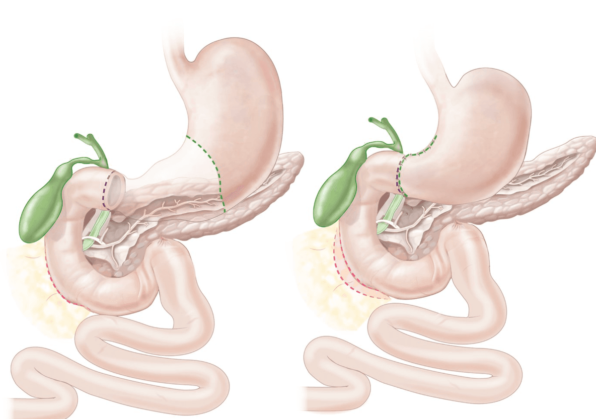
TAE: Perform angiography of coeliac trunk and SMA → evaluate for contrast extravasation → selective cannulation of bleeding vessels → angiographic coiling distal to proximal until extravasation ceased [9]
| Feature | Detail |
|---|---|
| When | Failed endoscopic therapy AND patient unfit for surgery [5]. Also used when surgery is high-risk (elderly, multiple comorbidities) |
| Technique | Femoral artery access → selective catheterisation (coeliac trunk / SMA) → identify extravasation → embolise with coils, gelatin sponge, particles, or NBCA |
| Efficacy | Equally effective compared with surgery in failed endoscopy patients, with fewer complications [9] |
| Advantage | Reduces the need for surgery without increasing overall mortality [9] |
| Risks | Contrast nephropathy, femoral haematoma, bowel ischaemia (especially if non-selective embolisation), rebleeding if collateral flow reconstitutes |
TAE vs Surgery — Decision Making
In the real world, the decision between TAE and surgery for failed endoscopic haemostasis depends on patient fitness and local expertise. TAE is preferred in the elderly and those with multiple comorbidities because it avoids general anaesthesia and laparotomy. Surgery is preferred when the bleeding is truly catastrophic and the patient is already in theatre, or when TAE is not available [5][9].
| Intervention | Detail |
|---|---|
| Sengstaken-Blakemore (SB) Tube | Emergency tamponade of gastro-oesophageal variceal bleeding unresponsive to medical and endoscopic treatment [12]. Gastric balloon inflated with 200–300 mL contrast + water (confirm position by CXR — displacement to oesophagus can be fatal). Oesophageal balloon inflated to 20–40 mmHg. Usually with ET intubation [12]. Temporary bridge (max 12–24 hours) to definitive therapy |
| Contraindications | Large hiatal hernia, unconfirmed variceal bleeding (e.g. Mallory-Weiss), oesophageal stricture, recent oesophageal surgery [12] |
| Complications | Pressure necrosis (deflate after 12h and re-inflate if ongoing bleed), oesophageal perforation, bleeding, pain [12] |
| TIPSS | Transjugular Intrahepatic Portosystemic Shunt — interventional radiology creates a channel between the hepatic vein and portal vein within the liver → decompresses the portal system → ↓ portal pressure → stops variceal bleeding. Definitive rescue for refractory variceal bleeding. Risk: hepatic encephalopathy (portal blood bypasses liver → ↑ ammonia) [3] |
| Surgical Shunts | Porto-caval or spleno-renal shunts — rarely used now due to TIPSS availability. Same principle: decompress portal system by creating a surgical anastomosis between portal and systemic venous systems [3] |
PHASE 5: Prevention of Recurrence (Secondary Prophylaxis)
| Measure | Detail | Why |
|---|---|---|
| H. pylori eradication | Triple therapy: PPI + amoxicillin + clarithromycin (or metronidazole) × 14 days | Removing H. pylori eliminates the chronic inflammatory drive → ulcer heals permanently. Eradication reduces PUD recurrence from ~70% to < 5% per year |
| NSAID management | Switch to less ulcerogenic NSAID or COX-2 inhibitors; withdraw NSAIDs during PPI treatment [9] | Remove the aggressive factor (COX-1 inhibition) |
| Long-term PPI | If NSAID/aspirin cannot be stopped (e.g. secondary cardiovascular prophylaxis) | Co-prescribe PPI to provide ongoing mucosal protection |
| Lifestyle | Smoking cessation, limit alcohol intake [9] | Smoking impairs ulcer healing; alcohol is a direct mucosal irritant |
| Follow-up OGD | Gastric ulcer: necessary until complete healing confirmed (rule out malignancy). Uncomplicated DU: unnecessary if asymptomatic. Complicated DU: necessary until healing confirmed [9] | Gastric ulcers carry malignancy risk — even a benign-looking ulcer needs follow-up biopsy to exclude sampling error [9] |
| Measure | Detail | Why |
|---|---|---|
| Non-selective β-blockers (NSBB) | Propranolol (or carvedilol/nadolol) | β1 blockade → ↓ cardiac output → ↓ portal inflow. β2 blockade → unopposed α-mediated splanchnic vasoconstriction → ↓ portal flow. Net effect: ↓ HVPG by ~20% |
| Repeat EBL | Band ligation sessions every 2–4 weeks | Until varices are obliterated — typically takes 3–5 sessions |
| Combination | NSBB + repeat EBL | Combination is superior to either alone for preventing variceal rebleeding |
| TIPSS | If rebleeding despite NSBB + EBL | Definitive portal decompression |
Special Scenarios
- Management: graft excision with extra-anatomical bypass [8]
- Definitive surgery is the only option — endoscopic/IR treatments are temporizing at best
- Pre-operative stabilisation with massive transfusion may be needed
- Endoscopic therapy: sclerosant injection, endoscopic clips [5]
- If endoscopy fails: surgical wedge excision of the gastric wall segment containing the aberrant vessel
| Drug | Route | Indication | Mechanism | Key Notes |
|---|---|---|---|---|
| Esomeprazole / Pantoprazole | IV 80 mg stat → 8 mg/h × 72h | Non-variceal ulcer bleeding (Forrest Ia–IIb) | Irreversible H⁺/K⁺-ATPase inhibition → ↑ gastric pH → clot stabilisation | Pre-endoscopic PPI only if early endoscopy cannot be arranged [3]. Post-OGD: 72h infusion |
| Terlipressin | IV | Suspected/confirmed variceal bleeding | V1 agonist → splanchnic vasoconstriction → ↓ portal pressure | Caution if IHD/stroke [3] |
| Octreotide | IV | Suspected/confirmed variceal bleeding | Somatostatin analogue → inhibits glucagon → splanchnic vasoconstriction | Fewer CV side effects than terlipressin [3] |
| Ceftriaxone | IV 1g daily × 7 days | All cirrhotic patients with GI bleeding | Broad-spectrum antibiotic for SBP prophylaxis | Alternatives: augmentin, levofloxacin [3] |
| Vitamin K1 | IV 10 mg daily × 3 days | Cirrhotic patients | γ-carboxylation of factors II, VII, IX, X | Probably useless in parenchymal liver disease but given routinely [2] |
| Tranexamic acid | — | — | Antifibrinolytic | NOT recommended in UGIB — insufficient evidence of benefit, potential ↑ thromboembolic risk [2] |
Tranexamic Acid in UGIB
Tranexamic acid is NOT recommended for UGIB [2]. The HALT-IT trial (2020) showed no mortality benefit and a potential increase in venous thromboembolic events. Do not prescribe it reflexively.
High Yield Summary — Management of UGIB
Resuscitation (ABC): Airway (cuffed ET tube if massive haematemesis/low GCS), Breathing (O2), Circulation (2 large-bore IV, NS 2L fast, transfuse if Hb < 7 or < 9 in IHD, withhold anticoagulants, FFP/platelets for coagulopathy)
Variceal pathway: IV terlipressin/octreotide + IV ceftriaxone + IV vitamin K → Emergency OGD → EBL for oesophageal varices, Histoacryl glue for gastric varices → If persistent: Sengstaken-Blakemore tube → TIPSS/surgical shunt
Non-variceal pathway: IV PPI 80 mg stat → 8 mg/h → OGD within 24h → Forrest Ia–IIb: dual endoscopic therapy (adrenaline + heater probe/clip) → IV PPI 72h infusion; Forrest IIc–III: oral PPI, start feeding, early discharge
Dual therapy = adrenaline injection + thermal or mechanical — injection monotherapy has unacceptable rebleeding rates
Post-OGD monitoring: Watch for rebleeding signs in first 3 days — ↑ pulse, haematemesis, fresh melena, blood in NG tube, ↓ Hb
Escalation if endoscopy fails: Surgery (DU: undersew GDA; GU: partial gastrectomy) OR TAE (if unfit for surgery) — TAE is equally effective with fewer complications
Secondary prophylaxis: H. pylori eradication + stop/switch NSAIDs + PPI; Variceal: NSBB + repeat EBL until obliteration
Do NOT use: Tranexamic acid (no benefit, ↑ thrombosis risk); IV PPI for variceal bleeds (only oral PPI for post-banding ulcers); NG tube in variceal bleeding
Active Recall - Management of UGIB
References
[1] Lecture slides: GC 198. Profuse vomiting of fresh blood and in shock severe upper GI bleeding.pdf (p19, p21, p24, p25, p28) [2] Senior notes: felixlai.md (Upper GI bleeding — Treatment: General management; Medications; Variceal hemorrhage — Initial management) [3] Senior notes: maxim.md (3.3 UGIB — Resuscitation; Pre-endoscopy management; OGD; Forrest classification; Post-OGD PPI; Antithrombotic management; Cirrhotic patients management) [5] Senior notes: maxim.md (3.6 Benign diseases of stomach — PUD complication hemorrhage; Surgical management; TAE) [6] Senior notes: maxim.md (Mallory-Weiss syndrome — Management) [8] Senior notes: maxim.md (Approach to UGIB with background of AAA repair) [9] Senior notes: felixlai.md (Peptic ulcer disease — Treatment; Bleeding complication; Endoscopic treatment; Surgical treatment; TAE; Follow-up endoscopy; Aspirin management) [10] Senior notes: maxim.md (Angiodysplasia — Management) [11] Senior notes: maxim.md (Transfusion and blood products; Management of antiplatelets and anticoagulants) [12] Senior notes: maxim.md (Sengstaken-Blakemore tube — indications, contraindications, complications)
Complications of Upper GI Bleed
Complications of UGIB can be organised into three categories:
- Complications of the bleeding itself (hypovolaemia, organ damage)
- Complications of the underlying disease (perforation, obstruction, malignancy)
- Complications of treatment (endoscopic, pharmacological, surgical, and interventional radiology)
Understanding each complication from first principles means tracing the chain from what went wrong → why it causes harm → how to recognise it → how to manage it.
1. Complications of the Bleeding Itself
These are consequences of acute blood loss and hypovolaemia. Every complication below flows from the same root problem: insufficient oxygen delivery to tissues.
| Feature | Detail |
|---|---|
| Mechanism | Acute haemorrhage → ↓ circulating blood volume → ↓ venous return → ↓ cardiac output → ↓ tissue perfusion → cellular hypoxia → anaerobic metabolism → lactic acidosis |
| Recognition | Tachycardia → hypotension → oliguria → confusion → obtundation (follows ATLS classification: Class I–IV) [3] |
| Key point | Blood pressure does NOT drop until > 30% of blood volume is lost (Class III) — tachycardia is the earliest compensatory sign. Relying on BP alone leads to false reassurance [3] |
| Management | Aggressive fluid resuscitation + blood transfusion + source control (endoscopic/surgical haemostasis) |
Why does shock kill? Prolonged tissue hypoperfusion leads to multi-organ failure — the organs most vulnerable are those with the highest metabolic demand (brain, kidneys, heart) and those dependent on high blood flow (splanchnic bed → gut mucosal barrier failure → bacterial translocation → sepsis).
This is the single most important complication to watch for and the main predictor of mortality in UGIB.
Signs of recurrence of bleeding: [9]
- Haematemesis / fresh melena
- Tachycardia
- Falling Hb trend (anaemia)
- Blood in the NG tube
Per lecture slides [1]:
After therapeutic endoscopy — close monitoring, look out for rebleeding (first 3 days):
- Increasing pulse rate
- Haematemesis
- Pass fresh melaena again
- Fresh blood aspirated from nasogastric tube
- Drop in haemoglobin level
Why does rebleeding occur?
- The endoscopic haemostasis creates a temporary seal (clot, coagulum, clip) — if the clot is lysed by pepsin (at low pH) or dislodged mechanically, the underlying vessel re-opens
- This is why IV PPI 80 mg stat then 8 mg/h for 72 hours is critical post-endoscopy — maintaining pH > 6 prevents pepsin activation and clot lysis [3]
- Rebleeding is most likely in the first 72 hours — hence the intensive monitoring period [1]
Risk factors for rebleeding (from earlier sections, repeated here for completeness) [1]:
Risk factors for recurrent bleeding:
- Shock on presentation
- Hb < 8.0 g/dL on presentation
- Transfusion requirement
- Age > 60 yrs
- Comorbidity
- Coagulopathy
- Patient already hospitalised
- Large ulcer
- Ulcer on posterior D1
- Ulcer on higher posterior lesser curve
Management of rebleeding:
| Feature | Detail |
|---|---|
| Mechanism | Hypovolaemia → ↓ renal perfusion → pre-renal AKI → oliguria, ↑ urea, ↑ creatinine |
| Why urea rises disproportionately | Two reasons: (1) ↓ GFR → ↑ urea reabsorption in proximal tubule. (2) Blood in the GI tract is digested → amino acids absorbed → hepatic urea synthesis ↑. This gives the characteristic elevated urea:creatinine ratio > 100:1 [3] |
| Prevention | Adequate fluid resuscitation, avoid nephrotoxins (NSAIDs, contrast dye if possible) |
| Management | Volume restoration; if progresses to ATN (acute tubular necrosis from prolonged ischaemia), may require renal replacement therapy |
| Feature | Detail |
|---|---|
| Mechanism | ↓ Haemoglobin → ↓ oxygen-carrying capacity → demand ischaemia in patients with pre-existing coronary artery disease. The heart needs to work harder (compensatory tachycardia) with less oxygen available |
| Who is at risk | Elderly patients, those with IHD, aortic stenosis |
| Recognition | New chest pain, ECG changes (ST depression/elevation), troponin rise |
| Prevention | This is why the transfusion threshold is Hb < 9 g/dL in high-risk patients (e.g. elderly, CAD) rather than the standard Hb < 7 [2] |
| Feature | Detail |
|---|---|
| Mechanism | Massive haematemesis → blood pools in the oropharynx → aspiration into the tracheobronchial tree → chemical pneumonitis → secondary bacterial infection |
| Who is at risk | Patients with impaired consciousness (hepatic encephalopathy in cirrhotics, severe shock), elderly, those without a protected airway |
| Prevention | Cuffed ET tube for airway protection in patients with massive haematemesis or GCS ≤ 8 [3]. Semi-upright positioning. NG decompression |
| Management | Broad-spectrum antibiotics, physiotherapy, mechanical ventilation if severe |
| Feature | Detail |
|---|---|
| Mechanism | Blood in the GI tract is a massive protein load → bacterial breakdown produces ammonia → cirrhotic liver cannot clear ammonia (impaired urea cycle) → ammonia crosses the blood-brain barrier → astrocyte swelling + altered neurotransmission → confusion, asterixis, coma |
| Why UGIB specifically triggers it | 250 mL of blood in the gut = ~50 g of protein = enormous ammonia production. Combined with impaired hepatic clearance in cirrhosis, this is a potent trigger |
| Recognition | Confusion, asterixis (liver flap), fetor hepaticus, drowsiness progressing to coma |
| Management | Lactulose (osmotic laxative → acidifies colonic lumen → traps NH₄⁺ → increases faecal ammonia excretion), rifaximin (non-absorbable antibiotic → reduces ammonia-producing gut bacteria), NG tube to remove blood from the stomach |
| Feature | Detail |
|---|---|
| Mechanism | Chronic slow blood loss (e.g. from erosive gastritis, Cameron lesions, angiodysplasia, occult malignancy) → gradual depletion of iron stores → microcytic hypochromic anaemia |
| Why iron specifically | Each mL of blood lost = ~0.5 mg of iron. The body has limited iron absorption capacity (~1–2 mg/day). Chronic losses exceeding absorption → iron stores depleted → haemoglobin synthesis impaired |
| Recognition | Fatigue, pallor, glossitis, koilonychia (spoon nails), pica. Lab: ↓ Hb, ↓ MCV, ↓ ferritin, ↑ TIBC |
| Significance | Iron-deficiency anaemia in any adult should prompt investigation for GI malignancy (upper AND lower GI endoscopy) — it may be the only presenting feature of an occult gastric or oesophageal cancer [9] |
2. Complications of the Underlying Disease
These are complications of the specific pathology causing the UGIB, not of the bleeding per se.
PUD has four classic complications — bleeding is just one of them. A patient presenting with UGIB from PUD is also at risk of the other three:
| Complication | Mechanism | Key Features | Management |
|---|---|---|---|
| Bleeding (most common) | Ulcer erodes into a submucosal/mural artery (posterior D1 → GDA; lesser curve → left gastric artery) | Haematemesis, melena, shock. Leading cause of death of peptic ulcer [5] | Endoscopic dual therapy → IV PPI 72h → surgery/TAE if fails [9] |
| Perforation | Ulcer erodes through the full thickness of the bowel wall → free communication with peritoneal cavity. MC site: anterior wall of D1 [5] | Sudden onset severe generalised abdominal pain (can pinpoint exact onset time). Board-like rigidity. CXR: pneumoperitoneum (free gas under diaphragm in 60–70%) [5] | NPO, IV fluids, NG decompression, IV antibiotics, IV PPI. Do NOT perform OGD (may convert sealed perforation to real perforation) [5]. Surgery: omental patch repair (Graham patch) ± vagotomy + pyloroplasty |
| Gastric outlet obstruction (GOO) | Acute: inflammation → oedema and duodenal spasm. Chronic: fibrosis and scarring from repeated ulceration → narrowing of pylorus/duodenum [9] | Epigastric pain after eating, early satiety, vomiting of undigested food, succussion splash. Metabolic: hypochloraemic, hypokalaemic metabolic alkalosis (loss of HCl from vomiting) ± paradoxical aciduria (secondary hyperaldosteronism → H⁺/K⁺ exchange in kidneys) [5] | NPO, NG decompression ("drip and suck"), IV fluids + KCl, IV PPI. Endoscopic balloon dilatation ± stenting. Surgery: vagotomy + antrectomy + Billroth reconstruction [9] |
| Penetration | Ulcer erodes into an adjacent organ without free perforation into the peritoneal cavity. Order of frequency: pancreas > lesser omentum > biliary tract > liver > greater omentum > colon > vascular structures [9] | Change in pain pattern: more intense, longer duration, shift from vague visceral discomfort to localised intense pain radiating to the back (pancreatic penetration). NOT relieved by food or antacids [9] | Medical management (PPI, H. pylori eradication). Surgery NOT recommended for penetration alone [9] |
Perforation vs Bleeding — Anatomical Logic
Anterior D1 ulcers tend to perforate — because anteriorly there is only the peritoneal cavity, so the ulcer breaks through into free space. Posterior D1 ulcers tend to bleed — because posteriorly lies the GDA, so the ulcer erodes into a major artery before reaching the retroperitoneum. This anterior-perforation / posterior-bleeding pattern is a classic exam point [5][9].
Patients with variceal bleeding are cirrhotic, and the bleed itself triggers a cascade of liver-specific complications:
| Complication | Mechanism | Management |
|---|---|---|
| Hepatic encephalopathy | Blood protein → ammonia (as above) | Lactulose, rifaximin, NG aspiration of blood |
| Spontaneous bacterial peritonitis (SBP) | Gut bacterial translocation (↑ during variceal bleeding from mucosal hypoperfusion) + impaired hepatic reticuloendothelial clearance → bacteria seed ascitic fluid | IV ceftriaxone 1g daily for 7 days (prophylactic) [3] |
| Hepatorenal syndrome (HRS) | Splanchnic vasodilation (portal hypertension) → ↓ effective circulating volume → renal vasoconstriction → functional renal failure with no structural renal damage | IV terlipressin + albumin. TIPSS. Liver transplant (definitive) |
| Coagulopathy | ↓ Hepatic synthesis of clotting factors (II, V, VII, IX, X) + thrombocytopenia from hypersplenism + possible DIC | FFP, platelets, cryoprecipitate. IV vitamin K (limited benefit in parenchymal disease) [2] |
An underlying upper GI malignancy (gastric cancer, oesophageal cancer) causing UGIB carries the additional risk of:
- Gastric outlet obstruction — tumour mass occludes pylorus/duodenum [9]
- Perforation — tumour necrosis → full-thickness bowel wall breakdown → peritonitis [9]
- Metastatic spread — by the time a tumour bleeds, it may already be locally advanced or metastatic
- Iron-deficiency anaemia — chronic occult blood loss is often the presenting feature of GI malignancy
3. Complications of Treatment
Complications of OGD: [13]
| Timing | Complication | Mechanism |
|---|---|---|
| Pre-operative (sedation-related) | Cardiopulmonary complications (most frequent): hypoxaemia, respiratory depression, aspiration pneumonia, hypotension, cardiac arrhythmia, AMI [13] | Sedatives (midazolam, propofol) depress respiratory drive and lower BP. Aspiration risk if stomach full of blood |
| Intraoperative | Perforation | Risk increased with therapeutic manoeuvres (dilatation, EMR/ESD) and in patients with oesophageal diverticula. Insufflation of air can also perforate a weakened/ulcerated wall [13] |
| Bleeding | Biopsy sites, post-polypectomy, thermal injury to vessel during haemostasis | |
| Post-operative | Delayed bleeding (typically 5–7 days) | Sloughing of an eschar covering a blood vessel or extension of zone of thermal necrosis to non-injured tissues → bleeding if it involves a blood vessel [13] |
| Post-banding ulcers (after EBL for varices) | Band strangulates tissue → necrosis → ulcer at banding site → can bleed. Managed with oral PPI [3] |
Delayed Post-Endoscopy Bleeding
A patient who had endoscopic haemostasis and was doing well suddenly rebleeds on day 5–7. This is delayed bleeding — the thermal coagulation creates an eschar (scab), which sloughs off as the tissue heals, unroofing the underlying vessel. This is distinct from rebleeding in the first 72h (which is failure of initial haemostasis). It requires re-endoscopy and repeat therapy [13].
Complications of Histoacryl glue: ulceration, stricture, mediastinitis, obviates use of subsequent EBL [3]
| Complication | Mechanism |
|---|---|
| Ulceration | Glue polymerises and forms a hard cast → mechanical irritation of adjacent mucosa → ulceration |
| Stricture | Inflammatory reaction from glue → fibrosis → luminal narrowing |
| Mediastinitis | If glue injected too superficially or perforates the oesophageal/gastric wall → mediastinal contamination |
| Systemic embolisation | Glue can travel through the varix into the systemic venous circulation → pulmonary embolism or even cerebral embolism (if PFO present). This is the most feared complication |
| Obviates subsequent EBL | The hardened glue mass changes the tissue architecture → banding becomes technically impossible |
Complications of SB tube: [12]
- Pressure necrosis (deflate after 12h and re-inflate if ongoing bleed)
- Oesophageal perforation
- Bleeding, pain
- Why pressure necrosis? The inflated balloons exert sustained pressure on the oesophageal/gastric wall → mucosal ischaemia → necrosis. Maximum inflation time is 12 hours before deflation is mandatory
- Why oesophageal perforation? If the oesophageal balloon migrates into the pharynx and is still inflated → mechanical rupture. Or if inflated in a weakened oesophagus (stricture, recent surgery). Confirm position by CXR before inflation [12]
3.4 Complications of Surgery
| Timing | Complication | Mechanism |
|---|---|---|
| Immediate | Bleeding, injury to nearby organs (pancreas, duodenum, spleen) | Operative technical complications |
| Early | Anastomotic leak (typically POD 5–10) [14] | Failure of surgical anastomosis to heal → enteric contents leak into peritoneal cavity → peritonitis, sepsis |
| Duodenal stump blowout (after Billroth II) | Duodenal stump closure fails → bilious/enteric leak → severe sepsis | |
| Wound infection, chest infection, post-op ileus | Standard post-operative complications | |
| Late | Dumping syndrome | Early: rapid delivery of hyperosmolar chyme into small bowel → fluid shift into lumen → hypovolaemia + distension → vasomotor symptoms (30 min post-meal). Late: rapid carbohydrate absorption → hyperglycaemia → excessive insulin release → reactive hypoglycaemia (1–3h post-meal) |
| Bile reflux gastritis | Loss of pylorus → bile refluxes into gastric remnant → chronic gastritis | |
| Afferent loop syndrome (Billroth II/Roux-en-Y) | Obstruction of the afferent limb → accumulation of bile/pancreatic secretions → distension, pain, bilious vomiting [14] | |
| Nutritional deficiencies | Vitamin B12 deficiency (loss of intrinsic factor from parietal cells), iron deficiency (↓ acid → ↓ iron absorption), calcium/vitamin D malabsorption | |
| Marginal (stomal) ulcer | Acid from gastric remnant acts on unprotected jejunal mucosa at the anastomotic site | |
| Gastric stump cancer | Long-term bile reflux → chronic atrophic gastritis → intestinal metaplasia → dysplasia → carcinoma (risk increases > 15 years post-gastrectomy) |
| Complication | Mechanism |
|---|---|
| Gastroparesis / Delayed gastric emptying | Truncal vagotomy denervates the pylorus → loss of coordinated gastric motility → food stasis (this is why pyloroplasty is always added as a drainage procedure) |
| Diarrhoea | Vagal denervation of small bowel → rapid transit → malabsorption → osmotic diarrhoea (occurs in ~20% post-truncal vagotomy) |
| Dumping syndrome | Combined effect of vagotomy + pyloroplasty → loss of pyloric regulation → rapid gastric emptying |
| Complication | Mechanism |
|---|---|
| Rebleeding | Collateral arterial flow reconstitutes after embolisation → re-perfuses the bleeding vessel |
| Bowel ischaemia | Non-selective embolisation occludes end-arteries supplying bowel segments → ischaemic necrosis. Risk ~20% with standard technique, reduced to 3–4% with super-selective embolisation |
| Contrast nephropathy | Iodinated contrast → direct tubular toxicity + renal vasoconstriction → AKI |
| Femoral haematoma / dissection | Vascular access site complication |
| Post-embolisation syndrome | Pain, fever, nausea — inflammatory response to tissue ischaemia |
| Complication | Mechanism |
|---|---|
| Hepatic encephalopathy (most significant) | The shunt diverts portal blood (laden with gut-derived ammonia) directly into the systemic circulation, bypassing the liver → ↑ systemic ammonia → encephalopathy. Occurs in ~30% of patients |
| Shunt stenosis / thrombosis | Neointimal hyperplasia or thrombus within the stent → reduced shunt patency → recurrent portal hypertension → rebleeding. Managed with shunt revision/dilatation |
| Heart failure | Sudden ↑ venous return (the shunt dumps large volume into the systemic circulation) → volume overload → cardiac decompensation (especially in patients with pre-existing cardiac disease) |
While IV PPI for 72 hours is unlikely to cause problems, many UGIB patients are placed on long-term oral PPI for secondary prophylaxis. Complications of chronic PPI use include:
| Complication | Mechanism |
|---|---|
| C. difficile infection | ↑ Gastric pH → loss of acid barrier → altered gut microbiome → C. diff colonisation |
| Community-acquired pneumonia | ↑ Gastric pH → bacterial colonisation of stomach → micro-aspiration |
| Hypomagnesaemia | Impaired intestinal magnesium absorption (mechanism incompletely understood) |
| Bone fractures | ↓ Calcium absorption (calcium salts need acid for dissolution) → ↓ bone mineral density |
| Vitamin B12 deficiency | Acid is needed to release B12 from food proteins; PPI reduces this |
| Rebound acid hypersecretion | Chronic PPI → compensatory ↑ gastrin → parietal cell hyperplasia → on withdrawal, ↑↑ acid secretion |
| Fundic gland polyps | Chronic acid suppression → cystic dilation of fundic glands (benign, but can be mistaken for neoplastic polyps) |
| Category | Complications |
|---|---|
| From bleeding itself | Hypovolaemic shock, rebleeding, AKI (pre-renal), myocardial ischaemia/infarction, aspiration pneumonia, hepatic encephalopathy (cirrhotics), iron-deficiency anaemia (chronic) |
| From underlying disease | PUD: perforation, GOO, penetration. Varices: SBP, HRS, coagulopathy. Malignancy: obstruction, perforation, metastasis |
| From endoscopic treatment | Sedation complications (respiratory depression, aspiration, hypotension, arrhythmia), perforation, delayed bleeding (POD 5–7), post-banding ulcers, glue embolisation |
| From SB tube | Pressure necrosis, oesophageal perforation |
| From surgery | Anastomotic leak, dumping syndrome, bile reflux, afferent loop syndrome, nutritional deficiency, stump cancer, gastroparesis (vagotomy) |
| From TAE | Rebleeding, bowel ischaemia, contrast nephropathy, femoral haematoma |
| From TIPSS | Hepatic encephalopathy, shunt stenosis, heart failure |
| From long-term PPI | C. diff, pneumonia, hypomagnesaemia, fractures, B12 deficiency |
High Yield Summary — Complications of UGIB
Rebleeding is the most dangerous complication and the main predictor of mortality. Watch for it in the first 72 hours (↑ pulse, haematemesis, fresh melena, blood in NG tube, ↓ Hb). Risk factors: shock on presentation, Hb < 8, age > 60, posterior D1 ulcer, large ulcer, coagulopathy.
Anterior D1 ulcers perforate; posterior D1 ulcers bleed — anatomical logic (anterior = peritoneal cavity; posterior = GDA).
PUD has 4 complications: Bleeding (MC cause of PUD death), perforation, GOO, penetration.
Cirrhotic patients with variceal bleeding are at risk of hepatic encephalopathy (blood = protein load → ammonia), SBP (bacterial translocation), HRS, and coagulopathy.
OGD complications: sedation (most frequent = cardiopulmonary), perforation, delayed bleeding at POD 5–7 from eschar sloughing.
Histoacryl glue: ulceration, stricture, mediastinitis, systemic embolisation, prevents future EBL.
SB tube: pressure necrosis (deflate after 12h), oesophageal perforation.
TIPSS: hepatic encephalopathy (~30%), shunt stenosis, heart failure from volume overload.
Long-term PPI: C. diff, pneumonia, hypomagnesaemia, fractures, B12 deficiency.
Active Recall - Complications of UGIB
References
[1] Lecture slides: GC 198. Profuse vomiting of fresh blood and in shock severe upper GI bleeding.pdf (p25, p26–27) [2] Senior notes: felixlai.md (Upper GI bleeding — Treatment: General management; Variceal hemorrhage — Initial management) [3] Senior notes: maxim.md (3.3 UGIB — Resuscitation; Cirrhotic patients management; Post-OGD PPI; Variceal bleeding therapeutic) [5] Senior notes: maxim.md (3.6 Benign diseases of stomach — PUD complications: hemorrhage, perforation, GOO) [9] Senior notes: felixlai.md (Peptic ulcer disease — Complications: Bleeding, Perforation, GOO, Penetration; Treatment; Prevention) [12] Senior notes: maxim.md (Sengstaken-Blakemore tube — complications) [13] Senior notes: felixlai.md (Complications of OGD — Pre-operative, Intraoperative, Post-operative) [14] Senior notes: maxim.md (Post-gastrectomy complications — General and Specific)
High Yield Summary
Definition: UGIB = bleeding proximal to the ligament of Treitz (oesophagus, stomach, duodenum)
Key Presentations: Haematemesis (frank/coffee-ground), melena, haematochezia (if massive)
Most Common Causes (in order): Duodenal/gastric ulcer > Gastritis > Oesophageal/gastric varices > Mallory-Weiss > Gastric tumour
Critical Classification: Variceal vs Non-variceal — entirely different management pathways
PUD Risk Factors: H. pylori, NSAIDs, stress, excess acid (Zollinger-Ellison)
Dangerous Ulcer Locations: Posterior D1 (GDA) and posterior lesser curve (left gastric artery)
Variceal Bleeding: HVPG ≥ 12 mmHg → bleeding risk; Laplace's law → larger varices = higher risk
Forrest Classification: Ia/Ib/IIa = high risk → need endoscopic therapy; IIc/III = low risk → PPI only
ATLS Shock: BP doesn't drop until Class III ( > 30% loss) — tachycardia is the earlier sign
Risk Factors for Recurrent Bleeding: Shock on presentation, Hb < 8, age > 60, coagulopathy, posterior D1 ulcer, large ulcer, prior hospitalisation
Pre-endoscopy Workup: CBC, clotting, cross-match, LRFT, VBG, urea:Cr ratio ( > 100:1 = UGIB), CXR (pneumoperitoneum), CT aortogram if prior aortic graft
GBS = pre-endoscopy (need for intervention); Rockall = post-endoscopy (mortality prediction)
Pre-endoscopic PPI: Esomeprazole 80 mg IV bolus → 8 mg/h infusion (if early OGD unavailable)
High Yield Summary — Differential Diagnosis of UGIB
Most common causes (in order): PUD (MC) > Gastritis > Varices > Mallory-Weiss > Gastric tumour
Anatomical site approach: Oesophagus (varices, oesophagitis, Mallory-Weiss, CA) → Stomach (PUD, gastritis, Dieulafoy, PHG, GAVE, CA) → Duodenum (DU, duodenitis, aorto-enteric fistula, haemobilia) → Any site (angiodysplasia)
"More likely severe" causes: Varices, aorto-enteric fistula, stomal ulcer, Dieulafoy's lesion
Cirrhotic patient bleeding DDx: Varices (commonest) + PHG + coagulopathy + PUD + Mallory-Weiss
Aorto-enteric fistula: suspect in any patient with prior AAA graft + GI bleed → CT aortogram + OGD to D4
Obscure GI bleeding: negative top-and-tail → think small bowel (angiodysplasia, Meckel's, GIST, Crohn's, melanoma metastasis) → capsule endoscopy / double-balloon enteroscopy
Always exclude pseudo-UGIB: haemoptysis, swallowed epistaxis, iron/bismuth ingestion
High Yield Summary — Diagnosis of UGIB
Confirming UGIB: Haematemesis / coffee-ground vomiting / melena + elevated urea:Cr ratio ( > 100:1) + positive NG aspirate (but negative aspirate does NOT exclude UGIB)
Gold standard: OGD — simultaneously diagnoses, risk-stratifies, and treats
Risk stratification: GBS (pre-endoscopy, lab-based, identifies need for intervention; GBS 0 = outpatient) → Rockall (post-endoscopy, predicts mortality; < 3 good, > 8 high mortality)
Forrest classification (must know): Ia = spurting (~100% rebleed), Ib = oozing, IIa = visible vessel (50%), IIb = adherent clot (30–35%, flush to unmask), IIc = flat spot (5–8%), III = clean base ( < 3%). Classes Ia–IIb need endoscopic therapy + IV PPI 72h; IIc–III need oral PPI only
Pre-OGD essentials: CBC, clotting, LRFT, VBG, cross-match, erect CXR (exclude perforation), CT aortogram if AAA graft history
If OGD negative: Colonoscopy → CT angiogram (0.3–0.5 mL/min) → mesenteric angiogram (0.5–1 mL/min, therapeutic) → red cell scan (0.1 mL/min, most sensitive for intermittent bleeding) → capsule endoscopy / DBE for obscure bleeding
Never order barium study in acute UGIB — interferes with endoscopy, angiography, and surgery
High Yield Summary — Management of UGIB
Resuscitation (ABC): Airway (cuffed ET tube if massive haematemesis/low GCS), Breathing (O2), Circulation (2 large-bore IV, NS 2L fast, transfuse if Hb < 7 or < 9 in IHD, withhold anticoagulants, FFP/platelets for coagulopathy)
Variceal pathway: IV terlipressin/octreotide + IV ceftriaxone + IV vitamin K → Emergency OGD → EBL for oesophageal varices, Histoacryl glue for gastric varices → If persistent: Sengstaken-Blakemore tube → TIPSS/surgical shunt
Non-variceal pathway: IV PPI 80 mg stat → 8 mg/h → OGD within 24h → Forrest Ia–IIb: dual endoscopic therapy (adrenaline + heater probe/clip) → IV PPI 72h infusion; Forrest IIc–III: oral PPI, start feeding, early discharge
Dual therapy = adrenaline injection + thermal or mechanical — injection monotherapy has unacceptable rebleeding rates
Post-OGD monitoring: Watch for rebleeding signs in first 3 days — ↑ pulse, haematemesis, fresh melena, blood in NG tube, ↓ Hb
Escalation if endoscopy fails: Surgery (DU: undersew GDA; GU: partial gastrectomy) OR TAE (if unfit for surgery) — TAE is equally effective with fewer complications
Secondary prophylaxis: H. pylori eradication + stop/switch NSAIDs + PPI; Variceal: NSBB + repeat EBL until obliteration
Do NOT use: Tranexamic acid (no benefit, ↑ thrombosis risk); IV PPI for variceal bleeds (only oral PPI for post-banding ulcers); NG tube in variceal bleeding
High Yield Summary — Complications of UGIB
Rebleeding is the most dangerous complication and the main predictor of mortality. Watch for it in the first 72 hours (↑ pulse, haematemesis, fresh melena, blood in NG tube, ↓ Hb). Risk factors: shock on presentation, Hb < 8, age > 60, posterior D1 ulcer, large ulcer, coagulopathy.
Anterior D1 ulcers perforate; posterior D1 ulcers bleed — anatomical logic (anterior = peritoneal cavity; posterior = GDA).
PUD has 4 complications: Bleeding (MC cause of PUD death), perforation, GOO, penetration.
Cirrhotic patients with variceal bleeding are at risk of hepatic encephalopathy (blood = protein load → ammonia), SBP (bacterial translocation), HRS, and coagulopathy.
OGD complications: sedation (most frequent = cardiopulmonary), perforation, delayed bleeding at POD 5–7 from eschar sloughing.
Histoacryl glue: ulceration, stricture, mediastinitis, systemic embolisation, prevents future EBL.
SB tube: pressure necrosis (deflate after 12h), oesophageal perforation.
TIPSS: hepatic encephalopathy (~30%), shunt stenosis, heart failure from volume overload.
Long-term PPI: C. diff, pneumonia, hypomagnesaemia, fractures, B12 deficiency.
Peptic Ulcer Disease
Peptic ulcer disease is a condition characterized by mucosal defects in the stomach or duodenum that extend through the muscularis mucosae, resulting from an imbalance between gastric acid–pepsin aggression and mucosal defense mechanisms.
Bariatric Surgery
Bariatric surgery encompasses surgical procedures that modify the gastrointestinal tract to induce sustained weight loss in patients with severe obesity, primarily through restrictive, malabsorptive, or combined mechanisms.