
Hypertrophic Cardiomyopathy
Hypertrophic cardiomyopathy is a genetic myocardial disorder characterized by asymmetric left ventricular hypertrophy, often involving the interventricular septum, that can cause diastolic dysfunction, left ventricular outflow tract obstruction, and an increased risk of sudden cardiac death.
| Exam domain | One-glance essentials |
|---|---|
| Definition / diagnosis |
|
| Epidemiology / risks |
|
| Core mechanism |
|
| Clinical picture |
|
| Investigations |
|
| Management |
|
| Complications |
|
| Prevention / follow-up |
|
| Exam traps |
|
References
[1] Senior notes: Block A - Inherited Cardiac conditions.pdf (pp. 10–12) [2] Senior notes: Block A - Inherited Cardiac conditions.pdf (pp. 3–4, 10–13) [3] Senior notes: MBBS Final MB (Medicine) (Felix PY Lai).pdf (pp. 524–526 — HCM pathophysiology including histological findings and ischaemia mechanism) [4] Senior notes: Block A - Inherited Cardiac conditions.pdf (pp. 10–13) [5] Senior notes: Maksim Medicine Notes.pdf (p. 44 — Complications of HOCM and risk factors for SCD) [6] Senior notes: Ryan Ho Cardiology.pdf (pp. 167–169) [7] Lecture slides: GC 069. Inherited Cardiac conditions.pdf (p. 15 — Risk Factors of SCD in HCM, 2020 AHA/ACC Guidelines) [8] Lecture slides: GC 069. Inherited Cardiac conditions.pdf (p. 15 — Risk Factors of Sudden Cardiac Death in HCM, 2020 AHA/ACC Guidelines) [9] Senior notes: Block A - WCS32 Chest pain on exertion_ ischaemic heart disease; angina pectoris.pdf (p. 8 — cardiac causes of IHD without atherosclerosis) [10] Senior notes: MBBS Final MB (Medicine) (Felix PY Lai).pdf (p. 531 — Case 1: HOCM vs AS differentiation) [11] Senior notes: Block A - Introduction to CVS investigations (including ECG).pdf (p. 9 — Q waves and HOCM) [12] Senior notes: Block A - Inherited Cardiac conditions.pdf (pp. 7, 10 — amyloidosis ECG and investigation clues) [13] Senior notes: Maksim Medicine Notes.pdf (pp. 43–44, Cardiomyopathy and HCM sections) [14] Senior notes: Block A - Inherited Cardiac conditions.pdf (pp. 7–8 — phenocopy investigations, CMR LGE patterns) [15] Senior notes: Block A - Inherited Cardiac conditions.pdf (pp. 9–13) [16] Senior notes: Block A - WCS32 Chest pain on exertion_ ischaemic heart disease; angina pectoris.pdf (pp. 41 — beta-blocker and CCB indications, side effects, contraindications) [17] Guideline: 2024 AHA/ACC/AMSSM/HRS/PACES/SCMR Guideline for the Management of Hypertrophic Cardiomyopathy [18] Senior notes: Maksim Medicine Notes.pdf (pp. 43–44) [19] Senior notes: Ryan Ho Fundamentals.pdf (p. 210 — causes of exercise-related syncope) [20] Senior notes: MBBS Final MB (Medicine) (Felix PY Lai).pdf (p. 369 — nitrate contraindications including HOCM) [21] Senior notes: MBBS Final MB (Pediatrics) (Felix PY Lai).pdf (p. 220 — nitrate contraindications including HOCM) [22] Senior notes: Block A - WCS32 Chest pain on exertion_ ischaemic heart disease; angina pectoris.pdf (p. 8 — HCM as a cardiac cause of IHD) [23] Senior notes: Adrian Lui Pediatrics Notes.pdf (p. 184 — Noonan syndrome cardiac associations)
Hypertrophic Cardiomyopathy (HCM)
Hypertrophic cardiomyopathy (HCM) is defined as increased left ventricular (LV) wall thickness at end-diastole that cannot be explained by abnormal loading conditions such as hypertension or aortic stenosis [1][2].
In other words, HCM is an intrinsic or primary myocardial disease — the heart muscle itself is abnormally thick, not because it is working harder against pressure overload, but because the muscle fibres (myocytes) are inherently enlarged and disorganised due to a genetic defect. This is the cardinal distinction from "physiological" or "reactive" hypertrophy (e.g. an athlete's heart adapting to exercise, or LVH secondary to chronic hypertension).
Breaking down the name:
- Hypertrophic (Greek hyper = excessive + trophē = nourishment/growth) → abnormal thickening/growth of the myocardium
- Cardio (Greek kardia = heart) + myopathy (Greek myo = muscle + pathos = disease) → disease of the heart muscle
Key Diagnostic Premise – High Yield
The diagnosis of HCM is predicated on finding increased LV wall thickness that cannot be explained by abnormal loading conditions (hypertension, aortic stenosis, etc.) [1]. In adults: LV wall thickness ≥ 15 mm (or ≥ 13 mm if family history is positive or genetic test is positive). In children: body surface area (BSA)-adjusted z-score ≥ 2.5 SD above mean (or ≥ 2 SD if family history/genetic test is positive) [1].
2. Epidemiology
- 1 in 500 to 1 in 1000 individuals in the general population [1][2]. This makes HCM one of the most common inherited cardiac diseases — slightly less common than dilated cardiomyopathy (DCM).
- Many individuals are undiagnosed because 60–70% of patients with HCM will never experience any symptoms/presentation and live a completely normal life [1]. The disease is often discovered incidentally (e.g. an abnormal ECG during a routine check-up, or an echocardiogram done for another reason).
- HCM can present at any age, from infancy to the elderly. The hypertrophy typically develops during adolescence and early adulthood coinciding with the pubertal growth spurt — this is when the genetic programme for abnormal sarcomere proteins becomes phenotypically manifest.
- Male predominance in clinically diagnosed cases (roughly 2:1), though genetic prevalence is equal between sexes. The discrepancy likely reflects referral bias and the fact that women may present later with more symptomatic heart failure.
- In Hong Kong and East Asia, apical HCM accounts for 25–30% of all HCM cases (compared to only ~1–2% in Western populations) [3]. This is a crucial point for local exams.
Extremely High Yield
HCM is a major cause of premature sudden cardiac death (SCD) in young, apparently healthy athletes [1][2]. This is the classic exam question: "Which cardiomyopathy is the leading cause of sudden death in young athletes?" — the answer is HCM (in the US/Western data). In some registries, arrhythmogenic right ventricular cardiomyopathy (ARVC) or coronary anomalies are equally common causes, but HCM remains the prototypical answer.
- Why athletes? Exercise increases heart rate and contractility → worsens the dynamic LVOT obstruction (discussed below) → provokes ventricular arrhythmias (VT/VF) → sudden cardiac death. The disorganised myocardial architecture also creates re-entrant circuits that are arrhythmogenic.
3. Anatomy and Function — Relevant Normal Cardiac Anatomy
To understand HCM, you need to know normal ventricular anatomy:
- The LV is the thickest-walled chamber (normal wall thickness 6–11 mm in adults at end-diastole).
- The interventricular septum (IVS) separates the LV from the RV. In the most common form of HCM (asymmetric septal hypertrophy), the IVS becomes disproportionately thick (septum:free wall ratio > 1.3).
- The LVOT is the passage between the interventricular septum (anteriorly) and the anterior mitral valve leaflet (posteriorly) through which blood exits the LV into the aorta.
- This narrow corridor is the anatomical bottleneck where obstruction occurs in hypertrophic obstructive cardiomyopathy (HOCM).
- The anterior mitral valve leaflet is tethered by chordae tendineae to the papillary muscles attached to the LV wall.
- In HCM, hypertrophy displaces the papillary muscles anteriorly and inward, altering the geometry of the mitral valve apparatus, predisposing to systolic anterior motion (SAM).
- The myocardium is supplied by epicardial coronary arteries that branch into intramural arterioles.
- In HCM, these intramural vessels can be compressed by the hypertrophied muscle, and the increased muscle mass outstrips the vascular supply → demand-supply mismatch → ischaemia even without epicardial coronary artery disease.
4. Etiology
4.1 Genetic Basis — "A Disease of the Sarcomere"
HCM has autosomal dominant inheritance [1][2].
It is a genetically heterogeneous disease — many different genes/mutations can cause the same phenotype. As a result, HCM has variable penetrance, clinical presentation, and prognosis [1].
The majority of familial HCM in adults and adolescents is caused by mutations within the sarcomere protein genes [1][2].
The two most common genes (accounting for ~70% of genotype-positive cases):
- MYBPC3 — encodes myosin-binding protein C (the single most common gene, ~40% of familial cases)
- MYH7 — encodes β-myosin heavy chain (~30% of familial cases)
Other sarcomere genes (less common):
- TNNT2 — troponin T
- TNNI3 — troponin I
- TPM1 — α-tropomyosin
- MYL2, MYL3 — myosin light chains
- ACTC1 — cardiac actin
Why sarcomere mutations cause hypertrophy (first-principles explanation): The sarcomere is the fundamental contractile unit of the myocyte. Mutant sarcomere proteins result in inefficient contraction or altered calcium sensitivity → the myocyte responds by hypertrophying (growing bigger) as a compensatory mechanism, analogous to how skeletal muscle hypertrophies under stress. But unlike physiological hypertrophy, this is maladaptive: the myocytes become disorganised (myocardial disarray), there is excess interstitial fibrosis, and the architecture becomes chaotic — creating the substrate for diastolic dysfunction, obstruction, ischaemia, and arrhythmias.
40% of HCM is considered non-familial [1]:
-
5–10% associated with metabolic disorders / inborn errors of metabolism (phenocopies):
- Fabry disease (α-galactosidase A deficiency → glycosphingolipid accumulation) — X-linked, treatable with enzyme replacement therapy; very important to diagnose because it is one of the few treatable causes
- Danon disease (LAMP2 mutations)
- PRKAG2 syndrome (glycogen storage)
- Friedreich's ataxia (mitochondrial iron accumulation)
- Noonan syndrome (RASopathy) — in paediatric patients
- Amyloidosis — especially transthyretin (ATTR) amyloidosis in the elderly; increasingly recognised as a mimic of HCM
-
Remaining 25–30% are either unknown genes or other causes [1]
Phenocopies — Don't Miss Them
Always consider HCM phenocopies (especially Fabry disease and cardiac amyloidosis) because they are treatable. Clues: Fabry → corneal verticillata, angiokeratomas, neuropathic pain, renal failure. Amyloidosis → low voltage ECG (paradoxically, despite thick walls on echo — the thickness is from amyloid deposition, not true myocyte hypertrophy, and the amyloid dampens electrical signals).
A positive family history is defined as: ≥ 2 first- or second-degree relatives with HCM, OR one first-degree relative who has autopsy-proven HCM and sudden cardiac death < 50 years old [1].
5. Pathophysiology
This is the most important section for understanding HCM. There are 4 interrelated pathophysiological processes in HCM [1][3]:
LVOTO occurs in ~1/3 of HCM patients [3] and is a major determinant for HF symptoms and death [3].
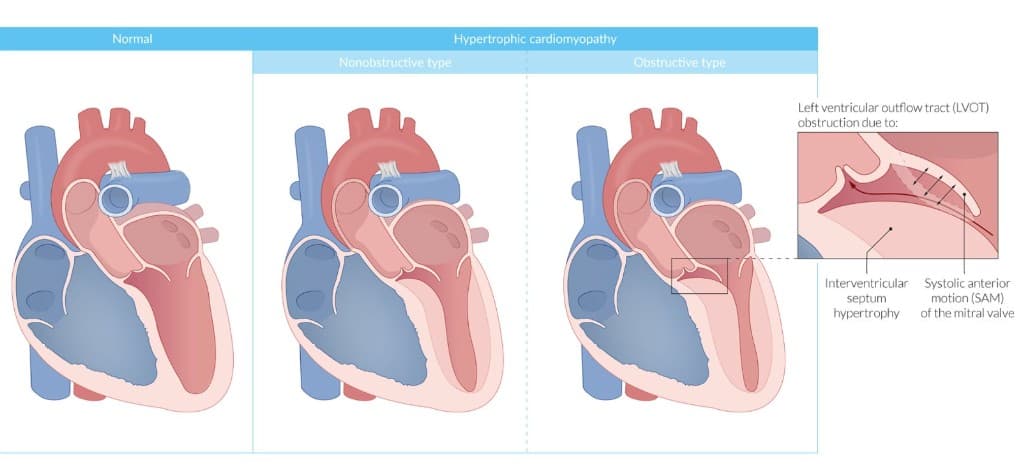
LVOTO is due to two mechanisms [1][3]:
-
Asymmetric septal hypertrophy (ASH) — The thickened septum encroaches on the LVOT, narrowing the outflow passage.
-
Systolic anterior motion (SAM) of the anterior mitral valve leaflet — This is the more interesting mechanism:
- The hypertrophied septum displaces the papillary muscles anteriorly
- During systole, blood accelerating through the narrowed LVOT creates a Venturi effect (or Bernoulli's principle [1]) — the high-velocity flow creates a low-pressure zone that sucks the anterior mitral valve leaflet toward the septum
- The anterior leaflet physically contacts the septum during mid-to-late systole → further obstructs the LVOT
Consequences of LVOTO [3]:
- ↑ Afterload → ↑ myocardial oxygen demand + ↓ cardiac output → myocardial ischaemia
- SAM → mitral regurgitation (because the mitral leaflet is pulled out of its proper coaptation position)
- Dynamic obstruction → jerky pulse (bisferiens or spike-and-dome pulse) [3]
Dynamic Nature of the Obstruction — Critical Concept
Unlike aortic stenosis (a fixed obstruction), LVOT obstruction in HOCM is dynamic — it changes with loading conditions. This is why the murmur and symptoms vary with manoeuvres:
| Manoeuvre | Effect on LV Volume | Effect on Obstruction | Murmur |
|---|---|---|---|
| Valsalva / Standing | ↓ preload → ↓ LV volume → septum and mitral valve closer together | Worsens | Louder |
| Squatting / Leg raise / Hand-grip | ↑ preload or ↑ afterload → ↑ LV volume → wider LVOT | Improves | Softer |
| Dehydration / Hypovolaemia | ↓ preload → ↓ LV volume | Worsens | Louder |
| Post-premature ventricular beat | ↑ contractility (post-extrasystolic potentiation) | Worsens | Louder |
Think of it like this: anything that makes the LV cavity smaller (less blood inside) brings the septum and mitral valve closer together, worsening obstruction. Anything that fills the LV up keeps the LVOT patent.
Diastolic dysfunction is a contributing factor in 80% of HCM patients [3].
Mechanisms:
- ↓ Compliance → ↓ diastolic filling → ↑ LV end-diastolic pressure → ↑ afterload [3]
- The hypertrophied, fibrotic myocardium is stiff and cannot relax properly during diastole
- Asymmetrical hypertrophy → non-uniform ventricular contraction/relaxation [3]
- Different segments of the LV relax at different rates, further impairing filling
- ↓ Diastolic filling → ↑ pulmonary venous pressure → accounts for exertional dyspnoea [3]
- Blood backs up into the pulmonary veins because the stiff LV cannot accommodate normal filling volumes
Why does this matter? Even without obstruction, diastolic dysfunction alone can cause significant symptoms. This is why many patients with non-obstructive HCM still have exertional dyspnoea and exercise intolerance.
Myocardial ischaemia often occurs without coronary artery disease [3].
Postulated mechanisms [3]:
- Hypertrophy → partial obliteration of intramural coronary arteries — the thickened muscle compresses the small vessels within it
- Inadequate vascular supply for the increased LV mass — the capillary density does not increase proportionally with muscle mass (demand-supply mismatch)
- Additionally: elevated LV end-diastolic pressure compresses the subendocardial vessels during diastole (when coronary flow normally occurs)
This explains why patients with HCM experience angina even with normal coronary arteries on angiography.
Cause: SAM of the mitral valve [3] — as the anterior leaflet is pulled toward the septum during systole, it fails to coapt properly with the posterior leaflet, creating a regurgitant jet directed posterolaterally.
Severity of MR is directly proportional to the degree of LVOT obstruction [3].
Consequence: ↑ pulmonary venous pressure → dyspnoea, orthopnoea [3].
Although not one of the "classic four" pathophysiological processes, the disorganised myocardial architecture (myocardial disarray + fibrosis) creates electrical heterogeneity → re-entrant circuits → substrate for ventricular tachycardia (VT) and ventricular fibrillation (VF) → sudden cardiac death.
Atrial fibrillation (AF) is also common (20–25% of HCM patients) due to left atrial enlargement from diastolic dysfunction and MR.
6. Classification
| Type | Resting LVOT Gradient | Features |
|---|---|---|
| Obstructive (HOCM) | ≥ 30 mmHg at rest | ~1/3 of patients |
| Latent/Labile obstruction | < 30 mmHg at rest but ≥ 30 mmHg with provocation | Another ~1/3 |
| Non-obstructive | < 30 mmHg at rest and with provocation | ~1/3 |
Treatment is required when the gradient is ≥ 50 mmHg [1].
LVOTO is assessed at rest and with provocation (infused epinephrine / exercise) — this is only for risk stratification and prognosis testing [1].
HOCM has worse survival compared to those without obstruction [1].
Three anatomical types of HCM [1][2]:
-
Asymmetric septal hypertrophy (ASH) — the most common form (~70%). The interventricular septum is disproportionately thickened compared to the free wall. This is the type most likely to cause LVOTO.
-
Symmetric (concentric) hypertrophy — uniform thickening of all LV walls. Could theoretically cause obstruction in very severe cases, but rarely seen [1].
-
Apical hypertrophy — hypertrophy predominantly affects the LV apex.
HCM sits within the broader WHO/ISFC classification of cardiomyopathies alongside dilated (DCM), restrictive (RCM), arrhythmogenic right ventricular cardiomyopathy (ARVC), and unclassified cardiomyopathies [2].
7. Clinical Features
HCM usually has slow clinical progression [3]. Symptoms depend on age of onset, degree, and location of hypertrophy [3].
| Symptom | Frequency | Pathophysiological Mechanism |
|---|---|---|
| Exertional dyspnoea (SOBOE) | > 90% [2][3] | Diastolic dysfunction → ↑ LV filling pressures → ↑ pulmonary venous pressure → pulmonary congestion. Also MR contributes. LVOTO worsens with exercise (↑ heart rate shortens diastolic filling time + ↑ contractility worsens obstruction) |
| Orthopnoea and PND | Less common | Same mechanism as above; redistribution of blood volume in supine position → further ↑ pulmonary pressures |
| Angina / exertional chest pain | 70–80% [3] | Myocardial ischaemia from: demand-supply mismatch (↑ mass, inadequate vasculature), compression of intramural vessels, ↑ LVEDP reducing coronary perfusion gradient. Note: often occurs without CAD [3] |
| Exertional syncope | 20% [3] | LVOT obstruction worsens with exertion [3] → inability to increase cardiac output → cerebral hypoperfusion. Also: exercise-induced ventricular arrhythmias |
| Presyncope (light-headedness, dizziness) | 50% [3] | Same mechanism as syncope but milder |
| Palpitations | Variable | Atrial fibrillation (from LA enlargement), supraventricular tachycardias, ventricular ectopics/NSVT |
| End-stage heart failure | ~5% [3] | ↓ LVEF due to adverse remodelling — diastolic dysfunction progresses so severely that systolic function eventually fails ("burnt-out" HCM). The ventricle may even dilate, resembling DCM |
Why Exertional Syncope in HCM is Ominous
Syncope in a young person with HCM — especially exertional syncope — is a red flag for sudden cardiac death risk. It suggests severe dynamic LVOTO or exercise-induced ventricular arrhythmias. An LVOT gradient > 40 mmHg is associated with angina and syncope risk [3]. Always take this seriously and risk-stratify urgently.
7.3 Signs (with Pathophysiological Basis)
- May appear completely normal (especially young, athletic individuals)
- Signs of heart failure if advanced disease (raised JVP, peripheral oedema, tachypnoea)
- Jerky pulse (bisferiens pulse / spike-and-dome) [3] — pathognomonic of HOCM
- Mechanism: The initial rapid ejection creates a spike, then the dynamic obstruction from SAM suddenly impedes flow (a dip), followed by a secondary push as the obstruction partially resolves → spike-and-dome morphology
- Contrast with aortic stenosis which gives a slow-rising (parvus et tardus) pulse
- Sustained, forceful, non-displaced (or minimally displaced) — reflects LV hypertrophy without significant dilatation
- May have a double impulse (palpable atrial kick + ventricular systole) reflecting the stiff, non-compliant LV that relies heavily on atrial contraction for filling
- Contrast with DCM where the apex beat is displaced and diffuse
The murmur that is pathognomonic for HCM is a crescendo-decrescendo holosystolic murmur best heard at the left sternal border [1].
- This is an ejection systolic murmur (ESM) due to LVOT obstruction — not a typical aortic stenosis murmur (which radiates to the carotids). The HOCM murmur radiates less to the neck because the obstruction is below the aortic valve (subvalvular).
The murmur classically increases with standing and Valsalva, while it decreases with squatting, hand-grip, and passive leg lift [1].
| Manoeuvre | Mechanism | Effect on Murmur |
|---|---|---|
| Standing / Valsalva | ↓ Preload → ↓ LV cavity size → ↑ obstruction | Louder |
| Squatting / Hand-grip / Leg raise | ↑ Preload or ↑ afterload → ↑ LV cavity size → ↓ obstruction | Softer |
May also have a concomitant mitral regurgitation murmur radiating to the apex [1] — a pansystolic murmur at the apex radiating to the axilla, due to SAM causing MR.
Other auscultatory findings:
- S4 gallop — due to forceful atrial contraction against a stiff, non-compliant LV (atrial gallop). This is common in HCM.
- S3 — usually only if the disease has progressed to the "burnt-out" dilated phase with systolic dysfunction
- Reversed splitting of S2 — if there is severe LVOTO, the LV takes longer to eject → delayed A2
Seven key tests for diagnosing HCM [1]:
| Investigation | Findings in HCM |
|---|---|
| ECG | LVH with strain (symmetrical or asymmetrical) [1]; precordial T-wave inversion (apical type) [1]; dagger-like Q waves in inferior/lateral leads [2]; AF or WPW [2]; Low voltage → amyloidosis (those toxic deposits + effusion dampens the voltage) [1] |
| 3-generation family history / genetic testing | Identifies familial cases; genetic testing is not mandatory [2] but recommended when diagnosis is uncertain or for cascade family screening |
| Echocardiography | Gold standard for diagnosis — measures wall thickness, identifies ASH, SAM, MR, LVOTO. LVOTO assessed at rest and with provocation (resting / provoked with infused epinephrine / exercise) [1]. Gradient ≥ 30 mmHg diagnostic of obstruction; ≥ 50 mmHg requires treatment [1] |
| Holter monitor | Non-sustained ventricular tachycardia (NSVT) → worse prognosis [1] |
| Cardiac MRI | Extensive late gadolinium enhancement (LGE) → worse prognosis [1]; also excellent for measuring wall thickness and identifying apical HCM that may be missed on echo |
| Exercise stress test | Provocative testing for LVOTO; also assesses for exercise-induced hypotension (a risk factor for SCD) and exercise-induced arrhythmias |
| Coronary angiography | To rule out concomitant coronary artery disease in patients with angina |
ECG Findings in Detail
ECG in HOCM typically shows [4]:
- LVH (Sokolow-Lyon criteria or Cornell voltage criteria positive)
- ST depression and T-wave inversion — "strain pattern" in lateral leads
- Dagger-like Q waves in inferior (II, III, aVF) and lateral leads (I, aVL, V5–V6) — these are not from MI but from septal depolarisation being abnormally prominent due to the thick septum
- Giant T-wave inversion in precordial leads — classic for apical HCM [3]
ECG findings suggestive of HCM for syncope workup [4]:
- LVH, ST depression, TWI [4]
ECG Clue: Giant T-Wave Inversions
Giant T-wave inversions (> 10 mm depth) across the precordial leads (V3–V5) are highly suggestive of apical HCM — this is the "Yamaguchi syndrome" pattern. On echo or cardiac MRI, you will see a spade-like configuration at end-diastole and obliteration of the distal cavity during systole [3]. Remember this is particularly common in Hong Kong (25–30% of local HCM).
This is one of the most commonly examined topics.
2020 AHA/ACC Guidelines — Risk Factors for SCD in HCM [5]:
| Risk Factor | Definition |
|---|---|
| Family history of sudden death from HCM | Sudden death judged attributable to HCM in ≥ 1 first-degree or close relatives who are < 50 years of age [5]. Close relatives would generally be second-degree; multiple SCDs in tertiary relatives should also be considered. |
| Massive LVH | Wall thickness ≥ 30 mm in any segment [5]. Borderline values ≥ 28 mm also considered at discretion. For paediatric patients, z-score ≥ 20 (or > 10 with other risk factors) [5]. |
| Unexplained syncope | ≥ 1 unexplained episode of transient loss of consciousness, unlikely neurocardiogenic and not attributable to LVOTO [5]. Especially relevant within 6 months of evaluation; events > 5 years ago may not be relevant [5]. |
| HCM with LV systolic dysfunction | EF < 50% by echo or CMR [5]. |
| LV apical aneurysm | Thin-walled, dyskinetic or akinetic segment at the most distal portion of the LV chamber, independent of size [5]. |
| Extensive LGE on CMR | Fibrosis ≥ 15% of LV mass [5]. The extent conferring risk has not been established in children. |
| NSVT on ambulatory monitor | Greater weight when runs are frequent (≥ 3), longer (≥ 10 beats), faster (≥ 200 bpm), usually over 24–48 hours of monitoring [5]. |
SCD Risk Stratification — Why It Matters
The purpose of identifying these risk factors is to decide who needs an implantable cardioverter-defibrillator (ICD). An ICD does not treat HCM but provides a lifeline if the patient develops VT/VF — it shocks the heart back into rhythm. The presence of one or more major risk factors in a patient with HCM warrants serious consideration for ICD implantation. The ESC uses a HCM Risk-SCD calculator (a validated 5-year risk score) to quantify the risk and guide ICD decisions.
10. Pathophysiology of Specific Clinical Scenarios
- During exercise → heart rate increases, contractility increases
- Increased contractility + shorter diastolic filling time → smaller LV cavity → septum and mitral valve closer together → worsened LVOT obstruction
- Simultaneously, exercise causes peripheral vasodilation → reduced afterload → further worsens the gradient
- Net effect: inability to increase cardiac output during exercise + cerebral hypoperfusion → syncope
- The stiff, hypertrophied LV depends heavily on atrial contraction ("atrial kick") for 30–40% of its diastolic filling (compared to ~15–20% in a normal heart)
- AF eliminates the atrial kick → sudden drop in LV filling → severe reduction in cardiac output
- Additionally, the rapid ventricular rate in AF → shortened diastolic filling time → further worsens filling
- Also: AF + stagnant blood in the dilated LA → high risk of thromboembolism/stroke
- Chronic ischaemia + fibrosis → progressive loss of functional myocytes
- Over decades, the hypertrophied myocardium "burns out" → the LV dilates, walls thin, and EF drops below 50%
- This is clinically indistinguishable from DCM and is managed as HFrEF
- Occurs in ~5% of HCM patients [3]
| Feature | HCM | Hypertensive Heart Disease | Aortic Stenosis | Athlete's Heart |
|---|---|---|---|---|
| Mechanism | Genetic (sarcomere mutation) | Pressure overload (↑ afterload) | Pressure overload (fixed valvular obstruction) | Physiological adaptation to exercise |
| Wall thickness | Often > 15 mm, asymmetric | Usually < 15 mm, concentric | Usually < 15 mm, concentric | Usually < 13 mm |
| Loading conditions | Normal BP, no valve disease | Hypertension present | Aortic valve abnormal | Normal |
| Diastolic function | Impaired | Impaired | Impaired (late) | Normal |
| LV cavity | Normal or small | Normal | Normal | Normal or mildly dilated |
| Regression with detraining | No | Partial (with BP control) | No | Yes (within 3–6 months) |
| SAM / LVOTO | Present in ~1/3 | Rare | No (obstruction is at valve) | No |
| Myocardial disarray on histology | Yes (hallmark) | No | No | No |
| SCD risk | High | Low (unless severe LVH) | Low (unless critical AS) | Extremely low |
High Yield Summary
Definition: Increased LV wall thickness at end-diastole not explained by abnormal loading conditions (≥ 15 mm in adults; ≥ 13 mm if FHx+/genetic+).
Epidemiology: 1 in 500 to 1 in 1000. Major cause of SCD in young athletes. 60–70% remain asymptomatic lifelong. Apical HCM: 25–30% in HK/Japan.
Genetics: Autosomal dominant, genetically heterogeneous, variable penetrance. Majority: sarcomere genes (MYBPC3, MYH7). 40% non-familial (including 5–10% metabolic phenocopies).
Four Interrelated Pathophysiological Processes:
- LVOTO — ASH + SAM (Bernoulli's principle); dynamic obstruction; worsens with ↓ preload/↓ afterload/↑ contractility
- Diastolic dysfunction — stiff, non-compliant LV → ↑ filling pressures → pulmonary congestion
- Myocardial ischaemia — without CAD; due to ↑ mass, compressed intramural vessels, inadequate vasculature
- Mitral regurgitation — due to SAM; severity proportional to LVOTO
Symptoms: SOBOE (> 90%), angina (70–80%), presyncope (50%), exertional syncope (20%), palpitations, SCD.
Signs: Jerky pulse, sustained apex beat, ESM at left sternal edge (louder with Valsalva/standing, softer with squatting/hand-grip), S4, ± MR murmur.
ECG: LVH + strain, dagger Q waves, giant T-wave inversions (apical), AF/WPW. Low voltage → consider amyloidosis.
SCD Risk Factors (2020 AHA/ACC): FHx SCD < 50y, massive LVH ≥ 30 mm, unexplained syncope (within 6 months), EF < 50%, LV apical aneurysm, LGE ≥ 15% LV mass, frequent/fast NSVT.
Active Recall - Hypertrophic Cardiomyopathy
[1] Senior notes: Block A - Inherited Cardiac conditions.pdf (pp. 10–13) [2] Senior notes: Maksim Medicine Notes.pdf (p. 43, Cardiomyopathy section) [3] Senior notes: Ryan Ho Cardiology.pdf (pp. 167–169) [4] Senior notes: Ryan Ho Fundamentals.pdf (pp. 202, 209–210) [5] Lecture slides: GC 069. Inherited Cardiac conditions.pdf (p. 15 — Risk Factors of Sudden Cardiac Death in HCM, 2020 AHA/ACC Guidelines)
Differential Diagnosis of Hypertrophic Cardiomyopathy
When a patient presents with features suggestive of HCM — unexplained LV wall thickening on imaging, exertional syncope, exertional chest pain, a systolic murmur that changes with loading manoeuvres, or a worrying family history of sudden cardiac death — you need a systematic differential diagnosis. The DDx falls into two broad categories:
- Conditions that mimic LV hypertrophy on imaging (phenocopies and other causes of increased wall thickness)
- Conditions that share the same clinical presentation (exertional syncope, exertional chest pain, systolic murmur, sudden cardiac death in the young)
We will address both systematically.
A. Differential Diagnosis of Left Ventricular Hypertrophy (Distinguishing True HCM from Other Causes of Thick Walls)
The definition of HCM itself requires you to exclude abnormal loading conditions [1]. Therefore, the first step when you see a thick LV is to ask: is there a pressure-overload reason for this?
- The single most common cause of LVH worldwide and the most important condition to exclude before diagnosing HCM.
- Hypertension causes pressure overload → concentric LVH as a compensatory mechanism [1].
- How to distinguish from HCM:
- Document blood pressure: sustained hypertension ≥ 140/90 mmHg (or on antihypertensives)
- LVH is typically concentric (uniform thickening), rarely exceeds 15 mm, and regresses with BP control (unlike HCM which does not regress)
- No SAM, no LVOT obstruction, no myocardial disarray on MRI
- Absence of family history of HCM/SCD
- Pitfall: Elderly patients may have both hypertension AND HCM. A wall thickness grossly disproportionate to the degree of hypertension (e.g. septum 22 mm in a patient with mild hypertension) should raise suspicion for co-existing HCM.
- Aortic stenosis causes chronic pressure overload → concentric LVH [1][6].
- Valvular, subvalvular, or supravalvular obstruction forces the LV to generate higher pressures → compensatory hypertrophy.
- How to distinguish from HOCM:
| Feature | HOCM | Aortic Stenosis |
|---|---|---|
| Site of obstruction | Subvalvular (LVOT) | Valvular (or sub/supravalvular) |
| Murmur radiation | Left sternal border, does NOT radiate well to neck | Aortic area, radiates to the neck [6] |
| Effect of Valsalva | Louder (↓ preload worsens dynamic obstruction) | Softer (↓ preload = less flow across fixed valve) |
| Effect of squatting | Softer | Louder |
| Pulse character | Jerky / bisferiens | Slow-rising, low-volume (parvus et tardus) |
| S2 | Usually normal or paradoxically split | Soft or absent A2 [6] |
| Aortic valve | Structurally normal | Calcified / bicuspid / rheumatic |
| Echo | ASH + SAM, normal aortic valve | Thickened/calcified aortic valve with restricted opening |
↓ Preload by Valsalva manoeuvre or from squatting to standing will ↑ intensity of HOCM but will ↓ intensity of AS [6]. This is a classic clinical pearl and favourite exam question.
Exam Favourite: HOCM vs AS Murmur
The key differentiating bedside test is the Valsalva manoeuvre. In HOCM, the obstruction is dynamic — reducing LV volume (by decreasing preload) brings the septum and mitral valve closer together, worsening obstruction and making the murmur louder. In AS, the obstruction is fixed — reducing preload means less blood flows across the stenotic valve, so the murmur gets quieter. This is a must-know point for clinical exams and MCQs.
- Highly trained athletes (especially endurance athletes like cyclists, rowers) develop physiological LVH as an adaptation to chronic volume and pressure loading during exercise.
- Wall thickness can reach 13–16 mm, overlapping with the "grey zone" of mild HCM.
- How to distinguish from HCM:
| Feature | Athlete's Heart | HCM |
|---|---|---|
| Wall thickness | Usually < 13 mm; rarely up to 16 mm | ≥ 15 mm (or ≥ 13 mm with FHx/genetics) |
| LV cavity | Normal or mildly dilated (adapted to volume loading) | Normal or small |
| Diastolic function | Normal | Impaired (abnormal E/A ratio, tissue Doppler) |
| Deconditioning | Regresses within 3–6 months of detraining | Does not regress |
| ECG | Voltage criteria for LVH, normal repolarisation | LVH + strain, deep T-wave inversions, Q waves |
| LGE on MRI | Absent | May be present (fibrosis) |
| Family history / genetics | Negative | Often positive |
- This distinction is extremely important because misdiagnosing an athlete with HCM leads to unnecessary restriction from sport (and psychological harm), while missing HCM in an athlete can be fatal.
- Amyloid protein (light chain AL, or transthyretin ATTR) infiltrates the myocardium → increased wall thickness that mimics HCM.
- Low voltage ECG despite thick walls on echo → classical for amyloidosis [7]. This paradox is because the amyloid deposits are electrically inert and the pericardial effusion (common in amyloidosis) further dampens the voltage. In contrast, true HCM causes high voltage (more myocytes = more electrical signal).
- Other clues: biventricular thickening, "sparkling" myocardium on echo, diastolic heart failure, nephrotic syndrome (AL amyloidosis), peripheral neuropathy (ATTR), carpal tunnel syndrome (ATTR).
- Investigations: 99mTc-PYP scan for transthyretin (TTR) amyloidosis; serum protein / immunofixation electrophoresis for AL amyloidosis [7].
Low Voltage + Thick Walls = Think Amyloidosis
Low QRS voltages on ECG with very thickened LV wall is CLASSICAL FOR AMYLOIDOSIS [7]. This is because the deposits are non-contractile protein (not myocytes) and associated effusion dampens voltage. Do not mistake this for HCM — the management is completely different (tafamidis for ATTR, chemotherapy for AL). This is a common exam trap.
- X-linked lysosomal storage disease (α-galactosidase A deficiency) → accumulation of globotriaosylceramide (Gb3) in multiple organs including the heart → progressive concentric LVH.
- Typically presents in males aged 30–50 (females may have milder, later-onset disease due to random X-inactivation).
- Clues beyond cardiac: angiokeratomas (dark red papules in "bathing trunk" distribution), corneal verticillata (whorl-like opacities on slit-lamp), acroparesthesias (burning pain in hands/feet from small fibre neuropathy), proteinuria/renal impairment, stroke in young patients.
- Why it matters: Fabry disease is treatable with enzyme replacement therapy (agalsidase alfa/beta) or oral chaperone therapy (migalastat). Missing this diagnosis means missing a treatable cause of "HCM."
- Diagnosed by α-galactosidase A enzyme activity (low in males), confirmed by genetic testing (GLA gene).
- PRKAG2 syndrome: Autosomal dominant, causes glycogen accumulation in the myocardium → LVH + pre-excitation (WPW) + progressive conduction disease. The combination of LVH + WPW on ECG should raise suspicion.
- Pompe disease (acid maltase deficiency): Autosomal recessive, infantile form presents with massive cardiomegaly and hypertrophic cardiomyopathy.
- Danon disease (LAMP2 mutation): X-linked, severe HCM in young males, often with skeletal myopathy and intellectual disability.
- Noonan syndrome is associated with hypertrophic cardiomyopathy [8] — this is a paediatric phenocopy.
- Noonan syndrome is caused by mutations in the RAS-MAPK signalling pathway.
- Clinical features: Turner-like appearance (short stature, webbed neck, low hairline, shield chest, hypertelorism, ptosis, downslanting palpebral fissures, low-set ears, cryptorchidism in males) [8].
- Associated with right-sided cardiac lesions: valvular pulmonary stenosis with thick dysplastic valve cusps, peripheral branch pulmonary artery stenosis, ASD in addition to HCM [8].
- Autosomal recessive neurodegenerative disorder (GAA trinucleotide repeat in FXN gene) → deficiency of frataxin → mitochondrial iron accumulation → cardiac and neurological disease.
- ~65% develop concentric LVH mimicking HCM.
- Clues: progressive cerebellar ataxia (onset in adolescence), dysarthria, absent deep tendon reflexes (peripheral neuropathy), upgoing plantars (pyramidal tract involvement), pes cavus, scoliosis, diabetes mellitus.
- RCM causes increased myocardial stiffness → abnormal LV filling → diastolic dysfunction [1], which overlaps with the diastolic dysfunction seen in HCM.
- However, RCM does not have to be hypertrophied [1] — the walls may be of normal thickness but the myocardium is stiff (e.g. from infiltration, fibrosis, or endocardial disease).
- Distinguishing features: biatrial enlargement, normal or near-normal wall thickness (unless infiltrative), Kussmaul sign (JVP rises on inspiration), may need endomyocardial biopsy.
- Key causes: amyloidosis (most common), sarcoidosis, haemochromatosis, radiation, endomyocardial fibrosis, scleroderma.
B. Differential Diagnosis by Clinical Presentation
Causes of exercise-related syncope [9]:
- (1) LVOT obstruction: AS, HCMP
- (2) RVOT obstruction: pulmonary hypertension
- (3) Cardiomyopathy: DCMP, HCMP, ARVD
- (4) Coronary artery disease: atherosclerotic, anomalous origin of coronary arteries
- (5) Arrhythmogenic: VT, SVT, WPWS, LQTS
This list is extremely high yield. The approach to a young person with exertional syncope should systematically consider all of these.
For the specific case of exertional syncope with a family history of sudden death [6]:
Differential diagnosis includes: Hypertrophic obstructive cardiomyopathy, Aortic stenosis, Exercise-induced arrhythmia, Coronary artery disease [6].
Causes of exercise-induced syncope in HCM specifically [6]:
- Dynamic outflow obstruction
- Exercise-induced atrial or ventricular arrhythmia
- Reflex vasodilation
Primary cardiac causes of ischaemic heart disease without atherosclerotic coronary disease include [10]:
- Coronary artery anomalies
- Radiation-induced coronary disease
- Myocardial bridging
- Aortic stenosis
- Hypertrophic cardiomyopathy
- Dilated cardiomyopathy
- Tachycardia
The mechanism in HCM is demand-supply mismatch (as discussed in Part 1): increased muscle mass, compressed intramural vessels, inadequate capillary density, and elevated LVEDP reducing the coronary perfusion gradient.
| Condition | Murmur Characteristics | Key Distinguishing Features |
|---|---|---|
| HOCM | ESM at left sternal border; louder with Valsalva/standing | Jerky pulse, SAM on echo, dynamic gradient, family history of SCD |
| Aortic stenosis | ESM at right upper sternal border radiating to carotids; softer with Valsalva | Slow-rising pulse, calcified valve, ejection click (bicuspid) |
| Pulmonary stenosis | ESM at left upper sternal border with ejection click | Wide split S2, right-sided signs, louder with inspiration |
| Innocent flow murmur | Soft ESM, grade ≤ 2/6, no radiation | Normal echo, normal ECG, no symptoms, disappears on standing |
| Atrial septal defect | ESM at left upper sternal border (due to increased flow across pulmonary valve) | Wide fixed split S2, RV heave |
This is the scenario where HCM classically comes to mind. The DDx includes:
| Condition | Key Features |
|---|---|
| HCM | Most common structural cause of SCD in young athletes (US data); LVH on echo/ECG |
| ARVC | Fibro-fatty replacement of RV; epsilon waves and anterior TWI on ECG; RV dilatation on MRI. Specific ECG: anterior TWI, epsilon wave, complete/incomplete RBBB [11] |
| Long QT Syndrome (LQTS) | Prolonged QTc > 470 ms (males) or > 480 ms (females); peaked/abnormal T waves; polymorphic VT (torsades de pointes) |
| Brugada Syndrome | Partial RBBB, J (Osborne) wave [11]; coved ST elevation in V1–V3; provoked by fever, Na-channel blockers |
| WPW Syndrome | Short PR, delta waves [11]; risk of AF conducting rapidly via accessory pathway → VF |
| Coronary artery anomalies | Anomalous origin of LCA from right coronary sinus; exertional syncope/SCD |
| Commotio cordis | Blunt chest impact during vulnerable repolarisation window → VF; normal heart |
| Myocarditis | Preceding viral illness; elevated troponin; MRI shows oedema/LGE |
| Aortic dissection | Especially in Marfan syndrome; tearing chest/back pain, BP discrepancy |
| Catecholaminergic polymorphic VT (CPVT) | Exercise- or emotion-triggered bidirectional VT; normal resting ECG; normal heart structure |
Cardiomyopathy is listed as a primary cardiac cause of palpitations [12] — the hypertrophied, disorganised myocardium is a substrate for both atrial (AF, SVT) and ventricular arrhythmias (NSVT, VT, VF).
HCM (specifically HOCM) is listed as a cause of both systolic and diastolic heart failure [13]:
- HF due to systolic dysfunction: HOCM causes ↑ afterload [13]
- HF due to diastolic dysfunction: HOCM causes LVH → impaired relaxation [13]
The DDx of HFpEF includes hypertensive heart disease, HCM, RCM, constrictive pericarditis, and cardiac amyloidosis. All share the phenotype of dyspnoea/exercise intolerance with preserved EF but impaired diastolic filling.
| Investigation | What It Helps Distinguish |
|---|---|
| Blood pressure | Hypertensive LVH (elevated BP) vs HCM (normal BP, or BP disproportionate to wall thickness) |
| ECG | LVH + strain + Q waves (HCM) vs low voltage + thick walls (amyloidosis) vs LVH + WPW (PRKAG2) vs epsilon waves (ARVC) |
| Echocardiography | Wall thickness, distribution (ASH vs concentric vs apical), SAM, LVOT gradient, aortic valve morphology, cavity size |
| Cardiac MRI | Gold standard for wall thickness measurement; LGE pattern (mid-wall in HCM, subendocardial in amyloidosis, patchy in sarcoidosis); T1 mapping helps differentiate infiltrative causes |
| Genetic testing | Sarcomere gene mutations (HCM) vs GLA (Fabry) vs FXN (Friedreich) vs LAMP2 (Danon) vs PRKAG2 |
| α-galactosidase A activity | Low in Fabry disease (males) |
| 99mTc-PYP scan | Positive in ATTR amyloidosis |
| Serum/urine immunofixation | Monoclonal protein in AL amyloidosis |
| Deconditioning trial | Regression of LVH → athlete's heart; no regression → HCM |
High Yield Summary — Differential Diagnosis of HCM
When you see a thick LV, think:
- Loading conditions? Hypertension, aortic stenosis, coarctation → secondary LVH, not HCM
- Phenocopies? Fabry disease (treatable! — angiokeratomas, renal failure, corneal verticillata), cardiac amyloidosis (low voltage + thick walls), glycogen storage (LVH + WPW = PRKAG2), Noonan syndrome (dysmorphic child with right-sided lesions + HCM), Friedreich ataxia (ataxia + LVH)
- Physiological? Athlete's heart (regresses with detraining, normal diastolic function, mildly dilated cavity)
When you see exertional syncope in a young person, think:
- LVOT obstruction (HOCM, AS), RVOT obstruction (pulmonary HTN), cardiomyopathy (HCM, DCM, ARVC), coronary anomalies, arrhythmogenic causes (LQTS, WPW, CPVT) [9]
Key bedside test: Valsalva — louder in HOCM, softer in AS [6]
Key ECG clue for amyloid: Low voltage + thick walls [7]
Active Recall - DDx of Hypertrophic Cardiomyopathy
References
[1] Senior notes: Block A - Inherited Cardiac conditions.pdf (pp. 3–4, 10–13) [6] Senior notes: MBBS Final MB (Medicine) (Felix PY Lai).pdf (p. 531 — Case 1: HOCM vs AS differentiation) [7] Senior notes: Block A - Inherited Cardiac conditions.pdf (pp. 7, 10 — amyloidosis ECG and investigation clues) [8] Senior notes: Adrian Lui Pediatrics Notes.pdf (p. 184 — Noonan syndrome cardiac associations) [9] Senior notes: Ryan Ho Fundamentals.pdf (p. 210 — causes of exercise-related syncope) [10] Senior notes: Block A - WCS32 Chest pain on exertion_ ischaemic heart disease; angina pectoris.pdf (p. 8 — cardiac causes of IHD without atherosclerosis) [11] Senior notes: Ryan Ho Fundamentals.pdf (p. 209 — ECG changes in ARVD, LQTS, BrS, WPW) [12] Senior notes: Block A - Syncope and irregular heartbeat_ Cardiac arrhythmia; Heart blocks, Bradycardia.pdf (p. 7 — DDx of palpitations) [13] Senior notes: MBBS Final MB (Pediatrics) (Felix PY Lai).pdf (pp. 203–204 — causes of HF in adults and children)
Diagnostic Criteria, Diagnostic Algorithm, and Investigation Modalities for Hypertrophic Cardiomyopathy
A. Diagnostic Criteria
The diagnosis of HCM is fundamentally an imaging diagnosis supported by clinical context. There is no single "Modified Duke's-style" scoring system for HCM — instead, diagnosis rests on demonstrating unexplained LV wall thickening that meets specific thresholds.
The diagnosis of HCM is predicated on increased left ventricular wall thickness at end-diastole that cannot be explained by abnormal loading conditions such as hypertension or aortic stenosis [1].
This means you must satisfy two conditions simultaneously:
- Wall thickness exceeds the diagnostic threshold (see below)
- No identifiable loading condition adequately explains the degree of hypertrophy
| Population | Threshold for Diagnosis | Notes |
|---|---|---|
| Adults without family history or genetic confirmation | LV wall thickness ≥ 15 mm [1] | In any single myocardial segment; any imaging modality (echo or CMR) |
| Adults with positive family history or positive genetic test | LV wall thickness ≥ 13 mm [1] | Lower threshold because the pre-test probability is higher — you need less morphological evidence when there is genetic/familial corroboration |
| Children without family history or genetic confirmation | BSA-adjusted z-score ≥ 2.5 SD above mean [1] | Children's hearts grow with body size, so absolute mm measurements are unreliable — must use z-scores |
| Children with positive family history or positive genetic test | BSA-adjusted z-score ≥ 2.0 SD above mean [1] | Again, lower threshold when genetic context supports the diagnosis |
Why z-scores in children? A 6-year-old and a 16-year-old have vastly different normal wall thicknesses. Using absolute millimetres would either miss HCM in small children or over-diagnose it in large adolescents. Z-scores normalise the measurement to what is expected for that child's body surface area — a z-score of 2.5 means the wall is 2.5 standard deviations thicker than average for their body size.
The 'Grey Zone' Problem
Wall thickness of 13–15 mm in adults falls into a diagnostic grey zone — it could be mild HCM, athlete's heart, or hypertensive LVH. In this zone, additional features become critical: family history, genetic testing, ECG abnormalities (deep T-wave inversions, Q waves), diastolic dysfunction on echo, LGE on CMR, and response to deconditioning (athlete's heart regresses, HCM does not). Never diagnose HCM on wall thickness alone without clinical correlation.
A positive family history is defined as ≥ 2 first- or second-degree relatives with HCM, OR one first-degree relative who has autopsy-proven HCM and sudden cardiac death < 50 years old [1].
Not all HCM has obstruction, and defining obstruction has separate thresholds:
| Parameter | Threshold | Significance |
|---|---|---|
| LVOT gradient ≥ 30 mmHg [1] | Diagnostic of obstruction | Defines the patient as having HOCM rather than non-obstructive HCM |
| LVOT gradient ≥ 50 mmHg [1] | Requires treatment | This is the threshold at which haemodynamic consequences are significant enough to warrant specific intervention (medical or interventional) |
LVOTO can be assessed at rest and with provocation (infused epinephrine / exercise) [1]. About one-third of patients have obstruction at rest, another third have latent/provokable obstruction, and one-third are truly non-obstructive.
Why provocation? At rest, the LV is reasonably filled and the LVOT may be wide enough to avoid significant obstruction. During exercise or with catecholamines, heart rate increases, contractility increases, and preload may decrease — all of which shrink the LV cavity and worsen the dynamic obstruction. Patients with "latent obstruction" (normal at rest, obstructive with provocation) may still be very symptomatic during exercise.
The following algorithm represents the systematic approach to a patient with suspected HCM, from initial suspicion through to confirmed diagnosis and risk stratification.
Steps Explained
-
Clinical suspicion arises from: symptoms (exertional dyspnoea, angina, syncope, palpitations), family history of HCM or SCD, abnormal ECG found incidentally, or heart murmur on examination.
-
History and physical examination — assess for symptoms, family history (three generations), signs of HOCM (jerky pulse, ESM at left sternal border with dynamic features, S4), and signs of phenocopies (angiokeratomas for Fabry, ataxia for Friedreich, dysmorphism for Noonan).
-
12-lead ECG — nearly always abnormal in phenotypically expressed HCM (> 90% sensitivity). Provides clues to the type and severity (see Investigations section below).
-
Transthoracic echocardiography (TTE) — the first-line imaging modality and gold standard for measuring wall thickness, identifying SAM, quantifying LVOT gradient, and assessing diastolic function.
-
Assess wall thickness against diagnostic thresholds (above).
-
Exclude loading conditions — check blood pressure, assess aortic valve (for AS), and consider coarctation.
-
Consider phenocopies — especially in atypical presentations (concentric LVH, extracardiac features, low voltage on ECG despite thick walls, conduction disease + LVH).
-
Confirm diagnosis → further workup for risk stratification and management planning.
C. Investigation Modalities — Detailed
Seven key tests help arrive at a diagnosis of HCM [1]:
The ECG is abnormal in > 90% of patients with phenotypically expressed HCM. It is the most accessible screening tool and often the first clue to the diagnosis.
ECG findings in HCM [1][2][14]:
| Finding | Description | Pathophysiological Basis |
|---|---|---|
| LVH with strain [1] | High QRS voltages meeting Sokolow-Lyon or Cornell criteria + ST depression and T-wave inversion in lateral leads (I, aVL, V5–V6) | The hypertrophied muscle mass generates greater electrical forces → higher voltages. "Strain" pattern represents subendocardial ischaemia from the thick wall compressing its own blood supply |
| Precordial T-wave inversion [1] | Anterior leads T-wave inversion [2] — can be deep/giant (> 10 mm) | Classically seen in apical HCM; reflects abnormal repolarisation in the hypertrophied apex |
| Dagger-like Q waves in inferior/lateral leads [2] | Deep, narrow Q waves in II, III, aVF and/or I, aVL, V5–V6 | Q waves not specific for MI [14] — HOCM can cause Q waves [14]. The massively hypertrophied septum generates a dominant initial depolarisation vector towards the right (away from left-facing leads), creating deep Q waves. V1 to V3 looks at septum → V1 to V3 Q waves present for massive septal hypertrophy [14] |
| AF / WPW [2] | Atrial fibrillation (irregularly irregular with absent P waves) or WPW pattern (short PR, delta wave) | AF from LA enlargement secondary to diastolic dysfunction and MR; WPW is a known association (especially with PRKAG2 glycogen storage phenocopy) |
| Low voltage [1] | Small QRS complexes despite thick walls on echo | Classical for amyloidosis — those toxic deposits + effusion dampens the voltage [1]. This is the red flag that the LVH is not true HCM but an infiltrative phenocopy |
High Yield ECG Points from GC Lecture Slides
ECG in HOCM [15]:
- LVH, ST depression, TWI — these are the classic triad to look for on an ECG in the context of syncope workup
Q waves are NOT specific for MI [14]:
- HOCM can cause Q waves — septal hypertrophy creates abnormal initial depolarisation vectors
- V1 to V3 Q waves present for massive septal hypertrophy [14]
- Dilated cardiomyopathy can also cause Q waves (10–13% of cases) [14]
This is a favourite exam question: "Can you have Q waves without MI?" → Yes, HCM and DCM are important non-ischaemic causes.
Special ECG pattern — Apical HCM:
The cornerstone of HCM diagnosis and the first-line imaging modality.
| Finding | What to Look For | Significance |
|---|---|---|
| LV wall thickness | Measure maximal wall thickness in any segment at end-diastole using parasternal short-axis views | Diagnostic criterion: ≥ 15 mm (or ≥ 13 mm with FHx/genetics) |
| Asymmetric septal hypertrophy (ASH) [2] | Septum-to-free-wall ratio > 1.3 | Most common morphological pattern (~70%); predisposes to LVOTO |
| Systolic anterior motion of mitral valve (SAM) [2] | Anterior mitral leaflet moves toward septum during systole, visible on M-mode and 2D | Pathognomonic of HOCM; causes both LVOTO and MR |
| LVOT obstruction [1][2] | Measured by continuous-wave Doppler across the LVOT; resting gradient and provocative gradient | ≥ 30 mmHg = obstruction; ≥ 50 mmHg = requires treatment [1] |
| Mitral regurgitation | Colour Doppler shows a posterior-directed jet (SAM-related MR) | Severity proportional to LVOTO |
| Diastolic dysfunction | E/A ratio, deceleration time, tissue Doppler E' velocity | Impaired relaxation pattern; elevated E/e' ratio → elevated LV filling pressures |
| LA enlargement | LA volume index | Consequence of diastolic dysfunction and MR; risk factor for AF |
| LV cavity size | Usually normal or small (contrast with DCM which is dilated) | Small cavity + thick walls is the typical HCM pattern |
| Spade-like configuration (apical HCM) [3] | End-diastolic image shows a "spade" or "ace of spades" shape | Characteristic of apical HCM |
| Obliteration of distal cavity in systole (apical HCM) [3] | The hypertrophied apex obliterates the LV cavity at end-systole | Specific for apical variant |
Provocative testing during echo: LVOTO is assessed at rest and with provocation (infused epinephrine / exercise) — only for risk stratification and prognosis testing [1]. If the resting gradient is < 30 mmHg, provocation with Valsalva, amyl nitrite, exercise, or dobutamine infusion can unmask latent obstruction. This matters because latent obstruction still carries prognostic significance and may guide management decisions.
ECHO findings summary for HCM [2]:
- LVH, ASH, SAM of mitral valve, LVOT
Echo Is Not Just for Diagnosis — It Also Risk-Stratifies
Echo quantifies the LVOT gradient (which predicts symptoms and prognosis), identifies SAM and MR severity, and measures wall thickness (massive LVH ≥ 30 mm is a risk factor for SCD). It also assesses LVEF — a drop below 50% indicates progression to end-stage/burnt-out HCM, which is itself an SCD risk factor [5].
CMR is the gold standard for myocardial tissue characterisation and is superior to echo in several aspects.
Cardiac MRI findings in HCM [1]:
| Finding | Significance |
|---|---|
| Wall thickness measurement | More accurate than echo, especially for apical and anterolateral segments that may be poorly visualised by echo |
| Distribution of hypertrophy | Precise mapping of which segments are affected (ASH, concentric, apical); can detect focal hypertrophy missed by echo |
| Extensive late gadolinium enhancement (LGE) → worse prognosis [1] | LGE represents myocardial fibrosis — the gadolinium contrast agent washes out of normal myocardium but is retained in fibrotic tissue. ≥ 15% of LV mass is considered extensive and is a risk factor for SCD [5] |
| Apical aneurysm detection | CMR is superior to echo for detecting thin-walled, dyskinetic apical aneurysms (another SCD risk factor) |
| Tissue characterisation (T1/T2 mapping, ECV) | Helps distinguish true sarcomeric HCM from phenocopies: Fabry disease shows low native T1 (due to intracellular lipid), amyloidosis shows very high native T1 and elevated ECV, Anderson-Fabry shows a specific inferolateral LGE pattern |
Why LGE indicates worse prognosis: Fibrosis is the end result of chronic myocyte injury (ischaemia, disarray, mechanical stress). Fibrotic tissue is electrically inert, creating areas of slow conduction and conduction block — the perfect substrate for re-entrant ventricular arrhythmias (VT/VF). More fibrosis = more arrhythmogenic substrate = higher risk of SCD.
CMR LGE pattern can sometimes help differentiate the type of cardiomyopathy [16]: hereditary forms show a specific mid-wall pattern at the RV insertion points, myocarditis shows subepicardial LGE, and sarcoidosis shows patchy septal LGE.
Holter findings in HCM [1]:
| Finding | Significance |
|---|---|
| Non-sustained ventricular tachycardia (NSVT) → worse prognosis [1] | NSVT (≥ 3 consecutive ventricular beats at ≥ 120 bpm lasting < 30 seconds) on 24–48h Holter monitoring is a risk factor for SCD |
| Atrial fibrillation | Paroxysmal AF may be detected; has implications for anticoagulation and symptom management |
| Supraventricular tachycardia | SVT including AVNRT, AVRT (especially if WPW co-exists) |
Per the 2020 AHA/ACC guidelines, greater weight should be placed on NSVT as a risk marker when runs are frequent (≥ 3), longer (≥ 10 beats), and faster (≥ 200 bpm), usually occurring over 24–48 hours of monitoring [5].
Choice of ambulatory monitoring device [9]:
| Device | Duration | Indication |
|---|---|---|
| Holter monitor | 24–48 hours | Standard screening in HCM; daily or near-daily symptoms |
| Event (loop) monitor | 2–4 weeks | Weekly or monthly symptoms |
| Patch recorder (Zio patch) | 14 days | Continuous recording for extended period |
| Implantable loop recorder (ILR) | Up to 3 years | Very infrequent (< 1/month) symptoms |
ECG is only diagnostic when recorded during symptoms [9] — this is why choosing the right duration of monitoring matters. A 24-hour Holter will miss an arrhythmia that occurs once a month.
Exercise testing serves multiple purposes in HCM:
| Purpose | What to Look For |
|---|---|
| Provocative LVOTO assessment | LVOT gradient may increase dramatically with exercise; unmasks latent obstruction |
| Blood pressure response | Abnormal BP response to exercise [2] (failure to augment systolic BP by ≥ 20 mmHg, or a drop during exercise) is a risk factor for SCD, reflecting the inability to increase cardiac output |
| Exercise-induced arrhythmias | VT, SVT, or AF provoked by exercise |
| Functional capacity | Objective measurement of exercise tolerance (METs, VO2 max); guides activity recommendations |
| Symptom reproduction | Correlates symptoms (dyspnoea, chest pain, presyncope) with haemodynamic data |
Caution with Exercise Testing in HCM
Exercise testing in HCM should be performed under supervised conditions with resuscitation equipment available. The risk of exercise-provoked VT/VF is real. Exercise testing is not a diagnostic test for HCM itself — it is a risk stratification and provocative test.
3-generation family history / genetic testing [1]:
- A detailed three-generation pedigree is essential — look for sudden cardiac death, heart failure, pacemaker/ICD implantation, and known HCM diagnoses among relatives
- Genetic testing is NOT mandatory [2] for the diagnosis of HCM (the diagnosis is clinical + imaging), but it is strongly recommended when:
- The diagnosis is equivocal (grey zone thickness)
- Cascade screening of family members is planned (genotype-positive/phenotype-negative relatives need surveillance echocardiography)
- A phenocopy is suspected (Fabry, PRKAG2, etc.)
- Prognostic information is desired (certain mutations, e.g. TNNT2, carry higher SCD risk even with modest hypertrophy)
Cascade screening concept: If a patient is confirmed to have HCM and their causative mutation is identified, all first-degree relatives can be tested for the same mutation. Genotype-positive relatives (even if currently phenotype-negative — no LVH yet) require regular surveillance echocardiography (typically every 1–3 years in adults, annually in children/adolescents) because the hypertrophy may develop later due to variable penetrance [1].
- Used to exclude concomitant coronary artery disease in patients with angina, particularly those > 40 years old or with cardiovascular risk factors
- CT coronary angiogram can assess coronary arteries non-invasively; invasive coronary angiography reserved for those with high pre-test probability or planned septal reduction therapy
- Important because: myocardial ischaemia in HCM can be due to the disease itself (demand-supply mismatch) OR co-existing atherosclerotic CAD — the management differs
When clinical features suggest the LVH is not true sarcomeric HCM, targeted investigations include:
| Suspected Phenocopy | Investigation | Expected Finding |
|---|---|---|
| Fabry disease | α-galactosidase A enzyme activity (blood); GLA gene sequencing | Low enzyme activity in males; pathogenic GLA variant |
| Cardiac amyloidosis (ATTR) | 99mTc-PYP scan [16] | Grade 2–3 myocardial uptake (positive) |
| Cardiac amyloidosis (AL) | Serum protein / immunofixation electrophoresis [16]; serum free light chains; 24h urine immunofixation | Monoclonal protein; abnormal free light chain ratio |
| Sarcoidosis | FDG-PET scan [16] (requires low-carb, high-fat diet for 72h before); serum ACE; cardiac MRI (LGE at septum) [16] | FDG uptake in affected myocardium; elevated ACE; characteristic LGE pattern |
| PRKAG2 / glycogen storage | Genetic testing for PRKAG2; consider LAMP2 (Danon) | Pathogenic variant |
| Friedreich ataxia | FXN gene GAA repeat analysis | Expanded GAA repeats |
EPS is indicated for suspected arrhythmic aetiology in cardiomyopathy and CHF [9].
In HCM specifically:
- EPS has a limited role in routine SCD risk stratification (unlike in ischaemic heart disease where inducible VT is prognostic)
- May be useful for: mapping accessory pathways (if WPW co-exists), evaluating specific arrhythmias that remain undiagnosed after non-invasive testing
- NOT indicated if normal ECG, normal cardiac structure/function unless arrhythmic aetiology suspected [9]
- Rarely needed in HCM — the diagnosis is almost always made non-invasively
- May be considered when an infiltrative phenocopy is suspected and non-invasive tests are inconclusive (e.g. cardiac amyloidosis where 99mTc-PYP is negative but clinical suspicion remains high)
- Carries risk of cardiac perforation [17]
| Investigation | Key Findings in HCM | Primary Purpose |
|---|---|---|
| ECG | LVH + strain, dagger Q waves, giant TWI (apical), AF/WPW, low voltage (→ amyloidosis) | Screening, initial assessment |
| TTE (Echo) | Wall thickness, ASH, SAM, LVOT gradient (rest + provoked), MR, diastolic dysfunction | Diagnosis (gold standard for wall thickness) |
| Cardiac MRI | Wall thickness, LGE (fibrosis ≥ 15% = high risk), apical aneurysm, tissue characterisation | Risk stratification, phenocopy differentiation |
| Holter | NSVT (frequency, length, rate), AF | Risk stratification |
| Exercise stress test | Provocable LVOTO, abnormal BP response, exercise-induced arrhythmia | Risk stratification, functional assessment |
| Genetic testing | Sarcomere gene mutations (MYBPC3, MYH7, etc.) | Cascade screening, phenocopy exclusion |
| Coronary angiography | Exclude concomitant CAD | Evaluate angina in older patients |
| Specific phenocopy tests | α-gal A, 99mTc-PYP, immunofixation, FDG-PET | Exclude treatable mimics |
CXR is often normal in early-stage HCM [2] — this is because hypertrophy does not change the external cardiac silhouette the way dilatation does. The heart can be massively thick internally while looking normal-sized on CXR.
- CXR: normal (early stage), cardiomegaly (late stage) [2]
- Late-stage findings (if heart failure develops): cardiomegaly (↑ CT ratio), pulmonary venous congestion (upper lobe diversion, Kerley B lines, pulmonary oedema)
- CXR is more useful for excluding alternative diagnoses (e.g. pneumothorax, pleural effusion, lung pathology) than for diagnosing HCM itself
Think of HCM diagnosis as a three-step process:
- Detect — abnormal ECG or clinical findings raise suspicion → echo confirms LV wall thickness ≥ 15 mm
- Differentiate — exclude loading conditions (HTN, AS) and phenocopies (Fabry, amyloidosis, storage diseases) → arrive at true sarcomeric HCM
- Risk-Stratify — CMR (LGE), Holter (NSVT), exercise test (BP response), family history, genetics → determine SCD risk → decide on ICD
High Yield Summary — Diagnosis of HCM
Diagnostic thresholds: Adults: LV wall ≥ 15 mm (or ≥ 13 mm if FHx+/genetics+); Children: z-score ≥ 2.5 SD (or ≥ 2.0 SD if FHx+/genetics+) [1]
LVOTO thresholds: ≥ 30 mmHg = obstruction; ≥ 50 mmHg = requires treatment [1]. Assessed at rest and with provocation.
ECG: LVH + strain, dagger Q waves, giant TWI (apical), AF/WPW. Q waves can be caused by HOCM — not specific for MI [14]. Low voltage + thick walls = amyloidosis [1].
Echo: LVH, ASH, SAM of mitral valve, LVOT gradient [2]. Gold standard for diagnosis.
CMR: Extensive LGE (≥ 15% LV mass) = worse prognosis and SCD risk factor [5]. Superior for apical HCM, apical aneurysm, and tissue characterisation.
Holter: NSVT → worse prognosis [1]. Weight given to frequent (≥ 3), longer (≥ 10 beats), faster (≥ 200 bpm) runs [5].
Genetic testing: NOT mandatory [2] but recommended for cascade screening and phenocopy exclusion.
Investigations for cardiomyopathies overall: ECG, CXR, ECHO, exercise test / Holter monitoring (look for arrhythmias), coronary angiography (to rule out IHD), endomyocardial biopsy [2].
Active Recall - Diagnosis and Investigations of HCM
References
[1] Senior notes: Block A - Inherited Cardiac conditions.pdf (pp. 10–13) [2] Senior notes: Maksim Medicine Notes.pdf (pp. 43–44, Cardiomyopathy and HCM sections) [3] Senior notes: Ryan Ho Cardiology.pdf (pp. 167–169) [5] Lecture slides: GC 069. Inherited Cardiac conditions.pdf (p. 15 — Risk Factors of SCD in HCM, 2020 AHA/ACC Guidelines) [9] Senior notes: Ryan Ho Fundamentals.pdf (pp. 209–210) [14] Senior notes: Block A - Introduction to CVS investigations (including ECG).pdf (p. 9 — Q waves and HOCM) [15] Senior notes: Ryan Ho Fundamentals.pdf (p. 209 — ECG changes table for HOCM in syncope workup) [16] Senior notes: Block A - Inherited Cardiac conditions.pdf (pp. 7–8 — phenocopy investigations, CMR LGE patterns) [17] Senior notes: Block A - Three Cases of Dyspnea.pdf (p. 2 — endomyocardial biopsy risks)
Management of Hypertrophic Cardiomyopathy
The management of HCM is fundamentally different from that of most other cardiac diseases. Unlike dilated cardiomyopathy (where you follow the HFrEF algorithm) or coronary artery disease (where you revascularise), HCM management centres on three pillars:
- Symptom relief — treating obstruction, diastolic dysfunction, and angina
- Sudden cardiac death prevention — risk stratification and ICD implantation
- Complication management — atrial fibrillation, end-stage heart failure, stroke prevention
The critical conceptual point is that many standard heart failure drugs are contraindicated or harmful in obstructive HCM because they worsen the dynamic LVOT obstruction. Understanding why each drug helps or harms requires you to think about its effect on preload, afterload, contractility, and heart rate — and how those parameters influence the dynamic gradient.
B. General Measures and Lifestyle
Leisure-time activities only if high risk for life-threatening arrhythmias [1] — this principle from the DCM lecture applies equally to HCM.
- Competitive sports: Patients with HCM (especially HOCM) have traditionally been advised to avoid competitive vigorous sports — HCM is the leading cause of SCD in young athletes. The 2024 ESC guidelines and 2020 AHA/ACC guidelines now take a more individualised approach, but the core message remains: high-intensity competitive exercise is generally discouraged in patients with high-risk features.
- Recreational exercise: Low-to-moderate intensity recreational exercise is now considered safe and even beneficial for most patients with HCM. Complete sedentarism is harmful.
- Why exercise is dangerous in HOCM: Exercise increases heart rate (shortens diastole → less filling time), increases contractility, and causes peripheral vasodilation (decreases afterload) — all three mechanisms worsen the dynamic LVOT gradient, potentially triggering VT/VF.
- Dehydration reduces preload → smaller LV cavity → worsened LVOT obstruction
- Counsel patients to maintain adequate hydration, especially in hot weather or during illness with vomiting/diarrhoea
- This is also why excessive diuretic use is dangerous in HOCM
- Genetic testing is NOT mandatory [2] but genetic counselling should be offered to all patients
- First-degree relatives should undergo cascade screening with ECG + echocardiography
- Genotype-positive/phenotype-negative relatives require ongoing surveillance (every 1–3 years in adults, annually in children/adolescents)
C. Pharmacological Management
The key principle: in obstructive HCM, you want drugs that INCREASE preload, INCREASE afterload, DECREASE contractility, and DECREASE heart rate. Anything doing the opposite will worsen the dynamic gradient.
Beta-blockers are first-line monotherapy [18] for symptomatic HCM, whether obstructive or non-obstructive.
Mechanism of benefit in HCM (from first principles):
- Negative chronotropy (↓ heart rate) → prolongs diastole → more time for diastolic filling → larger LV cavity → less obstruction; also more time for coronary perfusion
- Negative inotropy (↓ contractility) → less vigorous systolic contraction → less dynamic narrowing of the LVOT → less SAM
- No vasodilation (non-vasodilating beta-blockers) → maintains afterload → maintains LV cavity size
Drug choice:
- Non-vasodilating beta-blockers are preferred: metoprolol, atenolol, bisoprolol, nadolol
- Carvedilol is less ideal — it has alpha-blocking (vasodilatory) properties that reduce afterload, which can worsen the gradient
- Titrate to resting heart rate ~60 bpm and maximum tolerated dose before declaring failure
Contraindications for beta-blockers [18]:
- AV block, sinus node dysfunction, bradycardia
- Bronchial asthma (can trigger bronchospasm — beta-2 blockade)
- Vasospasm angina, peripheral artery disease, Raynaud phenomenon
- Depression
Side effects of beta-blockers [18]:
- Bradycardia, syncope, hypotension
- Bronchial spasm
- Impaired glucose tolerance
Verapamil (and to a lesser extent diltiazem) is the main alternative to beta-blockers.
Mechanism of benefit:
- Negative chronotropy and inotropy — similar to beta-blockers, slows heart rate and reduces contractility
- Improves diastolic relaxation — verapamil has a specific beneficial effect on myocardial relaxation (lusitropy), making the stiff HCM ventricle more compliant during diastole
- Particularly useful for non-obstructive HCM where diastolic dysfunction is the primary problem, and for patients who cannot tolerate beta-blockers
Drug choice:
- Verapamil 120–480 mg/day in divided doses — the most studied agent in HCM
- Diltiazem — less evidence but used similarly
Contraindications for non-DHP CCBs [18]:
- AV block (verapamil/diltiazem slow AV conduction)
- Severe heart failure — negative inotropy can worsen systolic function in decompensated patients
- Should be used cautiously in patients with severe resting obstruction (gradient > 100 mmHg) or high resting pulmonary pressures — the vasodilatory component can occasionally worsen obstruction
Do NOT Combine Beta-Blockers with Verapamil/Diltiazem
Combining a beta-blocker with a non-DHP CCB (verapamil or diltiazem) carries a high risk of severe bradycardia and AV block due to additive negative chronotropy and dromotropy. These drugs should generally be used as alternatives, not together, in HCM. If combination is required, it must be done with extreme caution and monitoring.
Disopyramide is a class IA antiarrhythmic (sodium channel blocker) with potent negative inotropic properties.
Breaking down the name: "dis-" = apart, "pyramide" = from its chemical pyridine structure — not etymologically helpful, but remember it as the HCM-specific drug that directly reduces contractility.
Mechanism of benefit:
- Strong negative inotropy → reduces the vigour of systolic contraction → reduces LVOT gradient and SAM
- Antiarrhythmic properties may help prevent AF and ventricular arrhythmias
How it is used:
- Added to beta-blocker or verapamil when monotherapy fails
- Dose: 100–200 mg TDS (controlled-release formulation preferred for compliance)
Side effects: Significant anticholinergic effects — dry mouth, urinary retention, constipation, blurred vision. Because of its anticholinergic properties, it can paradoxically increase heart rate (by blocking vagal tone), which is why it should always be combined with a beta-blocker or verapamil (to counteract the tachycardia).
Contraindications:
- QT prolongation (disopyramide prolongs QT → risk of torsades de pointes)
- Glaucoma, prostatic hypertrophy (anticholinergic effects)
- Severe HF with reduced EF
Mavacamten (brand name: Camzyos) is a first-in-class cardiac myosin inhibitor approved by the FDA in 2022 and increasingly available globally. This represents the first disease-specific targeted therapy for HCM.
Breaking down the name: "mava-" = from the drug development programme, "-camten" = cardiac myosin targeted agent.
Mechanism of action:
- Mavacamten is a selective allosteric inhibitor of cardiac myosin ATPase
- It reduces the number of myosin heads that can interact with actin during each cardiac cycle → directly reduces the hypercontractility that drives LVOT obstruction and SAM
- Essentially, it treats the fundamental sarcomeric abnormality — excess cross-bridge formation — rather than just blocking downstream compensatory mechanisms
Clinical evidence:
- The EXPLORER-HCM trial (2020) showed significant reduction in LVOT gradient, improvement in functional capacity (peak VO2), and symptom relief in obstructive HCM
- The VALOR-HCM trial (2022) showed mavacamten reduced the need for septal reduction therapy
Indications:
- Symptomatic obstructive HCM (NYHA II–III) with resting or provokable LVOT gradient ≥ 50 mmHg, despite or intolerant of beta-blocker/verapamil therapy
- Increasingly considered before invasive septal reduction therapy
Risks and monitoring:
- Excessive reduction in LVEF — mavacamten can over-suppress contractility, potentially causing systolic heart failure (LVEF < 50%). Requires regular echocardiographic monitoring (every 12 weeks initially)
- Drug interactions: metabolised by CYP2C19 and CYP3A4 — significant interactions with strong inhibitors (e.g. fluconazole, clarithromycin)
- Available through a restricted prescribing programme (REMS) in the US due to the LVEF risk
Mavacamten — The New Kid on the Block
Mavacamten is the first drug that directly targets the sarcomeric dysfunction in HCM. Prior to this, all pharmacotherapy for HCM was essentially "borrowing" drugs from other indications (beta-blockers from angina/HF, verapamil from hypertension, disopyramide from arrhythmias). Mavacamten treats the root cause. However, it requires careful monitoring because it can cause excessive myocardial depression. Know this for exams as a "new targeted therapy" — it is increasingly being examined.
This is one of the most commonly examined topics. Understanding why each drug is contraindicated requires thinking about its haemodynamic effect on the dynamic gradient.
| Drug Class | Effect | Why It Worsens HOCM |
|---|---|---|
| Nitrates | Venodilation → ↓ preload | Contraindicated in HOCM [19][20] — reduced preload means smaller LV cavity → septum and mitral valve closer together → worsened obstruction. Also reduces coronary perfusion pressure |
| Dihydropyridine CCBs (amlodipine, nifedipine) | Arteriolar vasodilation → ↓ afterload | Reduced afterload → less resistance to ejection → faster LVOT flow velocity → more Venturi drag on mitral valve → worsened SAM and obstruction |
| ACEi / ARBs | Afterload reduction + some preload reduction | Same principle as DHP CCBs — reduce afterload → worsen gradient. Not part of the standard management of obstructive HCM (contrast with DCM/HFrEF where they are first-line) |
| Digoxin | Positive inotropy | Increases contractility → more vigorous systolic contraction → worse dynamic gradient. Digoxin also has no role in HCM unless AF with rapid ventricular rate requires rate control (even then, beta-blockers preferred) |
| Inotropes (dobutamine, milrinone) | Positive inotropy + vasodilation | Dramatically worsen obstruction. Avoid in acute decompensation of HOCM (use phenylephrine for hypotension instead) |
| High-dose diuretics | Volume depletion → ↓ preload | Same mechanism as nitrates — less filling → worse obstruction. Use diuretics cautiously and in the lowest effective dose |
Critical Drug Contraindications in HOCM — Must Know
Nitrates are contraindicated in HOCM [19][20]. The mechanism: nitrates cause venodilation → ↓ preload → ↓ LV volume → worsened dynamic LVOT obstruction. Also applies to PDE5 inhibitors (sildenafil/tadalafil) which have nitrate-like vasodilatory effects.
Severe aortic stenosis drug contraindications [21]:
- Beta-blockers → negative inotropes
- Non-dihydropyridine CCBs → negative inotropes
- Nitrates → compromises preload
Note: In AS, beta-blockers and non-DHP CCBs are harmful because the fixed obstruction requires maximum contractility to maintain output. In HOCM, the situation is opposite — beta-blockers and non-DHP CCBs are beneficial because you want to reduce contractility to decrease the dynamic gradient. This is a classic exam comparison point.
How to remember what's safe vs. dangerous in HOCM:
- Safe: Anything that makes the LV bigger (↑ preload, ↑ afterload) or less contractile (↓ inotropy, ↓ chronotropy)
- Dangerous: Anything that makes the LV smaller (↓ preload, ↓ afterload) or more contractile (↑ inotropy, ↑ chronotropy)
D. Management of Specific Symptoms
Management of HFpEF [22]:
- Treat underlying causes: BP control, body weight, therapy for CAD
- Treat precipitating factors: e.g. AF
- Relieve symptoms: cautious use of diuretics
- Slowing of HR to increase time for diastolic filling: beta-blocker, verapamil, diltiazem
- Regression of LVH: BP control
In non-obstructive HCM, the dominant pathophysiology is diastolic dysfunction. The management mirrors HFpEF principles — there are no disease-modifying drugs proven to reduce mortality, so the focus is on symptom relief and treating comorbidities.
- AF occurs in 20–25% of HCM patients and carries a high thromboembolic risk
- All HCM patients with AF should receive anticoagulation regardless of CHA₂DS₂-VASc score — the stroke risk is considered high enough to warrant anticoagulation in all cases (HCM itself counts as a risk factor)
- Drug choice: Direct oral anticoagulants (DOACs) (apixaban, rivarelbocaban, edoxaban, dabigatran) are now preferred over warfarin in non-valvular AF; warfarin is used if mechanical valve or mitral stenosis co-exists
- Rate control: beta-blockers or non-DHP CCBs (verapamil/diltiazem) are first-line for ventricular rate control
- Rhythm control: amiodarone is the preferred antiarrhythmic for maintaining sinus rhythm in HCM with AF (other antiarrhythmics like flecainide are generally avoided in structural heart disease)
- Historically, HOCM was considered an indication for endocarditis prophylaxis before dental procedures
- Current guidelines (2023 ESC, 2021 AHA) have narrowed the indications for prophylaxis — HOCM alone is no longer a standard indication unless there is a prosthetic valve, prior IE, or certain congenital heart conditions
- However, some centres still recommend prophylaxis in HOCM with significant obstruction — be aware of local practice
E. Non-Pharmacological Management — Sudden Cardiac Death Prevention
The ICD is the only proven intervention to prevent sudden cardiac death in HCM. It does not treat the underlying disease but provides a lifeline by detecting and terminating VT/VF with an electrical shock.
ICD indications in HCM:
| Indication | Context |
|---|---|
| Secondary prevention | Any patient who has survived a cardiac arrest due to VT/VF, or who has documented haemodynamically significant sustained VT — ICD is mandatory |
| Primary prevention | Patients with ≥ 1 major risk factor for SCD (as per the 2020 AHA/ACC criteria discussed in Part 1 [5]): FHx SCD < 50y, massive LVH ≥ 30 mm, unexplained syncope, EF < 50%, LV apical aneurysm, LGE ≥ 15%, NSVT |
The ESC approach uses the HCM Risk-SCD calculator — a validated model that estimates the 5-year risk of SCD based on age, maximal wall thickness, LA diameter, LVOT gradient, family history, syncope, and NSVT:
- ≥ 6% 5-year risk → ICD recommended (Class IIa)
- 4–6% → ICD may be considered (Class IIb)
- < 4% → ICD generally not indicated (Class III)
The AHA/ACC approach is more qualitative — based on the presence of individual risk factors with clinical judgement, rather than a numerical score.
ICD for primary prevention in HF [23]:
- Prophylactic ICD placement should be considered in patients with NYHA II–III (LVEF < 30%), may be considered in LVEF 31–35%
- In HCM specifically, the threshold for ICD is different from ischaemic cardiomyopathy — it is driven by the SCD risk factors above, not solely by LVEF
- Transvenous ICD: Standard device with lead(s) placed through the subclavian vein into the RV (± RA). Most established and effective.
- Subcutaneous ICD (S-ICD): Entire system placed subcutaneously without transvenous leads. Increasingly used in young HCM patients (avoids complications of long-term transvenous leads in young people). Cannot provide anti-tachycardia pacing (ATP) but can defibrillate.
F. Invasive Septal Reduction Therapy — For Refractory Obstructive HCM
These procedures are reserved for patients with symptomatic obstructive HCM refractory to optimal medical therapy (including beta-blocker, verapamil/diltiazem, disopyramide, and now mavacamten).
Indications for septal reduction therapy:
- NYHA III–IV symptoms (or recurrent exertional syncope) despite maximum medical therapy
- Resting or provocable LVOT gradient ≥ 50 mmHg
- Septal thickness sufficient to safely perform the procedure (typically ≥ 16–18 mm)
This is the gold standard septal reduction procedure.
Procedure: Open-heart surgery via median sternotomy with cardiopulmonary bypass. The surgeon excises a portion of the hypertrophied basal septum through the aortic valve, widening the LVOT.
Advantages:
- Most extensive experience (> 50 years)
- Excellent long-term outcomes: > 90% symptom relief, < 1% operative mortality in experienced centres
- Can address concomitant pathology (e.g. mitral valve repair if intrinsic mitral valve abnormalities contribute to MR, coronary bypass if concomitant CAD)
- Reduces LVOT gradient immediately and permanently
- Also reduces SAM and MR (by widening the LVOT, the Venturi effect is diminished)
Disadvantages:
- Requires open-heart surgery and cardiopulmonary bypass
- Risk of iatrogenic VSD (< 1%), complete heart block requiring permanent pacemaker (2–5%), and aortic valve injury (rare)
Important principle: Myectomy should be performed at experienced centres (high-volume centres with dedicated HCM programmes). Outcomes are strongly volume-dependent.
Procedure: Percutaneous catheter-based procedure. A catheter is advanced into the first septal perforator branch of the LAD coronary artery. After confirming the correct territory by myocardial contrast echocardiography (injecting echo contrast into the septal branch to verify it perfuses the relevant septum), 1–3 mL of absolute ethanol is injected. This creates a controlled infarction of the basal septum → the infarcted tissue thins and scars → the LVOT widens.
Advantages:
- Less invasive than open surgery (percutaneous, no sternotomy)
- Shorter recovery time
- Suitable for patients who are poor surgical candidates (elderly, significant comorbidities)
Disadvantages:
- Higher rate of complete heart block requiring permanent pacemaker (10–20% — higher than myectomy) — the conduction system runs through the septum and is damaged by the infarction
- Unpredictable extent of septal necrosis
- Cannot address concomitant mitral valve or coronary pathology
- Less experience and fewer long-term outcome data compared to myectomy
- Creates a myocardial scar that may itself be arrhythmogenic (theoretical concern for VT substrate)
Choosing between myectomy and ASA:
| Factor | Favours Myectomy | Favours ASA |
|---|---|---|
| Age | Younger patients (long-term outcomes better documented) | Elderly or high surgical risk |
| Comorbidities | Low surgical risk | High surgical risk |
| Septal thickness | Very thick septum (> 25 mm) | Moderate thickness |
| Concomitant pathology | Mitral valve disease, CAD (can address simultaneously) | Isolated septal hypertrophy |
| Suitable septal perforator | Not dependent on coronary anatomy | Requires a suitable first septal branch |
| Centre expertise | High-volume myectomy programme | If myectomy expertise unavailable |
When HCM progresses to the end-stage dilated phase with EF < 50% (~5% of HCM patients [3]):
- Management follows the same algorithm as HFrEF [1]:
- 4 pillars in the chronic phase (ARNI/ACEi, beta-blocker, MRA, SGLT2 inhibitor) + loop diuretics/inotropes in the acute phase
- ICD implantation for those with very poor LVEF
- Cardiac resynchronisation therapy (CRT) for ventricles which are dyssynchronous (QRS > 120 ms with LBBB morphology + LVEF ≤ 35%)
- LVAD / Heart transplant → for intractable ventricular arrhythmias / heart failure [1]
Note the conceptual shift: in the burnt-out phase, the dynamic LVOT obstruction typically resolves (because the septum has thinned and the LV has dilated), so the contraindications to vasodilators and ACEi/ARBs no longer apply. The management transitions from the "HCM-specific" approach to the standard "HFrEF" approach.
If a patient with HOCM presents with acute hypotension or haemodynamic collapse (e.g. during general anaesthesia, dehydration, or acute arrhythmia), the management is counterintuitive compared to other causes of shock:
| Standard Shock Management | HOCM-Specific Management |
|---|---|
| Volume resuscitation ✓ | Volume resuscitation ✓ — increase preload → increase LV cavity size → reduce obstruction |
| Vasopressors (noradrenaline) | Phenylephrine (pure alpha-1 agonist) ✓ — increases afterload without increasing contractility → widens LVOT |
| Inotropes (dobutamine, milrinone) | AVOID inotropes ✗ — will dramatically worsen the dynamic obstruction |
| Vasodilators | AVOID vasodilators ✗ — will reduce afterload and worsen obstruction |
| IABP | AVOID IABP ✗ — the balloon deflates during systole (reducing afterload) which worsens HOCM gradient |
Why phenylephrine? Phenylephrine is a pure alpha-1 adrenergic agonist — it causes vasoconstriction (increases SVR/afterload) without any beta-1 stimulation (no increase in contractility or heart rate). This increases afterload → increases LV cavity pressure → pushes the septum and mitral valve apart → reduces obstruction. It is the vasopressor of choice in HOCM-related hypotension.
| Scenario | Management |
|---|---|
| Asymptomatic, no risk factors | Surveillance: annual/biannual echo, ECG, Holter; lifestyle counselling; genetic counselling and family screening |
| Symptomatic, obstructive | Beta-blocker (first-line) → add disopyramide or switch to verapamil → consider mavacamten → invasive septal reduction (myectomy or ASA) if refractory |
| Symptomatic, non-obstructive | Beta-blocker or verapamil for diastolic dysfunction; cautious diuretics for congestion; treat comorbidities (HFpEF approach) |
| AF | Rate control (beta-blocker or verapamil) + anticoagulation (DOAC or warfarin) ± rhythm control (amiodarone) |
| High SCD risk | ICD implantation (secondary or primary prevention) |
| End-stage / burnt-out HCM | Standard HFrEF management: ARNI, beta-blocker, MRA, SGLT2i, diuretics; CRT if dyssynchronous; LVAD/transplant if refractory |
| Acute hypotension in HOCM | IV fluids + phenylephrine; AVOID inotropes, vasodilators, nitrates, IABP |
High Yield Summary — Management of HCM
Pharmacological — what works:
- Beta-blockers: first-line (negative chronotropy + inotropy → ↓ gradient, ↑ diastolic filling)
- Verapamil: second-line (improves diastolic relaxation; alternative if BB-intolerant)
- Disopyramide: add-on (strong negative inotropy; always combine with BB or verapamil to prevent reflex tachycardia)
- Mavacamten: cardiac myosin inhibitor — first disease-specific targeted therapy; reduces LVOT gradient; requires LVEF monitoring
Pharmacological — what is CONTRAINDICATED in HOCM:
- Nitrates → compromises preload [19][20][21]
- DHP CCBs (amlodipine) → reduces afterload
- ACEi/ARBs → reduce afterload (except in burnt-out phase)
- Digoxin/inotropes → increase contractility
- High-dose diuretics → reduce preload
Non-pharmacological:
- ICD: Only proven intervention for SCD prevention; indicated for high-risk patients (per SCD risk factors) [5]
- Surgical myectomy: Gold standard for refractory obstructive HCM; > 90% symptom relief
- Alcohol septal ablation: Less invasive alternative; higher rate of heart block
- LVAD/transplant: For end-stage/refractory HF [1]
Key concept: In obstructive HCM, anything that ↓ preload, ↓ afterload, or ↑ contractility worsens the gradient. The opposite helps.
Active Recall - Management of HCM
References
[1] Senior notes: Block A - Inherited Cardiac conditions.pdf (pp. 9–13) [2] Senior notes: Maksim Medicine Notes.pdf (pp. 43–44) [3] Senior notes: Ryan Ho Cardiology.pdf (pp. 167–169) [5] Lecture slides: GC 069. Inherited Cardiac conditions.pdf (p. 15 — Risk Factors of SCD in HCM, 2020 AHA/ACC Guidelines) [18] Senior notes: Block A - WCS32 Chest pain on exertion_ ischaemic heart disease; angina pectoris.pdf (pp. 41 — beta-blocker and CCB indications, side effects, contraindications) [19] Senior notes: MBBS Final MB (Medicine) (Felix PY Lai).pdf (p. 369 — nitrate contraindications including HOCM) [20] Senior notes: MBBS Final MB (Pediatrics) (Felix PY Lai).pdf (p. 220 — nitrate contraindications including HOCM) [21] Senior notes: Block A - Clinical Pharmacology of anti-HT and anti-HF medications.pdf (p. 5 — severe AS drug contraindications) [22] Senior notes: Block A - Shortness of breath on exertion_ heart failure.pdf (p. 40 — HFpEF management principles) [23] Senior notes: Block A - Shortness of breath on exertion_ heart failure.pdf (pp. 38–40 — ICD and CRT indications in HF)
Complications of Hypertrophic Cardiomyopathy
The complications of HCM follow logically from the four interrelated pathophysiological processes (LVOT obstruction, diastolic dysfunction, myocardial ischaemia, mitral regurgitation) and the inherent arrhythmogenic substrate created by myocardial disarray and fibrosis. Each complication can be traced back to a first-principles mechanism.
Complications of HOCM [2]:
- Arrhythmia: AF (most common), ventricular arrhythmias
- Diastolic heart failure: pressure overload due to LVOT obstruction
- Sudden cardiac death
- Angina / MI: increased muscle load and myocardial O₂ demand
- Infective endocarditis
- Systemic embolization
1. Sudden Cardiac Death (SCD)
This is the most feared and most examined complication of HCM.
HCM is a major cause of premature sudden cardiac death in young and apparently healthy athletes [1][24].
The hypertrophied, disorganised myocardium (myocardial disarray) with interspersed fibrosis creates regions of differing conduction velocities and refractory periods. This electrical heterogeneity provides the perfect substrate for re-entrant ventricular arrhythmias — a wavefront of depolarisation circles around a zone of slow conduction or block, perpetually re-exciting the tissue:
- Trigger: A premature ventricular complex (PVC) — often provoked by exercise (catecholamine surge) or ischaemia
- Substrate: Myocardial disarray + fibrosis → slow conduction + unidirectional block → re-entrant circuit
- Result: Non-sustained VT → sustained VT → ventricular fibrillation → cardiac arrest
The mechanism of sudden cardiac death is primarily ventricular tachycardia (VT) and ventricular fibrillation (VF) [24].
SCD typically occurs during or after vigorous physical activity [3] — this is why exercise restriction and pre-participation screening are so important in young athletes.
Risk factors for sudden cardiac death in HOCM [2][5]:
- History of syncope
- Family history of SCD
- Abnormal BP response to exercise
- Non-sustained VT
- Severe LVH with thickness > 30 mm
The full 2020 AHA/ACC criteria (discussed in detail in Part 1) also include: EF < 50%, LV apical aneurysm, and extensive LGE ≥ 15% of LV mass [5].
The ICD is the only proven intervention that prevents SCD. Beta-blockers reduce symptoms and may reduce arrhythmic triggers but have not been proven to prevent SCD in HCM. Amiodarone has some antiarrhythmic effect but is inferior to ICD for preventing death.
SCD in HCM — The Take-Home Message
SCD can be the first and only presentation of HCM — a seemingly healthy young person collapses during sport. This is why pre-participation screening (ECG ± echo) is advocated in many countries. Once HCM is diagnosed, every patient must be risk-stratified for SCD at initial evaluation and periodically thereafter. The ICD saves lives.
2. Atrial Fibrillation (AF)
AF is the most common arrhythmia complication of HCM [2], occurring in approximately 20–25% of patients over their lifetime.
- Diastolic dysfunction → elevated LV filling pressures → chronically elevated LA pressure → LA dilatation
- Mitral regurgitation → volume overload of the LA → further dilatation
- A dilated, stretched LA has altered electrophysiology — shortened refractory periods, fibrosis of atrial tissue, and heterogeneous conduction → substrate for AF
- Loss of atrial kick: The stiff, hypertrophied LV relies on atrial contraction for 30–40% of its diastolic filling (vs ~15–20% in normal hearts). AF eliminates this contribution → acute drop in cardiac output → can precipitate acute heart failure, syncope, or even cardiogenic shock
- Rapid ventricular rate: Tachycardia shortens diastolic filling time → further reduces filling of the stiff LV → worsens symptoms. In HOCM, tachycardia also increases contractility and worsens LVOT obstruction
- Thromboembolic risk: Stagnant blood in the dilated LA (especially the LA appendage) → thrombus formation → systemic embolisation (most commonly stroke)
- Rate control: Beta-blockers or verapamil/diltiazem (first-line). Digoxin only as add-on if rate control inadequate (remember digoxin is a positive inotrope — use cautiously in HOCM)
- Rhythm control: Amiodarone is the preferred antiarrhythmic; catheter ablation (pulmonary vein isolation) is increasingly used for drug-refractory AF
- Anticoagulation: All HCM patients with AF should be anticoagulated regardless of CHA₂DS₂-VASc score — HCM itself is considered a high-risk condition for thromboembolism. DOACs are preferred over warfarin in the absence of mechanical valves or mitral stenosis
3. Heart Failure
Heart failure in HCM presents along a spectrum, and the mechanism differs depending on the stage:
Diastolic heart failure results from pressure overload due to LVOT obstruction [2] and intrinsic myocardial stiffness.
Mechanism:
- Hypertrophied, fibrotic myocardium → impaired relaxation during diastole → ↓ LV compliance → ↑ LV end-diastolic pressure → transmitted back to the LA and pulmonary veins → pulmonary congestion → exertional dyspnoea, orthopnoea, PND
- In HOCM, the dynamic obstruction further raises afterload, increasing LV wall stress and oxygen demand
This is the most common form of HF in HCM and occurs while the LVEF is still preserved (typically 50–70%). Symptoms include progressive exertional dyspnoea, exercise intolerance, and fatigue.
End-stage HF with ↓ LVEF due to adverse remodelling occurs in ~5% of HCM patients [3].
Mechanism:
- Chronic ischaemia (from demand-supply mismatch) + ongoing fibrosis → progressive loss of viable myocytes → replacement with scar tissue → the hypertrophied walls eventually thin → the LV dilates → LVEF drops below 50%
- At this stage, the disease phenotypically resembles dilated cardiomyopathy
- The LVOT obstruction typically resolves (because the septum has thinned and the cavity has enlarged)
Management: Transitions to the standard HFrEF algorithm — the four pillars (ARNI/ACEi, beta-blocker, MRA, SGLT2i), loop diuretics, ICD, CRT if dyssynchronous, and ultimately LVAD/heart transplant if refractory.
This is a high-mortality phase — these patients often need heart transplant listing.
4. Myocardial Ischaemia and Angina
Angina / MI: increased muscle load and myocardial O₂ demand [2].
HCM causes myocardial ischaemia through demand-supply mismatch without epicardial coronary artery disease [3][10]:
| Demand Side (Increased) | Supply Side (Decreased) |
|---|---|
| Massive LV mass → ↑ oxygen consumption | Abnormally small and partially obliterated intramural coronary arteries due to hypertrophy [24] |
| LVOT obstruction → ↑ afterload → ↑ wall stress | Inadequate number of capillaries for the degree of increase in LV mass [24] |
| Tachycardia (e.g. during exercise) → ↑ metabolic demand | Elevated LVEDP → compresses subendocardial vessels during diastole → ↓ coronary perfusion gradient |
HCM is listed as a primary cardiac cause of ischaemic heart disease [10] — it produces angina through increased demand (not reduced supply from atherosclerosis).
- Recurrent episodes of ischaemia → myocyte necrosis and replacement fibrosis → contributes to progression to end-stage HCM
- Ischaemia also promotes ventricular arrhythmias — ischaemic myocytes release potassium, creating zones of altered repolarisation that can trigger VT/VF
- Results in life-threatening arrhythmia [24] — this links ischaemia directly to the risk of SCD
5. Mitral Regurgitation (MR)
MR is an integral part of the pathophysiology of HOCM, not merely a complication — but when severe, it significantly worsens the clinical course.
Systolic anterior motion (SAM) of mitral valve → mitral-septal contact → secondary MR [2]:
- During systole, the Venturi effect drags the anterior mitral valve leaflet toward the septum
- This pulls the leaflet out of its coaptation plane with the posterior leaflet → creates a regurgitant orifice
- The MR jet is characteristically posteriorly directed (because the anterior leaflet is displaced forward)
- Severity of MR is directly proportional to the degree of LVOT obstruction
Additionally, some HCM patients have intrinsic mitral valve abnormalities (elongated leaflets, anomalous papillary muscle insertion) independent of SAM → anterior-directed MR jet.
- Volume overload of LA → LA dilatation → predisposes to AF
- ↑ Pulmonary venous pressure → worsens dyspnoea, orthopnoea, PND
- Chronic MR → further LV remodelling and eventual LV dysfunction
- Reducing LVOT obstruction (with beta-blockers, disopyramide, mavacamten, or septal reduction therapy) usually reduces the SAM-mediated MR proportionally
- Surgical myectomy can address both the LVOT obstruction and MR (concomitant MV repair if needed)
- Isolated mitral valve surgery is occasionally needed if intrinsic MV disease is the primary driver
6. Stroke and Systemic Thromboembolism
Systemic embolization is listed as a complication of HOCM [2].
Stroke and thromboembolism due to AF [1] is one of the major clinical presentations of HCM.
- AF → loss of organised atrial contraction → blood stasis in the LA (especially the LA appendage) → thrombus formation → embolisation to the cerebral circulation → ischaemic stroke
- Even in sinus rhythm, severe LA dilatation and diastolic dysfunction can create conditions of relative blood stasis → risk of spontaneous echo contrast ("smoke") on echo → thrombus
- In HOCM with LV apical aneurysm, thrombus can form within the dyskinetic aneurysm → systemic embolisation
- Anticoagulation for all HCM patients with AF (as discussed above)
- Consider anticoagulation in patients with LV apical aneurysm even in sinus rhythm (high thromboembolic potential of a dyskinetic pouch)
7. Infective Endocarditis (IE)
Infective endocarditis is a recognised complication of HOCM [2].
- The turbulent flow across the LVOT (due to the narrowed outflow tract and SAM) creates high-velocity jets that impact the aortic valve leaflets, the anterior mitral valve leaflet, and the septal endocardium
- This mechanical trauma damages the endothelium → platelet-fibrin deposition → non-bacterial thrombotic endocarditis (NBTE) — a nidus on which circulating bacteria can adhere during transient bacteraemia
- Additionally, the MR jet from SAM traumatises the LA endocardium
- Aortic valve (from the LVOT turbulent jet impacting the ventricular surface of the aortic cusps)
- Mitral valve (from SAM-related endothelial damage)
- Contact lesions on the septal endocardium (the "jet lesion" where the high-velocity outflow impacts the septum)
- IE in HOCM is uncommon but when it occurs, it can be devastating — vegetations can worsen MR, cause embolic events, or form paravalvular abscesses
- Current guidelines (2023 ESC) do not routinely recommend antibiotic prophylaxis for HOCM alone, but good dental hygiene should be emphasised
Beyond SCD (which is the ultimate consequence), ventricular arrhythmias are common and clinically significant even when non-fatal:
- NSVT (≥ 3 beats at ≥ 120 bpm, lasting < 30 seconds) is detected on Holter monitoring in 20–30% of HCM patients
- Sustained VT (lasting > 30 seconds or requiring intervention) is less common but immediately life-threatening
- Mechanism: Myocardial disarray + fibrosis → electrical heterogeneity → re-entrant circuits → VT/VF
- Clinical impact: Palpitations, presyncope, syncope, or SCD
- Management: NSVT is a risk factor for SCD → feeds into the ICD decision. Sustained VT → ICD implantation (secondary prevention) is mandatory
- The hypertrophy, fibrosis, and calcification can involve the interventricular septum where the His bundle and bundle branches run → conduction disease
- Can present as: first-degree AV block, bundle branch block, or rarely complete heart block
- Particularly relevant after alcohol septal ablation — the controlled septal infarct may damage the conduction system → 10–20% rate of complete heart block requiring permanent pacemaker
- After surgical myectomy, the rate is lower (2–5%) but still a recognised risk
HCM in pregnancy deserves special mention:
- Pregnancy is generally well-tolerated in most women with HCM (the increased blood volume of pregnancy actually increases preload, which can reduce the LVOT gradient)
- However, complications can arise during delivery and the immediate postpartum period:
- Blood loss during delivery → reduced preload → worsened LVOT obstruction
- Epidural anaesthesia → vasodilation → reduced afterload → worsened obstruction
- Postpartum haemorrhage can be catastrophic in HOCM
- Pre-pregnancy counselling, genetic counselling (50% chance of transmitting the gene), and delivery planning at a tertiary centre are essential
| Complication | Frequency | Mechanism | Key Management Point |
|---|---|---|---|
| SCD | ~1%/year (higher with risk factors) | VT/VF from disarray + fibrosis | ICD (only proven prevention) |
| AF | 20–25% lifetime | LA dilatation from diastolic dysfunction + MR | Rate control + anticoagulation (all patients) |
| Diastolic HF (HFpEF) | Most common symptomatic presentation (> 90% of symptomatic patients) | ↓ LV compliance → ↑ filling pressures | Beta-blocker / verapamil; cautious diuretics |
| End-stage HF (HFrEF) | ~5% | Chronic ischaemia → fibrosis → LV thinning + dilatation | HFrEF algorithm; transplant listing |
| Angina | 70–80% | Demand-supply mismatch without CAD | Beta-blocker; exclude concomitant CAD |
| MR | Proportional to LVOTO | SAM → malcoaptation | Treat LVOTO (reduces MR proportionally) |
| Stroke / thromboembolism | Increased with AF | LA stasis → thrombus; apical aneurysm thrombus | Anticoagulation |
| IE | Rare | Turbulent flow → endothelial damage → NBTE nidus | Good dental hygiene; prophylaxis only per guidelines |
| Conduction disease | Variable; high post-ASA | Fibrosis/calcification involving conduction system | Pacemaker if complete heart block |
High Yield Summary — Complications of HCM
Six key complications of HOCM [2]:
- Arrhythmia: AF (most common), ventricular arrhythmias
- Diastolic heart failure: pressure overload due to LVOT obstruction
- Sudden cardiac death
- Angina / MI: increased muscle load and myocardial O₂ demand
- Infective endocarditis
- Systemic embolization
SCD: Major cause of premature sudden cardiac death in young athletes [1][24]. Mechanism: VT/VF from myocardial disarray + fibrosis. Only prevention: ICD.
AF: Most common arrhythmia. Particularly dangerous because loss of atrial kick in a stiff LV → acute haemodynamic deterioration. All HCM + AF patients need anticoagulation.
End-stage HCM: ~5% progress to a burnt-out dilated phase with EF < 50%. Management transitions to standard HFrEF algorithm.
Myocardial ischaemia: Abnormally small, obliterated intramural coronary arteries + inadequate capillary density for increased LV mass [24] → demand-supply mismatch without CAD → angina and arrhythmogenic substrate.
Key exam point: Many standard HF drugs (nitrates, vasodilators, inotropes) are contraindicated in obstructive HCM because they worsen the dynamic gradient — this is a complication-prevention issue as much as a management issue.
Active Recall - Complications of HCM
References
[1] Senior notes: Block A - Inherited Cardiac conditions.pdf (pp. 10–12) [2] Senior notes: Maksim Medicine Notes.pdf (p. 44 — Complications of HOCM and risk factors for SCD) [3] Senior notes: Ryan Ho Cardiology.pdf (pp. 167–169) [5] Lecture slides: GC 069. Inherited Cardiac conditions.pdf (p. 15 — Risk Factors of SCD in HCM, 2020 AHA/ACC Guidelines) [10] Senior notes: Block A - WCS32 Chest pain on exertion_ ischaemic heart disease; angina pectoris.pdf (p. 8 — HCM as a cardiac cause of IHD) [24] Senior notes: MBBS Final MB (Medicine) (Felix PY Lai).pdf (pp. 524–526 — HCM pathophysiology including histological findings and ischaemia mechanism)
High Yield Summary
Definition: Increased LV wall thickness at end-diastole not explained by abnormal loading conditions (≥ 15 mm in adults; ≥ 13 mm if FHx+/genetic+).
Epidemiology: 1 in 500 to 1 in 1000. Major cause of SCD in young athletes. 60–70% remain asymptomatic lifelong. Apical HCM: 25–30% in HK/Japan.
Genetics: Autosomal dominant, genetically heterogeneous, variable penetrance. Majority: sarcomere genes (MYBPC3, MYH7). 40% non-familial (including 5–10% metabolic phenocopies).
Four Interrelated Pathophysiological Processes:
- LVOTO — ASH + SAM (Bernoulli's principle); dynamic obstruction; worsens with ↓ preload/↓ afterload/↑ contractility
- Diastolic dysfunction — stiff, non-compliant LV → ↑ filling pressures → pulmonary congestion
- Myocardial ischaemia — without CAD; due to ↑ mass, compressed intramural vessels, inadequate vasculature
- Mitral regurgitation — due to SAM; severity proportional to LVOTO
Symptoms: SOBOE (> 90%), angina (70–80%), presyncope (50%), exertional syncope (20%), palpitations, SCD.
Signs: Jerky pulse, sustained apex beat, ESM at left sternal edge (louder with Valsalva/standing, softer with squatting/hand-grip), S4, ± MR murmur.
ECG: LVH + strain, dagger Q waves, giant T-wave inversions (apical), AF/WPW. Low voltage → consider amyloidosis.
SCD Risk Factors (2020 AHA/ACC): FHx SCD < 50y, massive LVH ≥ 30 mm, unexplained syncope (within 6 months), EF < 50%, LV apical aneurysm, LGE ≥ 15% LV mass, frequent/fast NSVT.
High Yield Summary — Differential Diagnosis of HCM
When you see a thick LV, think:
- Loading conditions? Hypertension, aortic stenosis, coarctation → secondary LVH, not HCM
- Phenocopies? Fabry disease (treatable! — angiokeratomas, renal failure, corneal verticillata), cardiac amyloidosis (low voltage + thick walls), glycogen storage (LVH + WPW = PRKAG2), Noonan syndrome (dysmorphic child with right-sided lesions + HCM), Friedreich ataxia (ataxia + LVH)
- Physiological? Athlete's heart (regresses with detraining, normal diastolic function, mildly dilated cavity)
When you see exertional syncope in a young person, think:
- LVOT obstruction (HOCM, AS), RVOT obstruction (pulmonary HTN), cardiomyopathy (HCM, DCM, ARVC), coronary anomalies, arrhythmogenic causes (LQTS, WPW, CPVT) [9]
Key bedside test: Valsalva — louder in HOCM, softer in AS [6]
Key ECG clue for amyloid: Low voltage + thick walls [7]
High Yield Summary — Diagnosis of HCM
Diagnostic thresholds: Adults: LV wall ≥ 15 mm (or ≥ 13 mm if FHx+/genetics+); Children: z-score ≥ 2.5 SD (or ≥ 2.0 SD if FHx+/genetics+) [1]
LVOTO thresholds: ≥ 30 mmHg = obstruction; ≥ 50 mmHg = requires treatment [1]. Assessed at rest and with provocation.
ECG: LVH + strain, dagger Q waves, giant TWI (apical), AF/WPW. Q waves can be caused by HOCM — not specific for MI [14]. Low voltage + thick walls = amyloidosis [1].
Echo: LVH, ASH, SAM of mitral valve, LVOT gradient [2]. Gold standard for diagnosis.
CMR: Extensive LGE (≥ 15% LV mass) = worse prognosis and SCD risk factor [5]. Superior for apical HCM, apical aneurysm, and tissue characterisation.
Holter: NSVT → worse prognosis [1]. Weight given to frequent (≥ 3), longer (≥ 10 beats), faster (≥ 200 bpm) runs [5].
Genetic testing: NOT mandatory [2] but recommended for cascade screening and phenocopy exclusion.
Investigations for cardiomyopathies overall: ECG, CXR, ECHO, exercise test / Holter monitoring (look for arrhythmias), coronary angiography (to rule out IHD), endomyocardial biopsy [2].
High Yield Summary — Management of HCM
Pharmacological — what works:
- Beta-blockers: first-line (negative chronotropy + inotropy → ↓ gradient, ↑ diastolic filling)
- Verapamil: second-line (improves diastolic relaxation; alternative if BB-intolerant)
- Disopyramide: add-on (strong negative inotropy; always combine with BB or verapamil to prevent reflex tachycardia)
- Mavacamten: cardiac myosin inhibitor — first disease-specific targeted therapy; reduces LVOT gradient; requires LVEF monitoring
Pharmacological — what is CONTRAINDICATED in HOCM:
- Nitrates → compromises preload [19][20][21]
- DHP CCBs (amlodipine) → reduces afterload
- ACEi/ARBs → reduce afterload (except in burnt-out phase)
- Digoxin/inotropes → increase contractility
- High-dose diuretics → reduce preload
Non-pharmacological:
- ICD: Only proven intervention for SCD prevention; indicated for high-risk patients (per SCD risk factors) [5]
- Surgical myectomy: Gold standard for refractory obstructive HCM; > 90% symptom relief
- Alcohol septal ablation: Less invasive alternative; higher rate of heart block
- LVAD/transplant: For end-stage/refractory HF [1]
Key concept: In obstructive HCM, anything that ↓ preload, ↓ afterload, or ↑ contractility worsens the gradient. The opposite helps.
High Yield Summary — Complications of HCM
Six key complications of HOCM [2]:
- Arrhythmia: AF (most common), ventricular arrhythmias
- Diastolic heart failure: pressure overload due to LVOT obstruction
- Sudden cardiac death
- Angina / MI: increased muscle load and myocardial O₂ demand
- Infective endocarditis
- Systemic embolization
SCD: Major cause of premature sudden cardiac death in young athletes [1][24]. Mechanism: VT/VF from myocardial disarray + fibrosis. Only prevention: ICD.
AF: Most common arrhythmia. Particularly dangerous because loss of atrial kick in a stiff LV → acute haemodynamic deterioration. All HCM + AF patients need anticoagulation.
End-stage HCM: ~5% progress to a burnt-out dilated phase with EF < 50%. Management transitions to standard HFrEF algorithm.
Myocardial ischaemia: Abnormally small, obliterated intramural coronary arteries + inadequate capillary density for increased LV mass [24] → demand-supply mismatch without CAD → angina and arrhythmogenic substrate.
Key exam point: Many standard HF drugs (nitrates, vasodilators, inotropes) are contraindicated in obstructive HCM because they worsen the dynamic gradient — this is a complication-prevention issue as much as a management issue.
Infective Endocarditis
Infective endocarditis is an infection of the endocardial surface of the heart, most commonly involving the heart valves, caused by bacteria or other microorganisms forming vegetations that can lead to valvular destruction and systemic emboli.
Dilated Cardiomyopathy
Dilated cardiomyopathy is a myocardial disease characterized by ventricular chamber dilation and impaired systolic function, leading to progressive heart failure.