
Patent Ductus Arteriosus
Patent ductus arteriosus is a congenital heart defect, most common in premature neonates, in which the fetal ductus arteriosus fails to close after birth, resulting in persistent left-to-right shunting of blood from the aorta to the pulmonary artery.
Patent Ductus Arteriosus (PDA) in Paediatrics
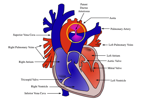
Patent ductus arteriosus (PDA) — literally "open arterial duct" — is the persistence of the ductus arteriosus (DA) after birth, maintaining a vascular communication between the descending aorta (distal to the left subclavian artery) and the pulmonary artery (at the junction of the main and left pulmonary arteries). It is classified as an acyanotic congenital heart disease (CHD) with a left-to-right shunt [1][2][3].
PDA is an acyanotic congenital heart disease with left-to-right shunt — in the same category as ASD, VSD, and AVSD [1][2].
In the fetus, the DA is a vital structure that diverts blood away from the high-resistance, fluid-filled lungs towards the systemic circulation. After birth, it should close functionally within 10–15 hours and anatomically by 2–3 weeks. When it fails to do so, the resulting left-to-right shunt causes pulmonary overcirculation and left ventricular volume overload [1][3][4].
Terminology Clarity
- Ductus arteriosus (DA): The normal fetal vascular channel — derived from the embryonic left 6th aortic arch.
- Patent ductus arteriosus (PDA): Pathological persistence of the DA after birth.
- Ligamentum arteriosum: The normal remnant after DA closure.
- In Chinese: 動脈導管未閉 [3].
| Feature | Detail |
|---|---|
| Incidence | 3–8 per 100,000 live births (isolated PDA in term infants) [2]; approximately 1 in 2,500–5,000 live births [3][4] |
| Proportion of CHD | ~5–10% of all congenital heart defects (6th commonest CHD) [3] |
| Sex ratio | Female predominance (M:F = 1:2) [2][3][4] |
| Prematurity | Extremely common — up to 60–70% of infants < 28 weeks' gestation have a haemodynamically significant PDA; incidence inversely proportional to gestational age and birth weight |
| Sibling recurrence | 2–4% risk in siblings [3][4] |
Preterm vs Term PDA — a crucial distinction
In preterm infants, the DA fails to close because the ductal smooth muscle is immature and less responsive to oxygen. This is a very common clinical problem in NICUs.
In term infants, an isolated PDA represents a true structural/congenital anomaly where normal closure mechanisms have failed despite mature ductal tissue.
Risk Factors
- Maternal rubella (especially first trimester infection — rubella embryopathy classically causes PDA + peripheral pulmonary artery stenosis) [3][4]
- Prematurity — the single most important risk factor [3][4]
- Birth at high altitude (chronic hypoxia prevents the normal postnatal oxygen-mediated ductal constriction) [3][4]
- Low birth weight
- Perinatal asphyxia (relative hypoxia delays closure)
- Excess fluid administration in neonates
High Yield — Rubella and PDA
Maternal rubella is the classic infectious cause of PDA. The rubella virus directly damages the ductal wall elastin and smooth muscle, preventing normal constriction. Rubella embryopathy = PDA + cataracts + sensorineural deafness + microcephaly. This is still relevant in Hong Kong exam settings even though rubella is now rare due to vaccination.
Anatomy and Normal Ductal Physiology
- The ductus arteriosus derives from the embryonic left 6th aortic arch [2]
- Aortic end: arises distal to the left subclavian artery [2][3]
- Pulmonary end: inserts at the junction of the main and left pulmonary arteries [2][3]
- The DA is a muscular artery (unlike the elastic aorta) — this is critical because its closure depends on smooth muscle contraction
In utero, the lungs are collapsed and filled with fluid → pulmonary vascular resistance (PVR) is very high. Only ~10% of right ventricular output goes to the lungs. The remaining ~60% of total cardiac output from the RV is diverted through the DA into the descending aorta to supply the systemic circulation (especially the lower body and placenta) [3][4].
This means in fetal life, the shunt is right-to-left (pulmonary artery → aorta), which is the physiological direction.
| Factor | Mechanism |
|---|---|
| Low PaO₂ | Fetal arterial PaO₂ is ~25 mmHg; hypoxia directly relaxes ductal smooth muscle via hypoxia-sensitive K⁺ channels |
| Circulating PGE₂ (from the placenta) | PGE₂ and PGI₂ are potent ductal vasodilators acting on EP₄ receptors on ductal smooth muscle [3][4] |
| Nitric oxide (NO) | Contributes to ductal patency |
| Low circulating endothelin-1 | Endothelin is a vasoconstrictor; its levels are low in fetal life |
At birth, two key changes trigger ductal constriction [3][4]:
-
↑PaO₂ (first breath → lungs expand → oxygen enters): Oxygen inhibits the voltage-gated K⁺ channels in ductal smooth muscle → membrane depolarisation → Ca²⁺ influx → smooth muscle contraction. The ductal smooth muscle is uniquely oxygen-sensitive (unlike systemic arteries).
-
↓Circulating PGE₂: Removal of the placenta (the major source of PGE₂) + increased pulmonary metabolism of PGE₂ by the now-functioning lungs → loss of the vasodilatory stimulus.
Timeline of closure:
- Functional closure: 10–15 hours after birth (smooth muscle constriction) [3][4]
- Anatomical closure: 2–3 weeks (intimal proliferation, fibrosis → becomes the ligamentum arteriosum) [3][4]
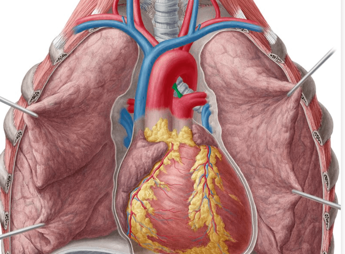
In preterm infants, the ductal smooth muscle is immature and has fewer oxygen-sensitive K⁺ channels + higher sensitivity to PGE₂ → this is why PDA is so common in preterms and why COX inhibitors (indomethacin/ibuprofen) work to close it (they reduce PGE₂ synthesis).
Key Concept — Oxygen and Prostaglandins
Think of ductal closure as a two-hit mechanism: (1) Oxygen constricts, (2) PGE₂ withdrawal allows constriction. In PDA, one or both of these mechanisms fail. This directly informs pharmacotherapy:
- To close PDA: use COX inhibitors (indomethacin, ibuprofen) → ↓PGE₂
- To keep PDA open (in duct-dependent lesions like TGA, critical coarctation): use PGE₁ (alprostadil) infusion
Aetiology and Pathophysiology
In term infants with PDA, the cause is usually:
- Structural abnormality of the ductal wall (deficient smooth muscle, abnormal elastic tissue)
- Genetic predisposition (TFAP2B mutations in Char syndrome)
- Environmental damage (rubella virus destroys ductal wall elastin)
In preterm infants, the cause is:
- Immature ductal smooth muscle with fewer oxygen-sensitive K⁺ channels
- Higher circulating PGE₂ levels
- Increased sensitivity to PGE₂
- Lower ability to generate endothelin-1 (a constrictor)
In PDA, the patent DA acts as a left-to-right shunt [3][4]:
Why left-to-right? After birth, systemic vascular resistance (SVR) is much higher than pulmonary vascular resistance (PVR). Since the DA connects the high-pressure aorta to the lower-pressure pulmonary artery, blood flows aorta → pulmonary artery (left-to-right).
Shunting occurs in PDA since the resistance and pressure in the pulmonary arterial bed < systemic arterial bed [2].
PDA present
  → L-to-R shunt (aorta → pulmonary artery)
    → ↑Pulmonary blood flow (pulmonary overcirculation)
      → ↑Pulmonary venous return to left atrium
        → ***LV volume overload*** [1][2]
          → LV dilatation → ↑stroke volume (Frank-Starling) → hyperdynamic circulation
            → ↑Pulse pressure (wide pulse pressure), bounding pulses
              → If large shunt persists: LV failure (HF)
By 6–8 weeks, ↓PVR leads to ↑shunting → ↑pulmonary venous return → LV volume overload + hyperdynamic circulation [3][4].
Why does it take 6–8 weeks?
At birth, PVR is still relatively high (the pulmonary arterioles are thick-walled from fetal life). Over the first 6–8 weeks, PVR progressively falls as pulmonary arteriolar smooth muscle regresses. As PVR drops, the pressure gradient across the PDA increases → more left-to-right shunting. This is why term infants with large PDA typically present with heart failure at 1–2 months of age [4]. In preterm infants, PVR falls much faster (within days), so they present earlier (within days) [4].
Unlike VSD (where the shunt is mainly systolic because the ventricles only generate pressure during systole), the PDA connects two great arteries. The aortic pressure exceeds pulmonary artery pressure in both systole AND diastole → flow through the PDA is continuous → this explains the characteristic continuous "machinery" murmur [4].
| Organ System | Effect | Mechanism |
|---|---|---|
| Lungs | Pulmonary overcirculation, congestion, ↑work of breathing, recurrent chest infections | ↑Pulmonary blood flow from L-to-R shunt |
| Left ventricle | Volume overload → dilatation → HF | ↑Venous return from pulmonary veins |
| Systemic circulation | "Steal" phenomenon — ↓diastolic BP, wide pulse pressure, bounding pulses | Blood "runs off" through PDA during diastole instead of perfusing systemic organs |
| Kidneys | ↓Renal perfusion → oliguria (especially in preterms) | Diastolic steal |
| Gut (preterms) | ↑Risk of necrotising enterocolitis (NEC) | Diastolic steal → mesenteric ischaemia |
| Brain (preterms) | ↑Risk of intraventricular haemorrhage (IVH) | Fluctuating cerebral blood flow from hyperdynamic circulation |
May lead to Eisenmenger syndrome after pulmonary arteriole remodelling with differential cyanosis [3][4].
If a large PDA is left untreated → chronic pulmonary overcirculation → pulmonary vascular remodelling (intimal hyperplasia, medial hypertrophy, fibrosis of pulmonary arterioles) → irreversible pulmonary hypertension → PVR eventually exceeds SVR → shunt reversal to right-to-left (deoxygenated blood from PA → aorta distal to left subclavian).
This causes differential cyanosis — a phenomenon unique to PDA-related Eisenmenger:
- Upper body (right hand, head): Normal saturation (supplied by aorta proximal to DA insertion, i.e., pre-ductal)
- Lower body (feet, left hand sometimes): Cyanosed (supplied by aorta distal to DA insertion, receiving deoxygenated blood from PA)
Differential cyanosis + differential clubbing = pathognomonic of Eisenmenger syndrome from PDA [3][4]. The feet are cyanosed and clubbed while the right hand is pink and normal.
Differential vs Reversed Differential Cyanosis
- Differential cyanosis (lower body cyanosed, upper body pink) = PDA with Eisenmenger
- Reversed differential cyanosis (upper body cyanosed, lower body pink) = occurs in TGA + PDA + coarctation (deoxygenated blood from RV→aorta goes to upper body; oxygenated blood from LV→PA→PDA goes to lower body). This is a rare but classic exam question.
Classification
This is the most clinically useful classification, based on Qp:Qs ratio (pulmonary-to-systemic blood flow ratio) [4]:
| Category | Qp:Qs | Clinical Impact |
|---|---|---|
| Small (silent/restrictive) PDA | < 1.5:1 | Minimal haemodynamic effect; may be incidentally detected |
| Moderate PDA | 1.5–2.2:1 | Some LV volume overload; may cause symptoms in adolescence/adulthood |
| Large (unrestrictive) PDA | > 2.2:1 | Significant LV volume overload; early HF, pulmonary hypertension risk |
| Type | Morphology |
|---|---|
| Type A (conical) | Most common; constriction at pulmonary end |
| Type B | Short, wide "window" PDA |
| Type C | Tubular |
| Type D | Complex with multiple constrictions |
| Type E | Elongated, conical |
This classification is mainly relevant for interventional catheter closure device selection.
| Context | Notes |
|---|---|
| Isolated PDA in term infant | True congenital anomaly; will not close spontaneously; requires intervention |
| PDA in preterm infant | Due to ductal immaturity; may respond to medical closure (COX inhibitors) or close spontaneously |
| PDA as part of a syndrome | Rubella embryopathy, Char syndrome, chromosomal abnormalities |
| Duct-dependent CHD | PDA is the patient's lifeline (e.g., in critical coarctation, interrupted aortic arch, transposition, pulmonary atresia) — must keep open with PGE₁ |
High Yield — Duct-Dependent Lesions
PDA is not always pathological. In duct-dependent CHD, closing the PDA is lethal:
- Duct-dependent systemic circulation: Critical coarctation of aorta, interrupted aortic arch, critical aortic stenosis, HLHS → PDA provides systemic blood flow
- Duct-dependent pulmonary circulation: Pulmonary atresia, critical pulmonary stenosis, severe TOF → PDA provides pulmonary blood flow
- Duct-dependent mixing: Transposition of great arteries (TGA) → PDA allows mixing of oxygenated and deoxygenated blood
In these cases, PGE₁ (alprostadil) infusion keeps the ductus open as a bridge to definitive surgery [1].
Clinical Features
The clinical presentation depends entirely on the size of the PDA and the degree of left-to-right shunting. Clinical features are determined by degree of L-to-R shunting [4].
Symptoms (with pathophysiological basis)
| Symptom | Pathophysiological Basis |
|---|---|
| Heart failure symptoms at 1–2 months (in term infants) [4] | As PVR drops over 6–8 weeks → ↑L-to-R shunt → LV volume overload → LV failure. Presents with tachypnoea, diaphoresis (especially with feeds), poor feeding, irritability |
| Earlier presentation in days in preterm (↓reserve) [4] | Preterm infants have immature myocardium with less contractile reserve + PVR falls faster (thin-walled pulmonary arterioles) → earlier onset of significant shunting |
| Failure to thrive / poor weight gain | Increased metabolic demands from ↑work of breathing + heart failure + poor caloric intake from feeding difficulties |
| Recurrent lower respiratory tract infections | Pulmonary overcirculation → pulmonary oedema → impaired mucociliary clearance and gas exchange → recurrent pneumonia |
| Dyspnoea / tachypnoea | Pulmonary congestion from volume overload → ↑lung water → ↓compliance → ↑work of breathing |
| Diaphoresis (sweating), especially during feeds | Sympathetic activation from HF; feeding is the infant equivalent of exercise |
| Symptom | Pathophysiological Basis |
|---|---|
| Differential cyanosis — cyanosis of lower extremities, pink upper extremities [3][4] | Reversed shunt (R-to-L) through PDA delivers deoxygenated blood to descending aorta (below the left subclavian artery). Pre-ductal blood remains oxygenated |
| Dyspnoea on exertion | Fixed pulmonary hypertension limits ability to increase pulmonary blood flow |
| Exercise-induced desaturation | Exercise → ↓SVR → worsens R-to-L shunting |
Signs (with pathophysiological basis)
| Sign | Pathophysiological Basis |
|---|---|
| Hyperdynamic circulation [4] | ↑Stroke volume (Frank-Starling compensation for LV volume overload) + low diastolic BP (diastolic runoff through PDA) → wide pulse pressure → visible/palpable hyperdynamic precordium |
| Wide pulse pressure (↑systolic, ↓diastolic BP) [4] | Blood "escapes" through PDA during diastole (aorta → PA) → diastolic pressure drops. Systolic pressure may be elevated from ↑stroke volume |
| Collapsing / bounding pulses (also called "water-hammer" pulse) [4] | Wide pulse pressure creates a rapidly rising and rapidly falling arterial waveform → felt as a bounding or collapsing pulse. Similar mechanism to aortic regurgitation |
| Tachycardia | Sympathetic compensation for ↓effective systemic output |
| Tachypnoea | Pulmonary congestion |
| Failure to thrive | ↑Metabolic demands + poor intake |
Collapsing pulse in a paediatric patient should make you think of PDA (or aortic regurgitation, which is much rarer in children).
| Sign | Pathophysiological Basis |
|---|---|
| Displaced, thrusting apex beat [4] | LV volume overload → LV dilatation → apex displaced laterally and inferiorly. "Thrusting" (volume-overloaded) rather than "heaving" (pressure-overloaded) |
| Hyperdynamic precordium | ↑Stroke volume → visible and palpable precordial impulse |
| Palpable thrill at left upper sternal border / left infraclavicular area | Turbulent continuous flow through the PDA |
This is the most important clinical sign and a classic exam question:
| Sign | Description | Pathophysiological Basis |
|---|---|---|
| Continuous murmur | Best heard at the left infraclavicular area / left upper sternal border (LUSB) [4] | The PDA connects aorta (high pressure) to PA (low pressure). Since aortic pressure exceeds PA pressure in BOTH systole AND diastole, blood flows continuously → murmur heard throughout the cardiac cycle |
| "Machine-like" or "machinery" murmur (Gibson's murmur) | Crescendo-decrescendo, peaking at S2, spilling into diastole | Maximal shunting occurs in late systole (when PA pressure is lowest relative to aortic); the murmur crescendos to S2 then diminuendos into diastole |
| Grade depends on PDA size | Large: 4/6, Moderate: 2–3/6, Small: < grade 3/6 [4] | Larger PDA = more turbulent flow = louder murmur (though very large unrestricted PDAs may paradoxically be quieter as flow becomes laminar) |
| In neonatal period: may be confined to systole only | Murmur confined to systole in the neonatal period before ↓PVR [4] | In the first days of life, PVR is still high → the pressure gradient across PDA is only present in systole (when aortic pressure exceeds PA pressure). As PVR drops over days to weeks, the diastolic gradient appears and the murmur becomes continuous |
The classic continuous "machinery" murmur of PDA at the left infraclavicular area is one of the most recognisable murmurs in paediatric cardiology [4].
High Yield — When the PDA Murmur Disappears
If a patient with known large PDA suddenly has a quieter murmur or the murmur loses its diastolic component, this is NOT a good sign. It may indicate:
- Equalisation of pressures due to severe pulmonary hypertension (approaching Eisenmenger) — the diastolic gradient disappears
- The murmur may eventually vanish entirely in Eisenmenger syndrome
A disappearing PDA murmur in an untreated patient = urgent concern for developing Eisenmenger physiology.
| Sign | Pathophysiological Basis |
|---|---|
| Loud P2 or single S2 (in large PDA with pulmonary hypertension) [4] | Pulmonary hypertension → forceful closure of pulmonary valve → loud P2. May become single S2 if PA pressure approaches aortic pressure (both valves close simultaneously) |
| Apical mid-diastolic rumble (functional mitral stenosis) | Large L-to-R shunt → ↑↑pulmonary venous return → relative mitral stenosis (too much blood flowing across a normal mitral valve) → low-pitched diastolic rumble at apex |
| S3 gallop | LV volume overload → rapid ventricular filling → S3 |
| Sign | Pathophysiological Basis |
|---|---|
| Differential cyanosis AND differential clubbing [3][4] | Deoxygenated blood from PA enters aorta below left subclavian → lower limbs cyanosed and clubbed; right hand/head remain pink. Left hand may be variably affected depending on exact DA insertion relative to left subclavian |
| Loud single S2 | Severe pulmonary hypertension → loud P2 merges with A2 |
| Absence of continuous murmur | No pressure gradient across PDA (PA pressure = aortic pressure) → no shunt murmur |
| Pulmonary regurgitation murmur (Graham Steell murmur) | Severe pulmonary hypertension → functional pulmonary valve regurgitation |
| Signs of right heart failure (hepatomegaly, raised JVP, peripheral oedema) | RV failure from chronic pressure overload |
| Small PDA (Qp:Qs < 1.5:1) | Moderate PDA (Qp:Qs 1.5–2.2:1) | Large PDA (Qp:Qs > 2.2:1) | |
|---|---|---|---|
| Symptoms | Asymptomatic murmur; infective endocarditis (uncommon) | ↓Exercise tolerance; HF in adolescence/adulthood | HF symptoms at 1–2 months (earlier in preterm); Eisenmenger syndrome |
| Pulses | Normal | Hyperdynamic, ↑pulse pressure | Collapsing pulse, hyperdynamic, ↑pulse pressure |
| Apex | Normal | Displaced thrusting apex | Displaced thrusting apex |
| Murmur | Continuous murmur at L infraclavicular < G3/6 | Continuous murmur at L infraclavicular/LUSB 2–3/6 | Continuous murmur at L infraclavicular/LUSB 4/6 |
| S2 | Normal | Normal or mildly loud P2 | Loud P2 or single S2 (if pHTN) |
| Other | — | — | ± Differential cyanosis/clubbing in Eisenmenger |
PDA in preterms deserves special mention because it is extremely common and presents differently:
| Feature | Preterm PDA | Term PDA |
|---|---|---|
| Incidence | Very high (up to 60–70% at < 28 weeks) | Uncommon (1/2500–5000) |
| Cause | Ductal immaturity | Structural anomaly |
| Timing of HF | Within days (PVR drops rapidly) [4] | 1–2 months |
| Murmur | May be systolic only or absent (high PVR in first days) | Classic continuous murmur |
| Additional complications | NEC, IVH, BPD (bronchopulmonary dysplasia), renal impairment | Less common |
| Medical closure | Responds to COX inhibitors (indomethacin/ibuprofen) or paracetamol | Unlikely to close with medical therapy |
| Spontaneous closure | May close spontaneously if gestational age > 28 weeks | Will NOT close spontaneously |
High Yield — Preterm PDA Clinical Signs
In a preterm infant, suspect a haemodynamically significant PDA if you see:
- Widened pulse pressure with bounding pulses
- Hyperdynamic precordium
- Systolic (not necessarily continuous) murmur at LUSB
- Worsening ventilator requirements (↑FiO₂, ↑ventilator pressures)
- Metabolic acidosis (from poor systemic perfusion)
- Oliguria (renal steal)
- Feed intolerance / abdominal distension (mesenteric steal → NEC risk)
High Yield Summary
Patent Ductus Arteriosus (PDA) — Key Points for Exams:
-
Definition: Persistence of the ductus arteriosus after birth → communication between descending aorta and PA → left-to-right shunt (acyanotic CHD)
-
Embryology: DA = left 6th aortic arch; aortic end distal to left subclavian; pulmonary end at junction of MPA and LPA
-
Normal closure mechanism: ↑PaO₂ + ↓PGE₂ → ductal constriction. Functional closure 10–15 hours; anatomical closure 2–3 weeks
-
Risk factors: Female sex, prematurity (most important), maternal rubella, high altitude, Char syndrome
-
Pathophysiology: L-to-R shunt → pulmonary overcirculation → LV volume overload → HF. Diastolic steal → wide pulse pressure, bounding pulses. Chronic overcirculation → Eisenmenger syndrome with differential cyanosis
-
Classic murmur: Continuous "machinery" (Gibson's) murmur at left infraclavicular area / LUSB; may be systolic-only in neonates; grade proportional to shunt size
-
Large PDA: HF at 1–2 months (days in preterms); collapsing pulse, displaced thrusting apex, loud P2
-
Small PDA: Asymptomatic; risk of infective endocarditis
-
Pharmacology link: COX inhibitors (indomethacin/ibuprofen) ↓PGE₂ → close PDA; PGE₁ (alprostadil) → keep PDA open in duct-dependent lesions
-
Eisenmenger in PDA = differential cyanosis (feet blue, right hand pink) — pathognomonic
Active Recall — Patent Ductus Arteriosus (PDA)
[1] Lecture slides: Block C - Heart failure and cyanosis in children: acyanotic and cyanotic congenital heart disease.pdf [2] Senior notes: MBBS Final MB (Pediatrics) (Felix PY Lai).pdf, Cardiovascular Diseases — PDA section (p251) [3] Senior notes: Adrian Lui Pediatrics Notes.pdf (p202) [4] Senior notes: Ryan Ho Cardiology.pdf (p189)
Differential Diagnosis of Patent Ductus Arteriosus (PDA)
When encountering a child with features suggestive of PDA — a continuous murmur, wide pulse pressure, bounding pulses, signs of heart failure in infancy, or differential cyanosis — you need a structured approach to the differential diagnosis. The DDx is best organised around the presenting clinical feature, because PDA can mimic (and be mimicked by) several conditions depending on what drew clinical attention in the first place.
A child with PDA can present in several ways. The DDx differs depending on which feature is dominant:
- Continuous murmur — the classic presentation
- Heart failure in infancy (acyanotic, with pulmonary overcirculation)
- Wide pulse pressure / bounding pulses / hyperdynamic circulation
- Differential cyanosis (Eisenmenger physiology)
- Unequal BP and pulses between upper and lower limbs
Let us work through each systematically.
A continuous murmur is one that extends through S2 without interruption — it occupies both systole and diastole. This is distinct from a "to-and-fro" murmur (which has two separate systolic and diastolic components with a gap at S2).
Patent ductus arteriosus is the classic and most common cause of a continuous murmur in paediatrics [1][5].
Why is the PDA murmur continuous? Because the aortic pressure exceeds pulmonary artery pressure throughout the entire cardiac cycle (both systole and diastole), creating uninterrupted flow through the duct [3][4][6].
Any other condition producing continuous flow between a high-pressure and low-pressure vascular bed can mimic this:
| Condition | Location of Murmur | Distinguishing Features | Why it's continuous |
|---|---|---|---|
| Patent ductus arteriosus | Left infraclavicular / LUSB [4][5] | Collapsing pulse, wide pulse pressure; machinery quality peaking at S2 | Aorta → PA pressure gradient persists in systole and diastole |
| Aortopulmonary window | LUSB / left 2nd–3rd ICS | Very rare; mimics large PDA clinically but murmur may be more "to-and-fro" than truly continuous; often severe HF early | Direct communication between ascending aorta and MPA (no duct involved); same continuous pressure gradient |
| Coronary arteriovenous fistula | Variable (often lower sternal border) | May have diastolic-dominant murmur; rare in children | Continuous flow from high-pressure coronary artery to low-pressure receiving chamber (RA, RV, PA, or CS) |
| Ruptured sinus of Valsalva aneurysm | Right sternal border (if ruptures into RV/RA) | Acute onset, severe HF; extremely rare in children (more common in adults, Marfan syndrome) | Continuous flow from aortic sinus into low-pressure right heart chamber |
| Venous hum | Supraclavicular (right > left), radiates to neck | Innocent murmur; abolished by turning head, lying supine, or compressing ipsilateral IJV; no other abnormal signs | Continuous turbulent flow in IJV; more prominent when upright |
| Surgical systemic-to-pulmonary shunt (e.g., Blalock-Taussig shunt) | Depends on shunt site | History of prior cardiac surgery for cyanotic CHD | Subclavian → PA anastomosis creates same physiology as PDA |
| Collateral vessels (e.g., in coarctation of aorta, pulmonary atresia) | Variable; often posterior (intercostal) | Associated features of underlying condition (e.g., rib notching in coarctation, cyanosis in PA) | Extensive collateral flow between high-pressure and low-pressure vascular beds |
| Mammary souffle | Over breast in adolescent / lactating | Disappears with firm stethoscope pressure; physiological in pregnancy/lactation | Increased blood flow through internal mammary/intercostal arteries |
| Peripheral pulmonary stenosis (physiological in neonates) | Bilateral axillae and back | Common in neonates; usually resolves by 6 months; soft, ejection quality with some spill into diastole | Turbulence at branch PA bifurcation points |
High Yield — Venous Hum vs PDA
The venous hum is the most common innocent continuous murmur in childhood and must be distinguished from PDA:
- Venous hum: supraclavicular, abolished by turning head / lying down / compressing IJV
- PDA: left infraclavicular / LUSB, not abolished by positional changes, associated with bounding pulses and wide pulse pressure
If a continuous murmur disappears when the child lies supine, it is almost certainly a venous hum — reassure and discharge.
CFB lecture slide [5]: Continuous murmur — AS/AR and Patent ductus arteriosus associated with cyanosis, left parasternal heave, collapsing pulse. Note: the CFB slide lists "AS/AR" alongside PDA as causes of continuous murmur. Strictly, combined AS + AR gives a "to-and-fro" murmur rather than a true continuous murmur (there is a brief gap at S2), but this is a recognised exam distractor.
When a term infant presents at 1–2 months with tachypnoea, feeding difficulty, diaphoresis, and failure to thrive — think of acyanotic CHD with left-to-right shunt causing volume overload [2][4]:
| Condition | Overloaded Chamber | Murmur | Key Distinguishing Feature |
|---|---|---|---|
| Large PDA | LV [2] | Continuous murmur at L infraclavicular area | Bounding pulses, wide pulse pressure, murmur maximal at S2 |
| Large VSD | LV [2] | Pansystolic at LLSB | Murmur loudest in smaller VSDs; large VSD may have soft murmur + loud P2; no wide pulse pressure (shunt only in systole) |
| Large ASD (secundum, uncommon to cause infant HF) | RV [2] | Ejection systolic at LUSB with fixed split S2 | Usually asymptomatic in infancy; HF rare unless very large or associated anomalies |
| AVSD (atrioventricular septal defect) | Both ventricles [2] | Pansystolic at LLSB ± apical | Strongly associated with Down syndrome; superior QRS axis on ECG |
| Aortopulmonary window | LV | Continuous or to-and-fro at LUSB | Very rare; often mistaken for large PDA. Diagnosed on echo |
| Total anomalous pulmonary venous return (TAPVR, non-obstructed) | RV | Ejection systolic at LUSB + fixed split S2 | All pulmonary veins drain to RA; RV volume overload. Obstructed TAPVR → severe cyanosis |
| Truncus arteriosus | Both ventricles | Ejection systolic + early diastolic (truncal regurgitation) | Single S2; cyanosis may be mild; VSD always present |
Large VSD and PDA = LV overloaded; Large ASD = RV overloaded; AVSD = Both ventricles overloaded [2][7].
Timing of HF Presentation — The PVR Clock
All large L-to-R shunts present with HF as PVR drops after birth. The timing gives a clue:
- Days to 1 week: Preterm PDA (PVR drops very fast in preterms)
- 1–2 months: Large VSD, large PDA in term infants
- Rarely causes HF in infancy: ASD (because the pressure difference between atria is small, so the shunt volume is modest)
If an infant has HF at 6 weeks with a continuous murmur + bounding pulses → PDA until proven otherwise. If an infant has HF at 6 weeks with a pansystolic murmur at LLSB → large VSD until proven otherwise.
The wide pulse pressure in PDA results from diastolic runoff (blood escaping through the duct during diastole) lowering the diastolic pressure, combined with increased stroke volume raising the systolic pressure. Other conditions causing a similar "runoff" physiology include:
| Condition | Mechanism of Wide Pulse Pressure | Distinguishing Features |
|---|---|---|
| PDA | Diastolic runoff through duct (aorta → PA) | Continuous murmur at L infraclavicular area |
| Aortic regurgitation (AR) | Diastolic runoff back into LV through incompetent aortic valve | Early diastolic decrescendo murmur at LUSB/Erb's point; associated with bicuspid aortic valve, rheumatic heart disease |
| Arteriovenous malformation (AVM) — e.g., cerebral AVM (Vein of Galen malformation in neonates), hepatic AVM | Diastolic runoff into low-resistance AV fistula | Location-dependent signs; Vein of Galen → high-output HF in neonate with cranial bruit; Hepatic AVM → hepatomegaly + bruit over liver |
| Severe anaemia | ↓Viscosity + ↑CO = hyperdynamic circulation; no true "runoff" but wide pulse pressure from ↑stroke volume | Pallor, tachycardia, flow murmur; no continuous murmur |
| Thyrotoxicosis | ↑CO + ↓SVR | Tachycardia, tremor, lid lag, goitre; rare in infants |
| Sepsis / high-output states | ↓SVR (warm shock) | Fever, ill-appearing, other sepsis signs |
| Truncus arteriosus | Runoff through common trunk to both systemic and pulmonary circulation | Single S2, mild cyanosis, VSD |
Differential cyanosis is pathognomonic of PDA with Eisenmenger syndrome [3][4] — deoxygenated blood enters the aorta distal to the left subclavian artery through the reversed (R-to-L) PDA. However, consider:
| Condition | Pattern | Mechanism |
|---|---|---|
| PDA with Eisenmenger | Lower body cyanosed, upper body pink (differential cyanosis) [3][4] | R-to-L shunt through PDA delivers deoxygenated blood post-ductally |
| Interrupted aortic arch / critical coarctation with PDA | Lower body cyanosed (if PDA shunts R-to-L) OR pink lower body (if PDA provides systemic flow from well-oxygenated LV blood) — depends on associated lesions | Complex physiology; duct-dependent systemic circulation |
| TGA + coarctation + PDA (reversed differential cyanosis) | Upper body cyanosed, lower body pink | RV → aorta (deoxygenated) supplies pre-ductal territory; LV → PA → PDA → descending aorta (oxygenated) supplies post-ductal territory |
| Persistent pulmonary hypertension of the newborn (PPHN) | Pre-ductal SpO₂ > post-ductal SpO₂ (similar to differential cyanosis) | R-to-L shunting through PDA due to elevated PVR in neonate; different aetiology (not Eisenmenger) |
CFB lecture slide [5]: PDA — more severe clubbing of feet than hands, because the ductus delivers deoxygenated blood distal to the left subclavian artery — this is a specific exam point for MCQs.
Felix Lai notes [2][7] list the differential diagnosis for unequal BP and pulses:
| Condition | Key Features | Paediatric Relevance |
|---|---|---|
| Coarctation of aorta | Upper limb hypertension, radio-femoral delay, weak femoral pulses; systolic murmur at left infra-scapular area | Most important DDx — must always check femoral pulses in any child with murmur or hypertension |
| Peripheral artery disease (atherosclerosis, arterial thrombosis) [2][7] | Essentially an adult condition | Very rare in children (unless iatrogenic — e.g., post-catheterisation femoral artery thrombosis) |
| Aortic dissection [2][7] | Acute chest/back pain, pulse discrepancy | Extremely rare in paediatrics (consider Marfan, Ehlers-Danlos) |
| Supravalvar aortic stenosis [2][7] | Higher BP in right arm than left (Coanda effect directs jet towards innominate artery); elfin facies | Associated with Williams syndrome |
| Interrupted aortic arch | Absent femoral pulses; duct-dependent systemic circulation in neonate | Presents in first days of life when PDA closes → cardiovascular collapse |
High Yield — Always Check Femoral Pulses
In any infant or child with a murmur, heart failure, or hypertension:
- Always palpate femoral pulses and compare with brachial/radial pulses
- Absent or weak femorals → coarctation of aorta until proven otherwise
- Bounding femorals with wide pulse pressure → PDA
- This simple examination step distinguishes two of the most important acyanotic CHDs
In the NICU, suspected haemodynamically significant PDA in a preterm must be differentiated from other causes of cardiorespiratory deterioration:
| Condition | Distinguishing Features |
|---|---|
| Haemodynamically significant PDA | Bounding pulses, wide pulse pressure, hyperdynamic precordium, systolic/continuous murmur at LUSB, worsening ventilatory needs, oliguria — confirmed on echo |
| Sepsis / pneumonia | Fever/hypothermia, ↑WBC or ↓WBC, ↑CRP, positive blood culture; may coexist with PDA |
| Necrotising enterocolitis (NEC) | Abdominal distension, bloody stools, pneumatosis intestinalis on AXR; may be a complication of PDA (mesenteric steal) |
| Intraventricular haemorrhage (IVH) | Sudden neurological deterioration, bulging fontanelle, ↓Hb; confirmed on cranial USS; may be complication of PDA (fluctuating CBF) |
| Bronchopulmonary dysplasia (BPD) | Chronic oxygen dependency; occurs in preterms with prolonged ventilation; PDA worsens BPD by increasing pulmonary blood flow |
| Presenting Feature | Most Likely DDx to Distinguish From PDA | Key Differentiator |
|---|---|---|
| Continuous murmur | Venous hum | Positional changes abolish venous hum |
| Continuous murmur + HF | Aortopulmonary window | Very rare; echo differentiates |
| HF in infancy | Large VSD | Pansystolic (not continuous) murmur at LLSB; no wide pulse pressure |
| Wide pulse pressure | Aortic regurgitation | Early diastolic murmur, not continuous machinery murmur |
| Differential cyanosis | PPHN (neonate) | PPHN is acute, in the neonatal period; responds to pulmonary vasodilators |
| Weak femoral pulses | Coarctation of aorta | In PDA, femorals are bounding not weak (unless Eisenmenger) |
High Yield Summary — DDx of PDA
-
Continuous murmur DDx: PDA (most common), venous hum (most common innocent cause — abolished by positional change), aortopulmonary window, coronary AV fistula, ruptured sinus of Valsalva, surgical BT shunt, collaterals
-
HF in infancy DDx: Large PDA vs large VSD vs AVSD — differentiated by murmur character and pulse features. Large VSD and PDA = LV overloaded; Large ASD = RV overloaded; AVSD = Both ventricles overloaded [2]
-
Wide pulse pressure DDx: PDA, aortic regurgitation, AVM, severe anaemia, thyrotoxicosis
-
Differential cyanosis (lower body blue, upper pink) = pathognomonic of PDA with Eisenmenger; reversed pattern = TGA + PDA + coarctation
-
PDA causes more severe clubbing of the feet than hands [5] — because deoxygenated blood enters aorta distal to L subclavian
-
Unequal limb pulses: Bounding femorals = PDA; Weak femorals = coarctation. Always check femoral pulses in any child with a murmur
-
Echocardiography is the definitive differentiator for all of these conditions
Active Recall — DDx of Patent Ductus Arteriosus
References
[1] Lecture slides: GC 147. Heart failure and cyanosis in children acyanotic and cyanotic congenital heart disease - Part 1.pdf [2] Senior notes: MBBS Final MB (Pediatrics) (Felix PY Lai).pdf, Cardiovascular Diseases — PDA section (p251, p262) [3] Senior notes: Adrian Lui Pediatrics Notes.pdf (p202) [4] Senior notes: Ryan Ho Cardiology.pdf (p189) [5] Lecture slides: CFB (MED06) Cardiovascular (II) Physical Examination.pdf (p74, p87) [6] Senior notes: Maksim Paediatric Notes.pdf (p58) [7] Senior notes: MBBS Final MB (Medicine) (Felix PY Lai).pdf (p564, p575)
Diagnostic Criteria, Investigation Modalities, and Diagnostic Algorithm for PDA
Unlike many conditions in medicine, PDA does not have a formal set of "diagnostic criteria" in the way that, say, rheumatic fever has the Jones criteria. Instead, the diagnosis rests on:
- Clinical suspicion — based on history and physical examination findings (continuous murmur, bounding pulses, wide pulse pressure, ± signs of HF)
- Echocardiographic confirmation — the definitive diagnostic modality in all age groups
The real clinical challenge is not "does this child have a PDA?" (echo answers that quickly) but rather "is this PDA haemodynamically significant?" — because this determines whether intervention is needed. This is especially important in preterm neonates where PDA is common and often an incidental echo finding.
Echocardiography is diagnostic and allows estimation of PDA size [4]. It is the gold-standard investigation for PDA in paediatrics.
Criteria for Haemodynamic Significance (Preterm & Term)
Because treatment decisions hinge on whether the PDA is causing meaningful cardiovascular compromise, you must understand what makes a PDA "haemodynamically significant" (hsPDA):
There is no universally agreed single definition, but the following echo and clinical parameters are used in combination:
| Parameter | Finding Suggesting hsPDA | Rationale |
|---|---|---|
| PDA diameter | > 1.5 mm (or > 1.4 mm/kg body weight) | Larger duct = larger shunt |
| LA:Ao ratio | > 1.4:1 (normal < 1.2:1) | LA enlarges from ↑pulmonary venous return (reflects volume of L-to-R shunt) |
| Direction of ductal flow | Unrestrictive L-to-R (low velocity, pulsatile) | Low velocity = no pressure gradient = large, unrestrictive duct |
| Diastolic flow reversal in descending aorta | Present (retrograde/absent diastolic flow) | Blood running backwards in diastole = significant diastolic steal through PDA |
| Absent or reversed end-diastolic flow in mesenteric/renal/cerebral arteries | Present | Organ steal → risk of NEC, renal failure, IVH |
| Qp:Qs ratio | > 1.5:1 (moderate) or > 2:1 (large) | Quantifies shunt volume |
| LV dilatation | Present | Volume overload |
Clinical signs of haemodynamic significance (complement echo findings):
- Widened pulse pressure (systolic − diastolic > 25 mmHg)
- Bounding pulses, hyperdynamic precordium
- Worsening ventilatory requirements
- Metabolic acidosis
- Oliguria, feed intolerance
| PDA Size | Echo & Clinical Criteria |
|---|---|
| Small PDA (Qp:Qs < 1.5:1) | CXR: normal; ECG: normal; Echo: small PDA, no chamber enlargement [4] |
| Moderate PDA (Qp:Qs 1.5–2.2:1) | LV dilatation on echo, mild LA enlargement, some ↑pulmonary flow |
| Large PDA (Qp:Qs > 2.2:1) | CXR: cardiomegaly (LV dilatation), pulmonary plethora; ECG: LVH, LAE, ± RVH or even RAE (if significant pHTN); Echo: large PDA with LV dilatation, LA enlargement, ± pHTN [4] |
Investigation Modalities
Let us work through each investigation systematically, with the key findings, their interpretation, and the underlying pathophysiological reasoning.
CXR is the first-line imaging and gives a quick overview of heart size and pulmonary vascularity.
| PDA Size | CXR Findings | Pathophysiological Explanation |
|---|---|---|
| Small PDA | Normal [4] | Shunt volume too small to cause any detectable chamber enlargement or pulmonary overcirculation |
| Moderate PDA | Mild cardiomegaly, mild ↑ pulmonary vascular markings | Some LV dilatation + moderate ↑ pulmonary blood flow |
| Large PDA | Cardiomegaly (LV dilatation), pulmonary plethora [4] | Large L-to-R shunt → significant ↑ pulmonary blood flow (plethora = engorged pulmonary vessels visible to the periphery of lung fields) + LV volume overload → LV and LA enlargement → overall cardiomegaly |
| Eisenmenger | Prominent main pulmonary artery (MPA) segment, "pruning" of peripheral pulmonary vessels, ± RV enlargement | Pulmonary hypertension causes proximal PA dilation but peripheral arteriolar obliteration ("pruning") |
How to read pulmonary vascularity on paediatric CXR:
- Pulmonary plethora = pulmonary arteries visible in the outer third of lung fields; vessels appear enlarged and numerous → suggests L-to-R shunt with ↑ pulmonary blood flow
- Oligaemic lung fields = paucity of pulmonary vessels → suggests ↓ pulmonary blood flow (e.g., TOF, pulmonary atresia)
- Pulmonary venous congestion = cephalisation (upper lobe vessel prominence), Kerley B lines, interstitial/alveolar oedema → suggests LV failure or obstructed pulmonary venous return
In PDA, you expect plethora (↑ flow), not congestion (↑ pressure from LV failure) — unless the patient is in frank LV failure, in which case both may coexist.
CXR Tip — Plethora vs Congestion
Both cause "increased markings" but they look different:
- Plethora: vessels are big and well-defined to the periphery; upper and lower lobes equally affected
- Congestion: cephalisation (upper > lower), hazy/fluffy markings, Kerley lines, pleural effusions
In an exam CXR, describe "pulmonary plethora" for L-to-R shunt lesions (PDA, VSD, ASD) and "pulmonary venous congestion" for left heart failure.
ECG reflects the haemodynamic burden on cardiac chambers. It does not diagnose PDA directly but gives information about volume/pressure overload patterns.
| PDA Size | ECG Findings | Pathophysiological Explanation |
|---|---|---|
| Small PDA | Normal [4] | No significant haemodynamic burden |
| Moderate PDA | LVH (tall R waves in V5-V6, deep S in V1) ± LAE (bifid P wave in II, negative terminal P wave in V1) | LV volume overload → LV hypertrophy. ↑ pulmonary venous return → LA stretching → LAE |
| Large PDA | LVH, LAE, ± RVH or even RAE (if significant pHTN) [4] | All of the above PLUS: if pulmonary hypertension develops → RV pressure overload → RVH (tall R in V1, RAD) and RA distension → RAE (peaked P waves in II) |
| Eisenmenger | RVH dominant (tall R in V1, RAD), RAE | Once PVR is suprasystemic, the RV is the predominant overloaded chamber |
Age-specific ECG interpretation in paediatrics — a critical point:
- Neonates normally have right-axis deviation and RV dominance (because in utero the RV is the dominant ventricle pumping against high PVR)
- Over the first 6 months, the axis gradually shifts leftward as LV becomes dominant
- Therefore, persistent RVH beyond 6 months or progressive RVH in a child with known PDA = developing pulmonary hypertension — a red flag
High Yield — ECG Pattern Progression in PDA
The ECG pattern in PDA evolves with disease severity:
- Normal → small PDA
- LVH + LAE → moderate-large PDA with volume overload
- Biventricular hypertrophy → large PDA with developing pHTN
- RVH dominant → Eisenmenger syndrome (irreversible pHTN)
If an ECG in a child with PDA shows RVH overtaking LVH, this is a worrying sign suggesting pulmonary hypertension — urgently re-evaluate with echo ± catheterisation.
Echo is diagnostic and allows estimation of size [4]. It is the single most important investigation in PDA. Transthoracic echocardiography (TTE) is used in essentially all paediatric cases (transoesophageal echo is rarely needed).
What Echo Assesses:
| Parameter | How It Is Measured | Clinical Significance |
|---|---|---|
| PDA presence and morphology | 2D imaging in parasternal short-axis and suprasternal views; colour Doppler shows turbulent flow jet in MPA | Confirms diagnosis; identifies anatomy for device/surgical planning |
| PDA diameter | Measured at narrowest point (usually pulmonary end) | Correlates with shunt volume; used for device sizing in transcatheter closure |
| Direction and pattern of ductal flow | Pulsed-wave (PW) and continuous-wave (CW) Doppler | L-to-R = standard; bidirectional or R-to-L = elevated PVR/Eisenmenger. Continuous L-to-R flow pattern = PDA with gradient in systole AND diastole |
| Peak velocity across PDA | CW Doppler | High velocity (> 2 m/s) = restrictive PDA (small/moderate). Low velocity = unrestrictive (large) PDA. Using modified Bernoulli equation: ΔP = 4V² → estimates pressure gradient |
| LA:Ao ratio | M-mode at aortic valve level | > 1.4:1 suggests significant L-to-R shunt. LA enlarges because of ↑ pulmonary venous return. Normal < 1.2:1 |
| LV dimensions and function | M-mode and 2D | LV end-diastolic dimension ↑ (volume overload); LV systolic function initially hyperdynamic (↑ EF/FS), later may deteriorate |
| Qp:Qs ratio | Calculated from Doppler flow across pulmonary and aortic valves | Quantifies shunt; < 1.5 = small, 1.5–2.2 = moderate, > 2.2 = large [4] |
| Estimated PA pressure | TR jet velocity (if tricuspid regurgitation present) → RVSP = 4V² + RAP; ductal flow gradient | Assesses for pulmonary hypertension |
| Diastolic flow reversal in descending aorta | PW Doppler in descending aorta | If present → significant diastolic steal through PDA; marker of haemodynamic significance (especially in preterms) |
| End-diastolic flow in renal / mesenteric / cerebral arteries (in preterms) | PW Doppler | Absent or reversed = organ steal → high risk of NEC, renal failure, IVH |
| Associated anomalies | Comprehensive structural assessment | Must exclude coarctation of aorta, VSD, ASD, aortic/pulmonary valve abnormalities. Important: a PDA may be masking coarctation — need to reassess after PDA closure |
Echo is the gold standard for PDA diagnosis, sizing, and haemodynamic assessment in all age groups [2][4].
High Yield — What to Look for on Echo
In an exam scenario where you are asked "What investigations would you order?", the answer is echocardiography as the key investigation. When describing what you expect, mention:
- Visualisation of the PDA on 2D + colour Doppler
- PDA diameter (for sizing)
- LA:Ao ratio (for shunt significance)
- LV dimensions (volume overload?)
- Estimated PA pressure (pulmonary hypertension?)
- Descending aorta diastolic flow (steal?)
- Exclusion of associated anomalies (especially coarctation)
Beware — PDA Masking Coarctation
A PDA may maintain flow to the descending aorta, effectively bypassing a coarctation. If the PDA is closed (spontaneously, medically, or surgically) without recognising the coarctation, the child can present with acute cardiovascular collapse (absent femoral pulses, shock, metabolic acidosis). Always look for coarctation on echo before and after PDA closure.
Pulse oximetry screening is now widely used for critical congenital heart disease (CCHD) in neonates. While PDA per se is not the target of pulse oximetry screening (the screen targets duct-dependent cyanotic lesions), oximetry is useful in PDA assessment:
| Measurement | Finding in PDA | Interpretation |
|---|---|---|
| Pre-ductal SpO₂ (right hand) | Normal (95–100%) | Blood from ascending aorta (pre-ductal) is normally oxygenated |
| Post-ductal SpO₂ (either foot) | Normal in isolated L-to-R PDA; ↓ if Eisenmenger (R-to-L) | In standard PDA, the shunt adds oxygenated blood to PA → post-ductal SpO₂ is normal. If R-to-L shunting → post-ductal SpO₂ < pre-ductal = differential cyanosis |
| Pre-post ductal difference > 3% | Significant | Suggests R-to-L ductal shunting → evaluate for PPHN, Eisenmenger, or duct-dependent cyanotic CHD |
In a neonate, a pre-post ductal SpO₂ difference > 3% is significant and warrants urgent echocardiography.
Cardiac catheterisation is not routinely needed for diagnosis of PDA in the current era (echo is sufficient). However, it is used in specific circumstances:
| Indication | Purpose |
|---|---|
| Assessment of pulmonary vascular resistance (PVR) when echo findings are equivocal for operability | Direct measurement of PA pressure, PVR, and response to pulmonary vasodilators (e.g., inhaled NO, oxygen). Determines whether Eisenmenger is irreversible |
| Therapeutic transcatheter closure | Combined diagnostic + interventional procedure (see Management section) |
| Assessment of associated complex CHD | When echo is insufficient for surgical planning |
Key catheterisation findings in PDA:
| Measurement | Finding | Interpretation |
|---|---|---|
| Step-up in oxygen saturation at PA level | PA O₂ sat > RA/RV O₂ sat | Oxygenated blood from aorta enters PA via PDA → O₂ step-up at PA (this localises the shunt to the great-artery level, distinguishing from VSD which shows step-up at RV) |
| Qp:Qs calculation | Calculated from Fick principle using O₂ saturations | Quantifies shunt precisely |
| PA pressure | Direct measurement | Determines severity of pulmonary hypertension |
| PVR | Calculated: PVR = (mean PAP − PAWP) / Qp | If PVR > 8–12 Wood units or PVR:SVR ratio > 0.67 → likely irreversible Eisenmenger → closure contraindicated [3] |
| Vasoreactivity testing | Response to inhaled NO / 100% O₂ / IV adenosine | If PVR drops significantly → some reversibility → may still be operable with careful peri-operative management |
Catheterisation — O₂ Saturation Step-Up Localises the Shunt Level
The location of the O₂ step-up on catheterisation tells you the level of the left-to-right shunt:
- RA step-up: ASD (oxygenated blood from LA enters RA)
- RV step-up: VSD (oxygenated blood from LV enters RV)
- PA step-up: PDA (oxygenated blood from aorta enters PA)
This is a classic exam concept.
Rarely needed in isolated PDA but may be used in:
- Complex anatomy where echo windows are suboptimal
- Adolescent/adult PDA with calcification (CT better delineates calcified duct)
- Assessment of associated aortic arch anomalies
- Delineation of major aortopulmonary collateral arteries (MAPCAs) in pulmonary atresia with VSD [3]
Not diagnostic for PDA but important in overall assessment:
| Test | Relevance |
|---|---|
| ABG / VBG | Metabolic acidosis (lactic acidosis from poor systemic perfusion in large PDA or preterm hsPDA) |
| BNP / NT-proBNP | Elevated in haemodynamically significant PDA; used as adjunct in preterm neonates to guide treatment. Correlates with shunt volume and ventricular strain |
| Renal function (Cr, urea) | Assess renal perfusion (steal phenomenon). Also baseline before giving indomethacin/ibuprofen (nephrotoxic) |
| Electrolytes | Hyponatraemia from dilutional effect (fluid retention in HF) or diuretic use |
| FBC | Polycythaemia (if chronic hypoxia/Eisenmenger); anaemia worsens symptoms |
| CRP / blood culture | If infective endocarditis (endarteritis) suspected |
| Investigation | Large PDA | Small PDA |
|---|---|---|
| CXR | Cardiomegaly (LV dilatation), pulmonary plethora | Normal |
| ECG | LVH, LAE, ± RVH or even RAE (if significant pHTN) | Normal |
| Echo | Diagnostic and estimation of size; large PDA with LV dilatation, LA enlargement, ↑ PA pressures, diastolic flow reversal in descending aorta | Diagnostic and estimation of size; small PDA without significant chamber changes |
Special Diagnostic Considerations by Age Group
- Echo is performed early (often day 2–3) in very preterm infants (< 28 weeks) because of the high incidence of hsPDA
- Clinical signs may be subtle initially — widened pulse pressure, hyperdynamic precordium, and worsening ventilatory requirements are often the first clues
- BNP/NT-proBNP can be used as an adjunct to guide whether to treat: markedly elevated levels correlate with hsPDA
- Cranial ultrasound should be done concurrently to screen for IVH (a complication of PDA-related fluctuating cerebral blood flow)
- The murmur may be confined to systole in the neonatal period before ↓PVR [4] — do not wait for a "classic continuous murmur" before investigating a preterm with suspected PDA
- CXR + ECG + Echo is the standard workup
- CXR: cardiomegaly with pulmonary plethora; ECG: LVH + LAE [4]
- Echo confirms PDA, quantifies shunt, and — critically — excludes coexisting coarctation of aorta and other anomalies
- Often detected during school health screening or sports pre-participation evaluation
- CXR and ECG may be normal [4] if the PDA is small
- Echo confirms the diagnosis even in an asymptomatic child with a continuous murmur
- If the presentation is a cyanotic neonate or a neonate in shock after duct closure → urgent echo to define the underlying anatomy
- PGE₁ infusion must be started empirically while awaiting echo if duct-dependent circulation is suspected [2][8]
- PGE₁/PGE₂ side effects: fever, hypotension, bradycardia, apnoea, seizure-like activities [2][8] — the baby must be monitored in a high-dependency or intensive care setting
Although not specific to PDA, this is relevant when a neonate presents with cyanosis and you need to determine if a duct-dependent cyanotic lesion is present:
| Cyanotic CHD | Respiratory Disease | |
|---|---|---|
| Baseline SpO₂ | Low (< 85%) | Low |
| Response to 100% O₂ for 10 min | PaO₂ remains < 150 mmHg (often < 100 mmHg) | PaO₂ rises > 150 mmHg |
| Interpretation | Fixed R-to-L shunt — oxygen cannot reach pulmonary blood because shunt bypasses lungs | V/Q mismatch or hypoventilation — supplemental O₂ can improve alveolar gas exchange |
An oligaemic lung field on CXR supports cyanotic CHD over respiratory disease as the cause of neonatal cyanosis [9].
High Yield Summary — Diagnosis of PDA
-
Echocardiography is the gold-standard diagnostic modality — confirms PDA, assesses size/morphology, quantifies shunt (Qp:Qs), estimates PA pressure, evaluates LA:Ao ratio, LV dimensions, and descending aortic diastolic flow
-
CXR findings scale with PDA size: small → normal; large → cardiomegaly + pulmonary plethora [4]
-
ECG findings scale with PDA size: small → normal; large → LVH, LAE, ± RVH/RAE if pHTN [4]
-
Cardiac catheterisation is reserved for assessing PVR when Eisenmenger is suspected or for therapeutic transcatheter closure. Key finding: O₂ saturation step-up at PA level localises the shunt
-
Pulse oximetry: pre-post ductal difference > 3% = R-to-L ductal shunting
-
Preterm PDA: echo + clinical signs + BNP together guide treatment decisions. Murmur may be systolic-only in first days
-
Always exclude coarctation of aorta on echo — PDA can mask coarctation
-
PGE₁ side effects: fever, hypotension, bradycardia, apnoea, seizure-like activities [2][8]
Active Recall — Diagnosis of PDA
References
[2] Senior notes: MBBS Final MB (Pediatrics) (Felix PY Lai).pdf, Cardiovascular Diseases — PDA section (p251, p281, p282) [3] Senior notes: Adrian Lui Pediatrics Notes.pdf (p202, p212, p215) [4] Senior notes: Ryan Ho Cardiology.pdf (p189) [8] Senior notes: MBBS Final MB (Medicine) (Felix PY Lai).pdf (p564–567, p594–595) [9] AOS material: AOS - Paeds.pdf (Question 6)
Management of Patent Ductus Arteriosus (PDA) in Paediatrics
Management depends on size, symptoms, shunt ratio (Qp:Qs), and pulmonary pressure [3][4]. The approach differs fundamentally between preterm neonates (where PDA is a problem of ductal immaturity and may respond to medical therapy) and term infants/older children (where PDA is a structural anomaly that will not close spontaneously and typically requires interventional or surgical closure).
The management also depends on whether the PDA is:
- An unwanted pathological shunt (isolated PDA) → aim to close it
- A life-sustaining connection in duct-dependent CHD → aim to keep it open
The duct in question is the ductus arteriosus — and whether you want it open or closed depends entirely on the underlying cardiac anatomy [1].
A. Management of Duct-Dependent Lesions — Keeping the PDA Open
Before discussing PDA closure, it is essential to recognise when the PDA is the patient's lifeline.
Ductal-dependent congenital heart diseases depend upon a patent ductus arteriosus to supply the pulmonary or systemic blood flow or to allow adequate mixing between parallel circulations [2][8].
| Type | Examples | Why the Duct Is Needed |
|---|---|---|
| Duct-dependent pulmonary circulation | Pulmonary atresia, critical PS, severe TOF | PDA provides the only route for blood to reach the lungs |
| Duct-dependent systemic circulation | Critical coarctation, interrupted aortic arch, HLHS | PDA provides the only route for blood to reach the descending aorta and lower body |
| Duct-dependent mixing | TGA (d-TGA) | PDA allows mixing of oxygenated and deoxygenated blood between the parallel circulations |
Continuous infusion of PGE₁/PGE₂ is useful for initial stabilisation of infants with a duct-dependent circulation prior to surgical interventions [2][8].
| Parameter | Detail |
|---|---|
| Drug | Alprostadil (PGE₁) — "alpros-tadil" → prostaglandin E₁ analogue |
| Mechanism | Binds EP₄ receptors on ductal smooth muscle → relaxation → maintains ductal patency. Reverses the normal postnatal constriction |
| Dose | Starting: 5–10 ng/kg/min IV. Maintenance: 2.5–5 ng/kg/min (titrate to effect). Can increase to 50–100 ng/kg/min if duct is closing |
| Route | Continuous IV infusion (ideally via central line but can use peripheral if central access unavailable) |
| Monitoring | Continuous SpO₂, HR, BP, respiratory monitoring. Must be in NICU/PICU |
Side effects of PGE₁ = Fever, Hypotension, Bradycardia, Apnoea, Seizure-like activities [2][8].
Mnemonic for PGE₁ side effects: "Feverish Hypotensive Baby Apnoeic Seizing"
High Yield — Apnoea from PGE₁
Apnoea is the most clinically dangerous side effect of PGE₁. It occurs in up to 12% of neonates, especially preterms. Always have intubation equipment ready at the bedside when starting PGE₁. Many centres electively intubate before starting PGE₁ in very small neonates or before inter-hospital transfer.
B. Management of PDA in Preterm Neonates
This is one of the most debated areas in neonatology. The approach has evolved considerably, moving away from aggressive early treatment towards a more conservative, individualised strategy.
For all preterm PDAs (including haemodynamically significant ones), initial conservative measures should be optimised:
| Measure | Rationale |
|---|---|
| Fluid restriction (typically 120–140 mL/kg/day rather than liberal volumes) | ↓Intravascular volume → ↓pulmonary overcirculation → ↓LV preload |
| Diuretics (furosemide) | ↓Pulmonary congestion and improve respiratory status. Furosemide 1 mg/kg IV/PO BD. Caution: chronic use → hypokalaemia, hypochloraemic alkalosis, nephrocalcinosis |
| Optimise respiratory support | Adequate CPAP/ventilator settings to maintain lung recruitment → ↑PVR slightly → ↓L-to-R shunting. Avoid over-ventilating |
| Optimise nutrition | Caloric-dense feeds to support growth despite fluid restriction |
| Correct anaemia | Anaemia worsens the hyperdynamic state; maintain adequate Hb/Hct |
Many preterm PDAs (especially in infants > 28 weeks) will close spontaneously with conservative management alone. The trend in current neonatology is towards patience.
Step 2: Pharmacological Closure
If the PDA remains haemodynamically significant despite conservative measures, pharmacological closure is attempted. Three agents are available:
IV Indomethacin can be administered to preterm babies for medical closure of ductus arteriosus [2][8].
| Parameter | Detail |
|---|---|
| Drug class | NSAID — prostaglandin synthase (COX) inhibitor [2][8] |
| Mechanism | Inhibits COX-1 and COX-2 → ↓PGE₂ synthesis → removes the vasodilatory stimulus on ductal smooth muscle → ductal constriction and closure |
| Why it works in preterms but not terms | Preterm ductal patency is PGE₂-dependent (immature smooth muscle relies on PGE₂ to stay open). In term infants with PDA, the problem is structural, not prostaglandin-dependent |
| Dose | 0.1–0.2 mg/kg/dose IV, given as 3 doses 12–24 hours apart (varies by postnatal age) |
| Timing | Effective when given early within first 2 weeks of life [2][8]. Less effective after 2 weeks (ductal tissue becomes less responsive) |
| Efficacy | ~70–80% closure rate in preterms when given early |
| Contraindications | Impaired renal function, NEC, and bleeding tendency [2][8] |
Why these contraindications?
- Impaired renal function: Indomethacin inhibits prostaglandin-mediated renal vasodilation → further ↓renal blood flow → ↓GFR → oliguria/renal failure. Must check Cr/urea and UOP before each dose
- NEC: Indomethacin ↓mesenteric blood flow (prostaglandins normally vasodilate mesenteric vessels) → worsens intestinal ischaemia
- Bleeding tendency: COX inhibition → ↓thromboxane A₂ → impaired platelet aggregation. Check platelet count before dosing
| Side Effect | Mechanism |
|---|---|
| Oliguria / transient renal impairment | ↓Renal prostaglandin-mediated vasodilation |
| ↓Mesenteric blood flow | ↓Mesenteric prostaglandin-mediated vasodilation |
| ↓Cerebral blood flow | ↓Cerebral prostaglandin-mediated vasodilation |
| Platelet dysfunction | ↓Thromboxane A₂ |
| Hyponatraemia | Impaired free water clearance |
| Parameter | Detail |
|---|---|
| Mechanism | Same as indomethacin — COX inhibitor → ↓PGE₂ |
| Dose | 10 mg/kg IV/PO loading, then 5 mg/kg/dose × 2 doses at 24-hour intervals |
| Advantage over indomethacin | Less reduction in renal, mesenteric, and cerebral blood flow. Better safety profile |
| Efficacy | Similar to indomethacin (~70–80%) |
| Contraindications | Same as indomethacin (renal impairment, NEC, bleeding tendency) |
| Current status | Many NICUs now use ibuprofen as first-line over indomethacin due to better renal safety profile |
| Parameter | Detail |
|---|---|
| Mechanism | Inhibits the peroxidase component of COX (specifically prostaglandin synthase) → ↓PGE₂ at the ductal level. Exact mechanism slightly different from NSAIDs — acts on the peroxidase site rather than the cyclooxygenase active site |
| Dose | 15 mg/kg IV or PO q6h for 3–7 days |
| Advantage | Does NOT impair renal, mesenteric, or cerebral blood flow. Does NOT affect platelet function. Can be used when NSAIDs are contraindicated |
| Efficacy | Growing evidence suggests similar efficacy (~70–80%) to indomethacin/ibuprofen |
| Current status (2025–2026) | Increasingly used as first-line in many centres due to superior safety profile. Recommended by recent Cochrane reviews as a reasonable alternative, particularly when NSAIDs are contraindicated |
| Caution | Hepatotoxicity (rare at standard doses); avoid in liver dysfunction |
High Yield — Pharmacological Agents for PDA Closure in Preterms
| Agent | Mechanism | Key Advantage | Key Risk |
|---|---|---|---|
| Indomethacin | COX inhibitor → ↓PGE₂ | Well-established, effective | ↓Renal, mesenteric, cerebral blood flow; C/I in renal impairment, NEC, bleeding [2][8] |
| Ibuprofen | COX inhibitor → ↓PGE₂ | Better renal safety than indomethacin | Same C/I as indomethacin but milder |
| Paracetamol | Peroxidase site of COX → ↓PGE₂ | No renal/platelet/mesenteric effects | Hepatotoxicity risk (rare); newer evidence base |
All three work by reducing PGE₂ — the molecule keeping the preterm duct open. The choice depends on local protocol and contraindications.
If pharmacological closure fails or is contraindicated, proceed to:
| Option | Detail |
|---|---|
| Surgical ligation | Left posterolateral thoracotomy → ligation ± division of PDA. Closed heart surgery only — does not require opening of the heart or cardiopulmonary bypass [2][8]. Historically the standard rescue after failed medical therapy |
| Transcatheter closure | Increasingly performed even in very small preterms (down to ~700g) using micro-devices (e.g., Amplatzer Piccolo Occluder). Growing evidence supports safety and efficacy in preterms |
C. Management of PDA in Term Infants and Older Children
PDA in term infants is a structural congenital anomaly — it will NOT close spontaneously and will NOT respond to COX inhibitors. Definitive closure is needed.
Indications for surgical/interventional closure [2][8]:
| Indication | Rationale |
|---|---|
| Refractory or contraindicated to HF treatment [2] | Medical therapy (diuretics, digoxin) has failed to control HF |
| Patients with ducts remaining large and HF persists [2] | Large shunt with ongoing volume overload |
| Patients with moderate or large PDA associated with symptoms of left-to-right shunting [2] | Justified for treating HF and eliminating the risk of developing pulmonary vascular diseases [2] |
| Patients with previous episode of infective endocarditis regardless of size of PDA and pulmonary hypertension [2] | Any history of PDA-related endarteritis/endocarditis is an indication for closure, even if the PDA is small |
| Moderate/large PDA with evidence of LV volume overload (echo: LA dilatation, LV dilatation) even if asymptomatic | Preventing future HF and irreversible pHTN |
The Controversy: Should Small, Asymptomatic PDAs Be Closed?
Closure of small ducts for the sole purpose of eliminating the risk of IE is recommended by most authorities but has been challenged [2].
- Arguments for closure: Lifetime risk of infective endarteritis (estimated ~0.45% per year); the procedure is low-risk; "why leave a lesion that serves no purpose?"
- Arguments against: Very low IE risk; procedure carries small but non-zero risk; some "silent" tiny PDAs may be physiologically insignificant
- Current (2025–2026) consensus: Most paediatric cardiologists recommend closure of all PDAs detectable on echocardiography (except truly "silent" PDAs detectable only by very sensitive Doppler). The AHA/ACC guidelines recommend closure of small PDAs with audible murmur. Truly silent PDAs (no murmur, incidental echo finding) are observed.
Furosemide + Digoxin [2]:
- Indicated in term infants < 5 kg who have symptomatic PDA until they are large enough to safely undergo percutaneous PDA occlusion [2]
| Drug | Mechanism in PDA | Paediatric Dose |
|---|---|---|
| Furosemide (loop diuretic) | ↓Intravascular volume → ↓pulmonary congestion → ↓LV preload → symptomatic relief of HF | 1–2 mg/kg/dose PO/IV BD |
| Digoxin (cardiac glycoside) | Inhibits Na⁺/K⁺-ATPase → ↑intracellular Ca²⁺ → ↑contractility (positive inotrope). Also ↑vagal tone (↓HR). Improves LV function in volume-overloaded state | Loading: 20–30 μg/kg (term neonate); Maintenance: 5–10 μg/kg/day divided BD. Always check digoxin level and K⁺ |
| ACE inhibitors (captopril/enalapril) | ↓Afterload → ↓SVR → but beware: ↓SVR may actually increase L-to-R shunting through PDA. Used cautiously, mainly for HF symptom control | Captopril: 0.1–0.5 mg/kg/dose TDS |
Why Not Just Use Medical Therapy Long-Term?
Medical therapy for PDA is purely symptomatic — it treats the HF but does not close the duct. The PDA will persist, the shunt will continue, and the risk of irreversible pulmonary vascular disease remains. Therefore, medical therapy is a bridge to definitive closure, not an endpoint.
Definitive Closure — Transcatheter vs Surgical
Transcatheter occlusion of PDA has surpassed surgical ligation as the treatment of choice in this era [2].
| Parameter | Detail |
|---|---|
| Approach | Percutaneous — typically via femoral vein/artery under fluoroscopic and echo guidance in the cardiac catheterisation laboratory |
| Devices | Commonly used devices are stainless steel coils (coil-occlusion) and a mushroom-shaped device made of nitinol (Amplatzer Duct Occluder) (device-occlusion) [2] |
| Coil occlusion | Suitable for small PDAs (< 2.5–3 mm). Gianturco coils or Cook detachable coils are deployed within the duct to cause thrombosis and fibrosis |
| Amplatzer Duct Occluder (ADO) | Nitinol mesh device; suitable for moderate-large PDAs. ADO I, ADO II, and ADO II-AS (additional sizes) available. Mushroom-shaped with a retention disc on the aortic side |
| Amplatzer Piccolo Occluder | Specifically designed for very small infants and preterms (down to ~700g). Self-expanding nitinol device |
| Success rate | > 95% complete closure at 1 year |
| Advantages | No thoracotomy, shorter hospital stay (often same-day discharge), no scar, lower morbidity, avoidance of general anaesthesia in some older children |
| Complications | Device embolisation (rare, < 1%), residual shunt, LPA stenosis (if device protrudes), haemolysis (rare), vascular access complications (femoral artery injury) |
| Age/weight requirements | Generally > 5–6 kg for standard devices; Piccolo device extends range to very low birth weight infants |
| Current recommendation | First-line for all PDAs where anatomy is suitable for device |
Surgical ligation of PDA is closed heart surgery only — correction of extra-cardiac abnormalities does not require opening of the heart or cardiopulmonary bypass [2].
| Parameter | Detail |
|---|---|
| Approach | Left posterolateral thoracotomy (muscle-sparing) or video-assisted thoracoscopic surgery (VATS). The PDA is ligated with sutures/clips ± divided |
| Key point | Closed heart surgery — no need for cardiopulmonary bypass (CPB) or sternotomy. The duct is an extracardiac structure between the aorta and PA, accessible from the left chest [2] |
| Indications (when surgery chosen over transcatheter) | Infant too small for transcatheter device (< 5 kg, though this threshold is decreasing); PDA anatomy unsuitable for device (very short/wide "window" type); failed transcatheter attempt; concurrent need for thoracotomy (e.g., other surgical condition) |
| Success rate | ~100% — direct visualisation ensures complete closure |
| Mortality | Very low (< 0.5%) |
| Complications | Recurrent laryngeal nerve injury (hoarseness — the left RLN loops under the aortic arch near the PDA), chylothorax (thoracic duct injury), pneumothorax, bleeding, recoarctation (if aorta inadvertently ligated instead of PDA — rare but catastrophic) |
| Timing | Usually performed early if infant is symptomatic and cannot wait for weight gain |
High Yield — Recurrent Laryngeal Nerve
The left recurrent laryngeal nerve loops under the aortic arch (ligamentum arteriosum/PDA) before ascending to the larynx. During PDA ligation, it is at risk of injury → hoarseness, weak cry, or feeding difficulties postoperatively. Always assess voice/cry after PDA surgery. This is a classic anatomy-surgery exam point.
Closure is CONTRAINDICATED in Eisenmenger syndrome [3]:
| Parameter | Detail |
|---|---|
| Definition | PAP suprasystemic or PVR > 12 Wood units → irreversible pulmonary vascular disease [3] |
| Why closure is contraindicated | If PDA is closed when PVR > SVR → the RV (which has been using the PDA as a "pop-off valve") faces an acute increase in afterload → acute RV failure + the LV loses the small amount of systemic contribution from the R-to-L shunt → ↓cardiac output → death |
| Caution zone | PAP 75–100% systemic, PVR 8–12 Wood units → associated with ↑risk of perioperative complications [3]. These patients may still be operable but require cardiac catheterisation with vasoreactivity testing and careful peri-operative pulmonary hypertension management |
If irreversible Eisenmenger physiology has developed, PDA closure is contraindicated. Management is medical:
| Treatment | Mechanism |
|---|---|
| Pulmonary vasodilators (bosentan, sildenafil, epoprostenol) | ↓PVR → improve symptoms and exercise capacity. Do not reverse the pathology but slow progression |
| Supplemental oxygen | Alleviate hypoxia-mediated symptoms (but does not reverse fixed PVR) |
| Phlebotomy (if Hct > 65% with hyperviscosity symptoms) | Chronic cyanosis → secondary erythrocytosis → hyperviscosity → headache, stroke risk. Phlebotomy with isovolaemic fluid replacement |
| Avoidance of dehydration, pregnancy, general anaesthesia | All can precipitate acute deterioration in Eisenmenger |
| Heart-lung transplantation | Last resort for end-stage disease |
| Infective endocarditis prophylaxis | These patients remain at risk |
The need for IE prophylaxis in PDA has been revised over the years:
| Situation | Prophylaxis? |
|---|---|
| Unrepaired PDA | IE prophylaxis recommended for dental and high-risk procedures (AHA 2021 — PDA with turbulent flow is a "moderate-risk" lesion; however, current 2025 ESC/AHA guidelines reserve antibiotic prophylaxis for highest-risk patients) |
| Repaired PDA < 6 months post-closure | Yes — until endothelialisation of device is complete |
| Repaired PDA > 6 months post-closure with no residual shunt | No longer needed |
| Repaired PDA with residual shunt | Continue prophylaxis |
Patient counselling points [2]:
| Topic | Information |
|---|---|
| Frequency | PDA = 6–10% of all congenital heart disease [2] |
| Recurrence risk in siblings | 2.5% [2] |
| Recurrence risk in offspring (mother affected) | 3.5–4% [2] |
| Recurrence risk in offspring (father affected) | 2.5% [2] |
| Prognosis after successful closure | Excellent — normal life expectancy if closed before irreversible pHTN develops |
| Follow-up | Echo at 1 month, 6 months, and 1 year post-closure to confirm complete occlusion and no device complications |
| Patient Group | Small Asymptomatic PDA | Moderate-Large Symptomatic PDA | Eisenmenger Syndrome |
|---|---|---|---|
| Preterm neonate | Conservative → observe (often closes spontaneously) | Conservative → pharmacological closure (ibuprofen/indomethacin/paracetamol) → surgical/transcatheter if failed | Rare in neonates (takes months-years to develop) |
| Term infant | Monitor vs elective transcatheter closure | Medical HF therapy (bridge) → transcatheter or surgical closure | Closure CONTRAINDICATED; medical pHTN therapy |
| Older child | Elective transcatheter closure | Transcatheter closure (first-line) or surgical ligation | Closure CONTRAINDICATED; pulmonary vasodilators |
High Yield Summary — Management of PDA
-
Preterm PDA: Conservative first → pharmacological closure with COX inhibitors (indomethacin, ibuprofen) or paracetamol → surgical ligation / transcatheter if medical therapy fails. Indomethacin is effective when given early within first 2 weeks of life; contraindicated in impaired renal function, NEC, and bleeding tendency [2][8]
-
Term PDA: Will NOT close spontaneously or respond to COX inhibitors → requires definitive closure. Transcatheter occlusion has surpassed surgical ligation as the treatment of choice [2]. Common devices: coils (small PDA) and Amplatzer Duct Occluder (nitinol mushroom-shaped device) [2]
-
Surgical ligation is closed heart surgery — does not require cardiopulmonary bypass [2]. Reserved for infants too small for device or unsuitable anatomy
-
Medical HF therapy (furosemide + digoxin) is a bridge to definitive closure in term infants < 5 kg [2]
-
Closure CONTRAINDICATED in Eisenmenger syndrome (PAP suprasystemic, PVR > 12 WU) [3] → risk of acute RV failure and death
-
Duct-dependent lesions: PGE₁ infusion to keep PDA open. Side effects: fever, hypotension, bradycardia, apnoea, seizure-like activities [2][8]
-
Recurrence risk: siblings 2.5%; offspring of affected mother 3.5–4%, affected father 2.5% [2]
Active Recall — Management of PDA
References
[1] Lecture slides: Block C - Heart failure and cyanosis in children: acyanotic and cyanotic congenital heart disease.pdf (p15) [2] Senior notes: MBBS Final MB (Pediatrics) (Felix PY Lai).pdf (p251–257, p281–282) [3] Senior notes: Adrian Lui Pediatrics Notes.pdf (p202) [4] Senior notes: Ryan Ho Cardiology.pdf (p189) [8] Senior notes: MBBS Final MB (Medicine) (Felix PY Lai).pdf (p565–570, p594–595)
Complications of Patent Ductus Arteriosus (PDA)
The complications of PDA arise from three fundamental pathophysiological mechanisms: (1) chronic left-to-right shunting causing volume overload and pulmonary overcirculation, (2) haemodynamic "steal" from the systemic circulation (especially in preterms), and (3) turbulent flow through the abnormal communication creating a nidus for infection. Complications can further be categorised as those of the untreated PDA and those related to treatment (surgical or device closure).
A. Complications of Untreated PDA
| Feature | Detail |
|---|---|
| Mechanism | L-to-R shunt → ↑pulmonary venous return → LV volume overload → LV dilatation and eventual failure [3][4]. The LV initially compensates via the Frank-Starling mechanism (↑stroke volume), but eventually decompensates when the myocardium can no longer cope |
| Who is affected? | Large PDA (Qp:Qs > 2.2:1): HF symptoms at 1–2 months in term infants; earlier in days in preterm (↓reserve) [4]. Moderate PDA: HF may not manifest until adolescence or adulthood [4] |
| Presentation in infants | Tachypnoea, tachycardia, hepatomegaly, diaphoresis with feeds, poor feeding, failure to thrive, recurrent chest infections |
| Why infants present differently from adults | Infants cannot report dyspnoea. Instead, feeding (the infant equivalent of exercise) becomes difficult → they sweat, tire during feeds, take frequent breaks, and fail to gain weight. Hepatomegaly is an earlier sign of RV/biventricular failure in infants than peripheral oedema (because infants have compliant abdominal walls and spend much time supine) |
Heart failure is the most common complication of a moderate-to-large PDA and is the primary indication for closure.
This is the most feared long-term complication of an unrepaired large PDA.
| Stage | Pathophysiology | Reversibility |
|---|---|---|
| Reactive pHTN | Chronic pulmonary overcirculation → ↑pulmonary arterial pressure due to ↑flow. Initially, the pulmonary vasculature is still responsive to vasodilators | Reversible — pHTN resolves after PDA closure |
| Obstructive pulmonary vascular disease | Sustained ↑flow and ↑pressure → pulmonary arteriolar remodelling (medial hypertrophy → intimal hyperplasia → fibrosis → plexiform lesions) | Partially reversible — may improve but not normalise after closure. Caution zone: PVR 8–12 Wood units [3] |
| Eisenmenger syndrome | Irreversible pulmonary vascular disease with ↑PVR → PVR exceeds SVR → shunt reversal (R-to-L) → systemic cyanosis [3][4] | Irreversible — PDA closure CONTRAINDICATED [3] |
Eisenmenger syndrome: triad of (1) congenital L-to-R shunt (2) pulmonary arterial disease (3) cyanosis [4].
Incidence: up to 80% in large VSD and PDA in infancy/early childhood if untreated [4].
Why does Eisenmenger develop faster in PDA than ASD?
- PDA exposes pulmonary arterioles to both high-pressure and high-flow shunting (aortic pressure is transmitted directly to the PA)
- ASD involves only low-pressure, high-flow shunting (RA → LA gradient is small)
- Therefore PDA and VSD cause Eisenmenger much sooner (years) than ASD (decades)
Complications of Eisenmenger Syndrome Itself [3][4]
Once Eisenmenger develops, the patient faces a cascade of secondary complications:
| Category | Complication | Mechanism |
|---|---|---|
| Cardiac | Progressive right heart failure [3][4] | RV faces chronically elevated afterload (suprasystemic PVR) → RV hypertrophy → eventual RV dilatation and failure |
| Arrhythmia [3][4] | Atrial and ventricular arrhythmias from chronic chamber dilatation and myocardial fibrosis | |
| IE (rare) [3][4] | Turbulent flow through residual shunt or damaged endocardium | |
| Pulmonary | Pulmonary artery thrombosis [3][4] | Sluggish flow in dilated pulmonary arteries + polycythaemia → in situ thrombosis |
| Massive haemoptysis [3][4] | Rupture of hypertensive pulmonary arterioles or bronchial collateral vessels; potentially fatal | |
| Systemic | Stunted growth [3][4] | Chronic hypoxia → ↓oxygen delivery to growing tissues → failure to thrive and short stature |
| Polycythaemia (reactive erythrocytosis) [3][4] | Chronic hypoxia → ↑EPO from kidneys → ↑RBC production. When Hct > 65% → hyperviscosity syndrome (headache, visual disturbance, stroke risk) | |
| Cerebral embolism / brain abscess [3][4] | R-to-L shunting allows venous thrombi or infected emboli to bypass the pulmonary capillary filter → paradoxical embolism to systemic circulation (brain, kidneys, etc.). Brain abscess from seeding of bacteria | |
| Differential cyanosis and clubbing | Deoxygenated blood enters descending aorta → lower limb cyanosis and clubbing; upper body spared |
Prognosis of Eisenmenger: 30–40% 10-year mortality with mean age of death at 37 years if transplant not done [3][4].
High Yield — Why Pregnancy is Contraindicated in Eisenmenger
Pregnancy is contraindicated in Eisenmenger syndrome due to maternal mortality of 30–50% [3][4]. Why? During pregnancy: SVR drops (physiological vasodilatation of pregnancy) → worsens R-to-L shunting → profound cyanosis. During delivery: bearing down → ↑intrathoracic pressure → ↓venous return → cardiovascular collapse. Postpartum: sudden ↑venous return after delivery of placenta → acute RV failure. This must be counselled to all female patients with Eisenmenger of reproductive age.
| Feature | Detail |
|---|---|
| Mechanism | Turbulent, high-velocity flow through the PDA damages the endothelium on the pulmonary artery side → forms a "jet lesion" → platelet-fibrin deposition → nidus for bacterial colonisation during transient bacteraemia. Technically an endarteritis (infection of the arterial wall) rather than endocarditis (infection of a valve), but the same pathogenic principles and clinical criteria (modified Duke's criteria) apply [10] |
| Risk | Present even in small PDAs [4] — infective endocarditis (uncommon) but the lifetime risk is non-trivial (estimated 0.45% per year cumulative) |
| Common organisms in children | Streptococcus viridans (most common after dental procedures), Staphylococcus aureus (most virulent), HACEK group organisms |
| Clinical features | Prolonged fever, new/changing murmur, splenomegaly, petechiae, splinter haemorrhages, Osler nodes, Janeway lesions, haematuria (immune-complex glomerulonephritis). In children: often presents as unexplained prolonged fever + known CHD |
| Diagnosis | Modified Duke's criteria: Definite = 2 major, OR 1 major + 3 minor, OR 5 minor [10]. Major criteria: (1) persistent bacteraemia with typical organisms, (2) endocardial involvement on echo. Minor criteria: predisposing lesion, fever > 38°C, embolic phenomena, immunological phenomena, atypical organisms in blood culture, other echo features |
| Complications of IE | Stroke (septic emboli or mycotic aneurysm rupture), heart failure (valve destruction), renal failure (immune-complex GN), septic emboli to spleen/kidneys/lungs [10] |
This is one of the main justifications for closure of even small PDAs — to eliminate the lifetime risk of endarteritis.
| Feature | Detail |
|---|---|
| Mechanism | Two factors: (1) ↑Metabolic demands from ↑cardiac work (heart failure, tachypnoea, ↑work of breathing) → caloric expenditure exceeds intake. (2) Poor feeding — infants with large PDAs tire during feeds, take small volumes, and may have associated GI complications (NEC in preterms). Combined effect: weight gain falters, then length/height falls |
| Clinical assessment | Plot weight, length, and head circumference on age- and sex-appropriate growth charts at every visit. Crossing of centile lines = growth faltering → warrants urgent assessment |
| Relevance | FTT is one of the standard indications for PDA closure (refractory to medical therapy) |
| Feature | Detail |
|---|---|
| Mechanism | Pulmonary overcirculation → pulmonary congestion → ↑interstitial fluid in lungs → impaired mucociliary clearance + compression of small airways → atelectasis + mucus plugging → ideal environment for bacterial colonisation → recurrent pneumonia and bronchiolitis |
| Clinical relevance | Parents report that the child gets "chest infections every month" or requires frequent hospitalisation for pneumonia. This is a key feature in the history of a child with undiagnosed moderate-large PDA |
Preterm infants face unique complications due to the diastolic steal phenomenon — blood that should perfuse end-organs during diastole instead runs through the PDA back into the pulmonary artery:
| Complication | Mechanism | Clinical Significance |
|---|---|---|
| Necrotising enterocolitis (NEC) | Diastolic steal → ↓mesenteric blood flow → intestinal ischaemia → bacterial translocation → bowel wall necrosis | NEC is one of the most devastating complications in preterms. Pneumatosis intestinalis on AXR is pathognomonic. A haemodynamically significant PDA is an independent risk factor for NEC |
| Intraventricular haemorrhage (IVH) | Hyperdynamic circulation → wide fluctuations in cerebral blood flow → fragile germinal matrix vessels in preterm brain rupture | IVH grading I–IV; grade III–IV associated with neurodevelopmental disability. PDA increases IVH risk particularly in extremely low birth weight infants |
| Bronchopulmonary dysplasia (BPD) | Chronic pulmonary overcirculation → pulmonary oedema → prolonged need for mechanical ventilation and supplemental oxygen → ventilator-induced + oxygen-induced lung injury → BPD | BPD is defined as ongoing oxygen requirement at 36 weeks' corrected gestational age. PDA is a modifiable risk factor |
| Renal impairment | Diastolic steal → ↓renal perfusion → ↓GFR → oliguria, rising creatinine | Contributes to fluid overload, electrolyte imbalances; also relevant because COX inhibitors (used for PDA closure) further impair renal perfusion |
| Periventricular leukomalacia (PVL) | Fluctuating cerebral blood flow → ischaemia in watershed zones of the immature brain → white matter necrosis | Long-term consequence: cerebral palsy (spastic diplegia), cognitive impairment |
Preterm PDA — Organ Steal Cascade
Think of the large PDA in a preterm as a "thief" that steals diastolic blood flow from every organ:
- Gut → NEC
- Brain → IVH, PVL
- Kidneys → oliguria, AKI
- Lungs → pulmonary oedema → worsens BPD
This is why haemodynamically significant PDA in preterms demands prompt attention.
| Feature | Detail |
|---|---|
| Mechanism | Rare complication; the ductal wall may become aneurysmal, especially in partially closed or spontaneously closing PDAs. Ductal tissue is inherently fragile (smooth muscle rather than elastic tissue) |
| Risk | Thrombosis within the aneurysm (can embolise), compression of adjacent structures (left bronchus, recurrent laryngeal nerve), rarely rupture |
| Diagnosis | Incidental finding on echo or CT; may present as a mediastinal mass on CXR |
B. Complications of PDA Treatment
| Drug | Complication | Mechanism |
|---|---|---|
| Indomethacin / Ibuprofen | Transient renal impairment (oliguria, ↑Cr) | COX inhibition → ↓prostaglandin-mediated renal vasodilation → ↓renal blood flow |
| ↓Mesenteric blood flow → ↑NEC risk | COX inhibition → ↓prostaglandin-mediated mesenteric vasodilation | |
| ↓Cerebral blood flow | COX inhibition → ↓prostaglandin-mediated cerebral vasodilation | |
| Platelet dysfunction / bleeding | ↓Thromboxane A₂ → impaired platelet aggregation | |
| GI perforation (rare) | Mucosal ischaemia from ↓mesenteric flow | |
| Paracetamol | Hepatotoxicity (rare at standard doses) | Accumulation of toxic metabolite NAPQI if dose excessive or liver immature |
| All agents | PDA reopening after initial closure | Incomplete structural closure; ductal tissue re-relaxes after drug clearance. Occurs in 20–35% after first course |
| Complication | Mechanism | Incidence |
|---|---|---|
| Device embolisation | Device dislodges from its position and migrates (usually to PA or aorta) | < 1%; may require surgical retrieval |
| Left pulmonary artery (LPA) stenosis | Device protrusion into LPA narrows the lumen | More common with larger devices in small children; assessed by echo post-procedure |
| Aortic obstruction | Device (especially aortic retention disc) protrudes into the aortic lumen | Rare; more concerning in small infants with small aortas |
| Residual shunt | Incomplete occlusion of PDA | Usually resolves spontaneously as device endothelialises; if persistent > 6 months → consider repeat intervention |
| Haemolysis | Turbulent high-velocity jet through residual leak strikes the device → mechanical RBC destruction | Presents with anaemia, jaundice, dark urine, ↑LDH, ↑reticulocytes, ↓haptoglobin |
| Infective endarteritis | Foreign body (device) before complete endothelialisation | Risk highest in first 6 months; prophylaxis recommended |
| Vascular access complications | Femoral artery spasm, thrombosis, or pseudoaneurysm from catheter insertion | More common in small children with small femoral vessels |
| Complication | Mechanism | Notes |
|---|---|---|
| Recurrent laryngeal nerve (RLN) injury | The left RLN loops under the aortic arch at the level of the ligamentum arteriosum / PDA → vulnerable during dissection and ligation | Presents as hoarseness, weak cry, or stridor postoperatively. Most injuries are neuropraxic (recovers in weeks-months); rarely permanent |
| Chylothorax | Thoracic duct injury during left thoracotomy → chyle (lymphatic fluid rich in triglycerides) leaks into pleural space | Presents as milky chest drain output; managed with medium-chain triglyceride (MCT) diet or parenteral nutrition; may need surgical duct ligation if refractory |
| Phrenic nerve injury | Rare; phrenic nerve runs near the surgical field | Diaphragmatic paresis → respiratory compromise, especially in small preterms |
| Pneumothorax / haemothorax | From thoracotomy | Managed with chest drain |
| Inadvertent ligation of wrong structure | Catastrophic if aorta or LPA is ligated instead of PDA | Extremely rare; avoidable with careful identification of anatomy. Ligation of descending aorta → lower body ischaemia, paraplegia. Ligation of LPA → left lung ischaemia |
| Post-ligation cardiac syndrome (preterms) | After duct ligation, the LV suddenly loses the "pop-off" valve (PDA). The LV afterload acutely increases → LV dysfunction → low cardiac output state | Occurs in 30–45% of very low birth weight infants post-ligation. Presents with hypotension requiring inotropic support 6–12 hours post-surgery. Managed with milrinone (inodilator) |
| Wound-related | Infection, scar, scoliosis (from thoracotomy in growing child) | Long-term cosmetic and musculoskeletal considerations |
| Recanalization | Incomplete ligation → PDA reopens | Rare (< 1%) with ligation + division |
High Yield — Post-Ligation Cardiac Syndrome in Preterms
After PDA ligation in a very low birth weight preterm, the LV suddenly faces ↑afterload (loss of the low-resistance pulmonary run-off). The immature myocardium may not cope → acute LV dysfunction → systemic hypotension. This is called post-ligation cardiac syndrome and occurs in up to 30–45% of VLBW infants. It is treated with milrinone (PDE3 inhibitor: ↑inotropy + ↓afterload). Prophylactic milrinone infusion may be started before ligation in high-risk cases.
| Category | Complication | Key Mechanism |
|---|---|---|
| Untreated — Cardiac | Heart failure | LV volume overload from chronic L-to-R shunting |
| Eisenmenger syndrome | Irreversible pulmonary vascular remodelling → shunt reversal | |
| Endarteritis / IE | Turbulent flow → endothelial damage → bacterial colonisation | |
| Arrhythmia | Chamber dilatation and myocardial fibrosis | |
| Untreated — Pulmonary | Recurrent LRTI | Pulmonary congestion → impaired clearance |
| Pulmonary artery thrombosis | Sluggish flow in dilated PAs + polycythaemia | |
| Massive haemoptysis | Rupture of hypertensive pulmonary vessels | |
| Untreated — Systemic | Failure to thrive | ↑Metabolic demands + poor feeding |
| Stunted growth | Chronic hypoxia (Eisenmenger) | |
| Cerebral embolism / abscess | Paradoxical embolism via R-to-L shunt | |
| Polycythaemia | Chronic hypoxia → ↑EPO | |
| Preterm-specific | NEC, IVH, BPD, renal impairment, PVL | Diastolic steal from systemic circulation |
| Treatment — Medical | Renal impairment, ↓mesenteric/cerebral flow, platelet dysfunction | COX inhibition |
| Treatment — Device | Device embolisation, LPA/aortic obstruction, haemolysis, residual shunt | Device-related mechanical complications |
| Treatment — Surgical | RLN injury, chylothorax, post-ligation cardiac syndrome, inadvertent wrong-structure ligation | Surgical proximity of vital structures; acute haemodynamic change |
High Yield Summary — Complications of PDA
-
Heart failure is the most common complication of moderate-large PDA, presenting at 1–2 months in term and days in preterm infants
-
Eisenmenger syndrome develops in up to 80% of large untreated PDA/VSD, involving irreversible pulmonary vascular disease with shunt reversal, differential cyanosis/clubbing, and 30–40% 10-year mortality [3][4]. Complications include progressive RHF, arrhythmia, PA thrombosis, massive haemoptysis, polycythaemia, cerebral embolism/abscess, and stunted growth [3][4]. Pregnancy is contraindicated (maternal mortality 30–50%) [3][4]
-
Endarteritis can occur even with small PDA — one of the arguments for closure of all detectable PDAs
-
Preterm-specific: NEC, IVH, BPD, renal impairment — all from diastolic steal through the PDA
-
Treatment complications: COX inhibitors → renal/mesenteric/cerebral ischaemia; transcatheter devices → embolisation, LPA stenosis, haemolysis; surgical ligation → recurrent laryngeal nerve injury (hoarseness), chylothorax, post-ligation cardiac syndrome in VLBW preterms (managed with milrinone)
-
After successful PDA closure before irreversible pHTN, the prognosis is excellent with normal life expectancy
Active Recall — Complications of PDA
References
[3] Senior notes: Adrian Lui Pediatrics Notes.pdf (p193, p202) [4] Senior notes: Ryan Ho Cardiology.pdf (p186, p189) [10] Senior notes: Block A - Cardiology Interactive Tutorial.pdf (p3 — Modified Duke's Criteria) [2] Senior notes: MBBS Final MB (Pediatrics) (Felix PY Lai).pdf (p251, p257) [8] Senior notes: MBBS Final MB (Medicine) (Felix PY Lai).pdf (p564, p570)
High Yield Summary
Patent Ductus Arteriosus (PDA) — Key Points for Exams:
-
Definition: Persistence of the ductus arteriosus after birth → communication between descending aorta and PA → left-to-right shunt (acyanotic CHD)
-
Embryology: DA = left 6th aortic arch; aortic end distal to left subclavian; pulmonary end at junction of MPA and LPA
-
Normal closure mechanism: ↑PaO₂ + ↓PGE₂ → ductal constriction. Functional closure 10–15 hours; anatomical closure 2–3 weeks
-
Risk factors: Female sex, prematurity (most important), maternal rubella, high altitude, Char syndrome
-
Pathophysiology: L-to-R shunt → pulmonary overcirculation → LV volume overload → HF. Diastolic steal → wide pulse pressure, bounding pulses. Chronic overcirculation → Eisenmenger syndrome with differential cyanosis
-
Classic murmur: Continuous "machinery" (Gibson's) murmur at left infraclavicular area / LUSB; may be systolic-only in neonates; grade proportional to shunt size
-
Large PDA: HF at 1–2 months (days in preterms); collapsing pulse, displaced thrusting apex, loud P2
-
Small PDA: Asymptomatic; risk of infective endocarditis
-
Pharmacology link: COX inhibitors (indomethacin/ibuprofen) ↓PGE₂ → close PDA; PGE₁ (alprostadil) → keep PDA open in duct-dependent lesions
-
Eisenmenger in PDA = differential cyanosis (feet blue, right hand pink) — pathognomonic
High Yield Summary — DDx of PDA
-
Continuous murmur DDx: PDA (most common), venous hum (most common innocent cause — abolished by positional change), aortopulmonary window, coronary AV fistula, ruptured sinus of Valsalva, surgical BT shunt, collaterals
-
HF in infancy DDx: Large PDA vs large VSD vs AVSD — differentiated by murmur character and pulse features. Large VSD and PDA = LV overloaded; Large ASD = RV overloaded; AVSD = Both ventricles overloaded [2]
-
Wide pulse pressure DDx: PDA, aortic regurgitation, AVM, severe anaemia, thyrotoxicosis
-
Differential cyanosis (lower body blue, upper pink) = pathognomonic of PDA with Eisenmenger; reversed pattern = TGA + PDA + coarctation
-
PDA causes more severe clubbing of the feet than hands [5] — because deoxygenated blood enters aorta distal to L subclavian
-
Unequal limb pulses: Bounding femorals = PDA; Weak femorals = coarctation. Always check femoral pulses in any child with a murmur
-
Echocardiography is the definitive differentiator for all of these conditions
High Yield Summary — Diagnosis of PDA
-
Echocardiography is the gold-standard diagnostic modality — confirms PDA, assesses size/morphology, quantifies shunt (Qp:Qs), estimates PA pressure, evaluates LA:Ao ratio, LV dimensions, and descending aortic diastolic flow
-
CXR findings scale with PDA size: small → normal; large → cardiomegaly + pulmonary plethora [4]
-
ECG findings scale with PDA size: small → normal; large → LVH, LAE, ± RVH/RAE if pHTN [4]
-
Cardiac catheterisation is reserved for assessing PVR when Eisenmenger is suspected or for therapeutic transcatheter closure. Key finding: O₂ saturation step-up at PA level localises the shunt
-
Pulse oximetry: pre-post ductal difference > 3% = R-to-L ductal shunting
-
Preterm PDA: echo + clinical signs + BNP together guide treatment decisions. Murmur may be systolic-only in first days
-
Always exclude coarctation of aorta on echo — PDA can mask coarctation
-
PGE₁ side effects: fever, hypotension, bradycardia, apnoea, seizure-like activities [2][8]
High Yield Summary — Management of PDA
-
Preterm PDA: Conservative first → pharmacological closure with COX inhibitors (indomethacin, ibuprofen) or paracetamol → surgical ligation / transcatheter if medical therapy fails. Indomethacin is effective when given early within first 2 weeks of life; contraindicated in impaired renal function, NEC, and bleeding tendency [2][8]
-
Term PDA: Will NOT close spontaneously or respond to COX inhibitors → requires definitive closure. Transcatheter occlusion has surpassed surgical ligation as the treatment of choice [2]. Common devices: coils (small PDA) and Amplatzer Duct Occluder (nitinol mushroom-shaped device) [2]
-
Surgical ligation is closed heart surgery — does not require cardiopulmonary bypass [2]. Reserved for infants too small for device or unsuitable anatomy
-
Medical HF therapy (furosemide + digoxin) is a bridge to definitive closure in term infants < 5 kg [2]
-
Closure CONTRAINDICATED in Eisenmenger syndrome (PAP suprasystemic, PVR > 12 WU) [3] → risk of acute RV failure and death
-
Duct-dependent lesions: PGE₁ infusion to keep PDA open. Side effects: fever, hypotension, bradycardia, apnoea, seizure-like activities [2][8]
-
Recurrence risk: siblings 2.5%; offspring of affected mother 3.5–4%, affected father 2.5% [2]
High Yield Summary — Complications of PDA
-
Heart failure is the most common complication of moderate-large PDA, presenting at 1–2 months in term and days in preterm infants
-
Eisenmenger syndrome develops in up to 80% of large untreated PDA/VSD, involving irreversible pulmonary vascular disease with shunt reversal, differential cyanosis/clubbing, and 30–40% 10-year mortality [3][4]. Complications include progressive RHF, arrhythmia, PA thrombosis, massive haemoptysis, polycythaemia, cerebral embolism/abscess, and stunted growth [3][4]. Pregnancy is contraindicated (maternal mortality 30–50%) [3][4]
-
Endarteritis can occur even with small PDA — one of the arguments for closure of all detectable PDAs
-
Preterm-specific: NEC, IVH, BPD, renal impairment — all from diastolic steal through the PDA
-
Treatment complications: COX inhibitors → renal/mesenteric/cerebral ischaemia; transcatheter devices → embolisation, LPA stenosis, haemolysis; surgical ligation → recurrent laryngeal nerve injury (hoarseness), chylothorax, post-ligation cardiac syndrome in VLBW preterms (managed with milrinone)
-
After successful PDA closure before irreversible pHTN, the prognosis is excellent with normal life expectancy
Ventricular Septal Defect
A ventricular septal defect is a congenital heart malformation, present from birth, in which an abnormal opening in the wall between the left and right ventricles allows oxygen-rich blood to shunt into the pulmonary circulation, commonly detected in infancy and representing the most frequent congenital heart defect in children.
Atrioventricular Septal Defect
Atrioventricular septal defect is a congenital heart malformation, commonly associated with Down syndrome, in which deficient development of the endocardial cushions results in a combined atrial and ventricular septal defect with a common atrioventricular valve, typically presenting in infancy with heart failure and failure to thrive.