
Transposition Of The Great Arteries
Transposition of the great arteries is a congenital heart defect present at birth in which the aorta arises from the right ventricle and the pulmonary artery from the left ventricle, creating two parallel circulations incompatible with life unless a communication such as a septal defect or patent ductus arteriosus allows mixing of oxygenated and deoxygenated blood, typically presenting with severe cyanosis in the newborn period.
Transposition of the Great Arteries (TGA) — Paediatric Cardiology
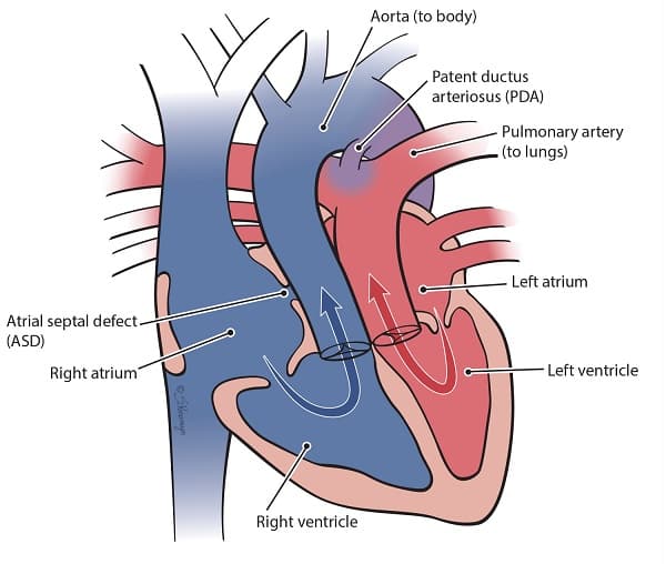
Transposition of the great arteries (TGA) is a ventriculoarterial discordant congenital heart defect in which the aorta arises from the right ventricle (RV) (anteriorly and to the right) and the pulmonary artery (PA) arises from the left ventricle (LV) (posteriorly and to the left) [1][2][3].
Breaking the name down:
- "Transposition" = trans (across) + position → the great arteries have swapped positions
- "Great arteries" = the aorta and pulmonary trunk — the two major outflow vessels of the heart
The term d-TGA (dextro-TGA) refers to the classical complete transposition where the aorta is anterior and rightward relative to the PA. This is distinct from l-TGA (levo-TGA / congenitally corrected TGA), where there is both atrioventricular AND ventriculoarterial discordance (a "double discordance" that is physiologically corrected).
This topic focuses exclusively on d-TGA (complete TGA), the common paediatric surgical emergency.
Key Concept — Parallel Circulations
In normal circulation, the systemic and pulmonary circuits function in series — deoxygenated blood goes to the lungs, gets oxygenated, returns to the left heart, and is pumped systemically. In TGA, the two circuits function in parallel — deoxygenated systemic venous blood returns to the RV and is pumped straight back out to the body via the aorta, while oxygenated pulmonary venous blood returns to the LV and is pumped back to the lungs via the PA. Without intercirculatory mixing, this is incompatible with life [1][2][3].
| Feature | Detail |
|---|---|
| Incidence | ~4.7 per 10,000 live births [1] |
| Proportion of all CHD | ~5% of all congenital heart disease [1][3] |
| Sex | Male predominance (M:F ≈ 2–3:1) |
| Rank | Second most common cyanotic CHD (after Tetralogy of Fallot in older infants); most common cyanotic CHD presenting in the neonatal period |
| Age at presentation | Typically within hours to days of birth |
| Without intervention | Fatal in ~90% by 1 year of age without surgical correction |
Hong Kong Context
- Congenital heart disease affects approximately 8–10 per 1,000 live births in Hong Kong
- TGA is one of the "critical" CHDs targeted by pulse oximetry screening in newborn nurseries in Hong Kong public hospitals
- Antenatal detection rates have improved with routine 18–22 week anomaly scans, but TGA remains one of the harder lesions to detect antenatally because the four-chamber view appears normal; the outflow tract views are essential
- Associated with fewer noncardiac anomalies ( < 10%) compared with other CHD defects [2][3]
TGA is NOT associated with any one common genetic abnormality [2][3]. Most cases are sporadic, but recognised risk factors include:
| Category | Risk Factors |
|---|---|
| Maternal | Gestational diabetes mellitus (strongest association), advanced maternal age, maternal rubella exposure, alcohol use, phenylketonuria |
| Genetic/Syndromic | Associated with DiGeorge syndrome (22q11.2 deletion) — as TGA is a conotruncal abnormality [1]; however, this association is less strong than with TOF or truncus arteriosus |
| Environmental | Herbicides, rodenticides, some teratogens |
| Familial | Low recurrence risk (~1–2% in siblings); multifactorial inheritance suspected |
Why DiGeorge?
DiGeorge syndrome (22q11.2 microdeletion) affects neural crest cell migration into the pharyngeal arches and outflow tract → conotruncal malformations. TGA, TOF, interrupted aortic arch, truncus arteriosus, and double-outlet RV are all conotruncal abnormalities. "CATCH-22" mnemonic: Cardiac anomaly, Abnormal facies, Thymic aplasia, Cleft palate, Hypocalcaemia — chromosome 22.
4. Anatomy and Embryology
To understand TGA, you need to understand how the single primitive outflow tract (truncus arteriosus and conus) normally septates:
- The truncus arteriosus is a single outflow channel in the early embryonic heart
- Neural crest cells migrate into the outflow tract and contribute to the aorticopulmonary (AP) septum
- The AP septum normally grows in a spiral (helical) fashion → this spiral septation divides the single outflow into:
- The aorta (posteriorly and to the left initially, then crossing to the right)
- The pulmonary trunk (anteriorly and to the right initially, then crossing to the left)
- The spiral arrangement ensures the aorta connects to the LV and the PA connects to the RV — i.e., the great arteries "wrap around" each other
- Abnormal septation of the conus arteriosus [1] → the AP septum grows straight (non-spirally) rather than spirally
- This results in the aorta arising anteriorly from the RV and the PA arising posteriorly from the LV — a direct swap
- The great arteries run parallel to each other rather than crossing
This is surgically critical:
- Coronary artery variations are common and must be delineated pre-operatively [2][3]
- The coronary arteries arise from the sinuses of the aorta that face the pulmonary artery (the "facing sinuses")
- Normal pattern (~65%): Left coronary artery (LCA) from left-facing sinus, right coronary artery (RCA) from right-facing sinus
- Variant patterns (~35%): Circumflex from right coronary, single coronary, intramural course, etc.
- Variant coronary anatomy increases surgical complexity of the arterial switch operation (ASO)
5. Pathophysiology
This is the conceptual core of TGA.
In TGA, systemic and pulmonary circulations run in parallel rather than in series [1][2][3]:
| Circuit | Normal | TGA |
|---|---|---|
| Systemic | LV → Aorta → Body → Vena cavae → RA → RV → PA | RA → RV → Aorta → Body → Vena cavae → RA (closed loop of deoxygenated blood) |
| Pulmonary | RV → PA → Lungs → Pulmonary veins → LA → LV → Aorta | LA → LV → PA → Lungs → Pulmonary veins → LA (closed loop of oxygenated blood) |
Key insight: Oxygenated blood from the lungs keeps recirculating through the lungs, and deoxygenated blood from the body keeps recirculating through the body. Without mixing between these two circuits, there is no way to deliver oxygen to the tissues → incompatible with postnatal life.
Anatomic sites for mixing are essential and must be present to be compatible with postnatal life [2][3]:
| Level of Mixing | Structure | Notes |
|---|---|---|
| Atrial level | PFO (patent foramen ovale), ASD | Most common site of mixing in neonates; bidirectional shunting |
| Ventricular level | VSD | If present, provides additional mixing |
| Arterial level | PDA | Critical in the first days of life; allows mixing between aorta and PA |
| Other | Bronchopulmonary collaterals | Minor contribution [2][3] |
The degree of intercirculatory mixing determines the severity of hypoxaemia [1]:
- More mixing sites / larger communications → better mixing → higher systemic oxygen saturation → less cyanosis
- Fewer/smaller mixing sites → poor mixing → profound cyanosis and metabolic acidosis
- In the fetus, the ductus arteriosus is patent → allows some mixing between circuits
- After birth, the ductus arteriosus begins to close (triggered by ↑PaO₂ and ↓circulating PGE₂ from the placenta)
- Severe systemic hypoxaemia occurs with closure of the duct, especially when the interatrial communication is small → can cause cardiac arrest [1]
- This is why TGA is classified as a ductal-dependent circulation — the neonate depends on the PDA for survival
Clinical Pearl — Ductal Closure = Emergency
A neonate with TGA and an intact ventricular septum (no VSD) who has only a small PFO is critically dependent on the PDA. When the duct closes (typically 24–72 hours), the baby develops sudden, profound cyanosis, metabolic acidosis, and cardiovascular collapse. This is a paediatric emergency requiring immediate prostaglandin E₁ (PGE₁) infusion and urgent balloon atrial septostomy.
5.4 Haemodynamic Consequences by Subtype
Simple TGA describes patients without another cardiac defect [2][3]:
- Only mixing via PFO/PDA → inadequate mixing → severe cyanosis in the neonatal period [1]
- High risk of neonatal hypoxia and death when PDA closes [1]
- The LV pumps blood into the low-resistance pulmonary circulation → high pulmonary flow [1]
- The RV supports the systemic circulation → pressure overloaded (working against systemic vascular resistance)
Complex TGA describes those with an additional cardiac defect [2][3]:
- VSD provides ventricular-level mixing → adequate mixing → minimal cyanosis [1]
- However, the combination of VSD + low PVR → large left-to-right shunt → pulmonary overcirculation
- Heart failure symptoms develop when PVR falls (typically within the first week of life) [1]
- If untreated, chronic pulmonary overcirculation → pulmonary vascular disease → Eisenmenger syndrome
- PS limits pulmonary blood flow → reduced pulmonary overcirculation (less HF)
- But PS also limits the amount of oxygenated blood available for mixing → more cyanosis
- Physiology resembles TOF more than simple TGA
High pulmonary flow is characteristic because the stronger LV is supplying the pulmonary circulation [1]:
- In TGA, the LV connects to the PA → pumps blood into the lungs against low PVR
- This means pulmonary blood flow is typically increased (unlike TOF where it is decreased)
- This is why TGA is classified as a cyanotic CHD with increased pulmonary blood flow
Classification of Cyanotic CHD by Pulmonary Blood Flow
| Increased Pulmonary Blood Flow | Decreased Pulmonary Blood Flow |
|---|---|
| TGA | Tetralogy of Fallot |
| Truncus arteriosus | Pulmonary atresia |
| Total anomalous pulmonary venous return (TAPVR) | Tricuspid atresia (without VSD) |
| Single ventricle (without PS) | Ebstein anomaly |
This distinction is critical for CXR interpretation and clinical reasoning [4].
A unique consideration in TGA:
- After birth, PVR falls → the LV (connected to PA) works against progressively lower resistance
- Over weeks, the LV deconditions (loses muscle mass/wall thickness) as it is no longer working against systemic pressure
- This has critical surgical implications: the arterial switch operation (ASO) must be performed within the first 2–3 weeks of life (ideally < 2 weeks) while the LV still has enough muscle mass to take over systemic workload
- If delayed beyond ~3–4 weeks, the LV may not be able to support systemic circulation after the switch → LV failure
6. Classification
| Type | Description |
|---|---|
| d-TGA (Complete TGA) | Atrioventricular concordance + ventriculoarterial discordance. RA→RV→Aorta, LA→LV→PA. The "classical" TGA. |
| l-TGA (Congenitally Corrected TGA / cc-TGA) | Atrioventricular discordance + ventriculoarterial discordance (double discordance). RA→LV→PA, LA→RV→Aorta. Physiologically "corrected" but the RV supports systemic circulation → eventual RV failure. |
| Category | Associated Defect | Frequency | Clinical Impact |
|---|---|---|---|
| Simple TGA | Intact ventricular septum (± PFO/PDA) | ~50% | Severe cyanosis, ductal-dependent |
| Complex TGA | VSD | ~50% | Better mixing but risk of HF and pulmonary vascular disease |
| LVOT obstruction / Pulmonary stenosis | ~25% | Can be anatomical (valvar PS, pulmonary atresia) or dynamic (systemic RV pressure causes interventricular septum to bow into LV cavity → dynamic obstruction between mitral valve and septum) [2][3] | |
| Mitral and tricuspid valve abnormalities | Variable | May complicate surgical repair | |
| Coronary artery variations | ~35% | Critical for surgical planning of ASO | |
| Coarctation of the aorta | ~5% | Adds systemic outflow obstruction |
Dynamic LVOT Obstruction in TGA
Why does this happen? In TGA, the RV is the systemic ventricle → generates systemic pressure → the interventricular septum bows toward the lower-pressure LV cavity. This septal bowing narrows the LVOT (the subpulmonary region), creating functional obstruction analogous to SAM in HOCM. This is important because it can worsen after birth as the LV pressure drops further.
7. Clinical Features
7.1 Symptoms
-
Cyanosis soon after birth [1] — often within the first hour of life
- Why? Parallel circulations with only a small PFO for mixing → very little oxygenated blood reaches the systemic circuit → profound desaturation (SpO₂ typically 60–80%)
- The cyanosis is central (involving mucous membranes, tongue, lips) and unresponsive to supplemental oxygen (the "hyperoxia test" is positive = stays cyanotic despite 100% FiO₂)
- Why doesn't oxygen help? The problem is not a ventilation/diffusion issue — oxygen is reaching the lungs perfectly well. The problem is that oxygenated blood cannot get to the systemic circulation because of the parallel circuits
-
Rapid clinical deterioration when the PDA begins to close (24–72h)
- Worsening cyanosis, tachypnoea, metabolic acidosis, lethargy, poor feeding
- Can cause cardiac arrest [1]
- Why? Loss of the last significant mixing point → near-complete separation of circuits
-
Tachypnoea without significant respiratory distress initially
- Why? The lungs are well-perfused (even over-perfused) → no parenchymal lung disease → no significant retractions or grunting at first. The tachypnoea is driven by chemoreceptor stimulation from hypoxaemia and acidosis
-
Minimal cyanosis — may appear only mildly dusky or even "pink" [1]
- Why? The large VSD allows significant bidirectional mixing → reasonable systemic oxygen saturation
-
Heart failure symptoms within the first week of life [1]
- As PVR falls, there is increasing left-to-right shunting through the VSD → pulmonary overcirculation
- Presents with: tachypnoea, feeding difficulty, diaphoresis (especially during feeds), poor weight gain, hepatomegaly
- Why feeding difficulty? Sucking and swallowing are aerobically demanding activities for a neonate; a failing heart cannot increase cardiac output adequately → the baby tires quickly, sweats, and takes smaller volumes
-
Later: failure to thrive from chronic heart failure and increased metabolic demands
- More significant cyanosis than TGA + VSD alone (because PS limits pulmonary blood flow)
- Less heart failure (because PS limits pulmonary overcirculation)
- Physiology resembles TOF
- Coarctation of aorta (CoA) or pulmonary hypertension (pHTN) → reverse differential cyanosis when PA pressure > aortic pressure [1]
- "Reverse differential" means the upper body is more cyanosed than the lower body — the opposite of what you see in PDA with Eisenmenger (where the lower body is more cyanosed)
- Why? If PA pressure exceeds aortic pressure, oxygenated blood from the LV (connected to PA) shunts through the PDA into the descending aorta → the legs get more oxygenated blood than the upper body (which receives purely deoxygenated blood from the RV via the aorta)
| Sign | Pathophysiological Basis |
|---|---|
| Cyanosis (central, unresponsive to O₂) | Parallel circulations → deoxygenated blood in systemic circuit |
| Heart failure signs (tachypnoea, hepatomegaly, gallop rhythm) — only if adequate mixing with VSD | Pulmonary overcirculation → volume overload |
| RV impulse (parasternal heave) | RV is the systemic ventricle → pressure overload as it supports systemic circulation against high SVR [1] |
| Loud and single S2 | Closure sound of the anteriorly positioned aortic valve — the aorta is anterior and closer to the chest wall → A2 is loud and easily heard. P2 is soft because the PA is posterior. Since A2 is loud and P2 is inaudible, S2 sounds single [1] |
| NO murmur or non-specific ESM/PSM | There is no obstruction or turbulent flow across a stenotic valve in simple TGA — the great arteries are just transposed, not stenotic. If a VSD is present → pansystolic murmur (PSM). If PS is present → ejection systolic murmur (ESM) [1] |
| Tachycardia | Compensatory response to hypoxaemia and/or heart failure |
| Poor perfusion / shock (late) | Metabolic acidosis from severe hypoxaemia; cardiovascular collapse |
Why is There No Murmur in Simple TGA?
Murmurs are caused by turbulent blood flow — usually from blood flowing across a stenotic or regurgitant valve, or through an abnormal communication. In simple TGA with intact ventricular septum, blood flows normally from RV to aorta and from LV to PA — there is no stenosis, no VSD, and no turbulence. The only problem is that the connections are swapped. Hence, the baby can be profoundly cyanotic with a completely silent precordium — a deceptively quiet examination that can catch you off guard.
Exam Pearl — Cyanotic Neonate with No Murmur
A cyanotic neonate with no murmur, loud single S2, and CXR showing increased pulmonary vascular markings = think TGA until proven otherwise. This is a classic OSCE/written exam scenario. Don't be fooled by the absence of a murmur — it doesn't mean the heart is normal!
| Feature | TGA | TOF | Tricuspid Atresia |
|---|---|---|---|
| Timing of cyanosis | Hours after birth | Weeks to months (progressive) | At birth |
| Pulmonary blood flow | Increased | Decreased | Decreased |
| Murmur | Often absent | ESM at LUSB | Variable |
| S2 | Loud, single | Single (soft P2) | Single |
| CXR heart shape | "Egg on side" | "Boot-shaped" | Normal/small heart |
| CXR lung fields | Plethoric | Oligaemic | Oligaemic |
| Hyperoxia test | Fails (stays cyanotic) | Fails | Fails |
| Tet spells | No | Yes | No |
| Age | Typical Presentation |
|---|---|
| Birth – 24h | Cyanosis detected on routine examination or pulse oximetry screening; may appear well otherwise |
| 24–72h | Worsening cyanosis as PDA closes; metabolic acidosis; tachypnoea |
| Days – 1 week | Heart failure symptoms if large VSD present (as PVR falls) |
| > 2 weeks (if untreated) | Chronic cyanosis, failure to thrive, progressive metabolic derangement |
8. Investigations (Pre-Diagnosis — Included for Completeness of Clinical Features Section)
While the full diagnostic workup will be covered in the next section, the characteristic investigation findings are closely tied to understanding the clinical features:
| Feature | Explanation |
|---|---|
| "Egg on side" appearance (also called "egg on string") | Oval/egg-shaped cardiac silhouette due to narrowed upper mediastinum [1] |
| Narrowed upper mediastinum | Two reasons: (1) Abnormal anteroposterior relationship of the aorta and pulmonary trunk — normally the great vessels create a wide shadow, but when they are parallel (not crossing), the mediastinal shadow narrows [1]; (2) Stress-induced thymic involution — severe neonatal hypoxaemia/stress causes the thymus to shrink [1] |
| Increased pulmonary vascular markings | The stronger LV is supplying the lungs → increased pulmonary blood flow → pulmonary plethora [1] |
- Typically normal for age [1] in the neonatal period
- Why normal? In the neonate, RV dominance is physiological → the TGA pattern (RV supporting systemic circulation) doesn't look different from normal neonatal RV dominance
- Over time: rightward axis deviation and RVH become apparent (as the RV hypertrophies to support systemic circulation)
- Diagnostic — the gold standard
- Demonstrates ventriculoarterial discordance, identifies associated defects (VSD, PS, CoA), and maps coronary artery anatomy
- Neonate placed on 100% FiO₂ for 10 minutes
- In cyanotic CHD (including TGA): PaO₂ remains < 150 mmHg (often < 100 mmHg)
- In respiratory disease: PaO₂ typically rises > 150 mmHg
- Oligaemic lung field on CXR supports cyanotic CHD over respiratory disease [4]
AOS Past Paper Point
"Radiograph showing an oligaemic lung field due to reduced pulmonary blood flow" supports cyanotic CHD over respiratory disease [4]. Note: This applies to cyanotic CHDs with REDUCED pulmonary blood flow (e.g., TOF). TGA characteristically has INCREASED pulmonary blood flow, so the lung fields would be plethoric, not oligaemic. However, the principle of using CXR lung fields to differentiate cardiac from respiratory cyanosis is key.
-
Growth: Without correction, infants with TGA develop failure to thrive due to:
- Chronic hypoxaemia → increased metabolic demands
- Heart failure → poor feeding tolerance → inadequate caloric intake
- Increased work of breathing → increased energy expenditure
-
Neurodevelopment: Chronic hypoxaemia and the haemodynamic instability of the neonatal period can cause neurological injury. Even after successful surgical repair, children with TGA have higher rates of:
- Mild motor delays
- Attention and executive function difficulties
- Learning disabilities
- These are likely related to perioperative brain injury (deep hypothermic circulatory arrest during surgery) and prenatal/postnatal cerebral hypoxia
- Breaking the diagnosis: Parents are often overwhelmed — the baby may appear "normal" apart from colour. Use simple language: "Your baby's heart tubes are connected the wrong way around, so the blood isn't getting enough oxygen"
- Urgency: Communicate the need for urgent intervention clearly but calmly
- Prognosis: After arterial switch operation, long-term survival is excellent (> 95% at 20 years) — this is a key reassurance point
- Consent: Informed consent for balloon atrial septostomy and subsequent surgery must come from parents/guardians. Explain risks, benefits, and alternatives in age-appropriate language
- Psychosocial: Connect families with cardiac support groups; address parental anxiety and bonding difficulties in the NICU setting
High Yield Summary
-
TGA = ventriculoarterial discordance: aorta from RV, PA from LV → parallel circulations → incompatible with life without intercirculatory mixing
-
Intercirculatory mixing occurs at atrial (PFO/ASD), ventricular (VSD), and arterial (PDA) levels — the degree of mixing determines clinical severity
-
Ductal-dependent circulation: PDA closure → sudden profound cyanosis → cardiac arrest → requires emergent PGE₁
-
Simple TGA (intact IVS): severe cyanosis within hours, NO murmur, loud single S2, CXR "egg on side" with pulmonary plethora, ECG normal for age
-
Complex TGA (with VSD): minimal cyanosis but heart failure symptoms ≤1 week as PVR falls
-
Cyanotic CHD with INCREASED pulmonary blood flow (unlike TOF which has decreased)
-
CXR: egg on side + narrow mediastinum (parallel great arteries + thymic involution) + increased pulmonary vascular markings
-
Embryology: failure of spiral septation of the conus → straight septation → transposed great arteries
-
LV deconditioning: ASO must be performed within first 2–3 weeks before LV loses capacity to support systemic circulation
-
Coronary artery variants (~35%) are critical for surgical planning
Active Recall - TGA Clinical Features and Pathophysiology
Differential Diagnosis of Neonatal Cyanosis — Approaching TGA
The clinical presentation of TGA is a cyanotic neonate. The differential diagnosis is therefore framed around the question: "Why is this newborn blue?" The approach must distinguish cardiac from non-cardiac causes, then narrow down among the cyanotic congenital heart diseases.
Central cyanosis becomes clinically visible when deoxyhaemoglobin exceeds 5 g/dL [6]. Because neonates are relatively polycythaemic (Hb ~15–17 g/dL), cyanosis may be detectable at higher SpO₂ values than in older children or anaemic patients. Check buccal mucosa instead of lip — lip colour can be misleading due to peripheral vasoconstriction [6].
GC High-Yield — DDx of Cyanosis in Neonates
The GC 147 lecture and Maksim paediatric notes list the following categories for the differential diagnosis of cyanosis in neonates [5][6]:
| Category | Examples |
|---|---|
| Cyanotic heart disease | Tetralogy of Fallot, transposition of great arteries, pulmonary atresia, tricuspid atresia, TAPVD, complex heart lesion (e.g. univentricular heart) |
| Respiratory disorders | Surfactant deficiency, meconium aspiration, pulmonary hypoplasia |
| Persistent pulmonary hypertension of the newborn (PPHN) | Primary or secondary |
| Infection | Group B Streptococcal septicaemia |
| Metabolic | Inborn error of metabolism → metabolic acidosis and shock |
The clinical approach to a cyanotic neonate follows a systematic algorithm. The goal is to rapidly identify whether the cause is cardiac or non-cardiac, because the management diverges immediately.
3. Cardiac Causes — Cyanotic Congenital Heart Disease
The major differential diagnoses for TGA are the other cyanotic CHDs. The key discriminating features are the pulmonary blood flow pattern (increased vs. decreased), the CXR silhouette, and the clinical behaviour.
These conditions share the feature of pulmonary plethora on CXR because too much blood is reaching the lungs.
| Condition | Pathophysiology | Key Differentiating Features from TGA |
|---|---|---|
| Transposition of Great Arteries (TGA) [1][2][3] | Ventriculoarterial discordance → parallel circulations → survival depends on intercirculatory mixing (PFO, ASD, VSD, PDA) | Cyanosis from birth (D1–2), egg-on-side CXR, narrow mediastinum, no murmur, loud single S2, ECG normal for age |
| Truncus arteriosus [3][7] | Aorta, pulmonary arteries, and coronary arteries all arise from a single great vessel → combined output of LV and RV exits through one trunk. Always has a large VSD (the truncal root overrides both ventricles). NOT ductal-dependent [3][7]. | Cyanosis at birth is mild (because mixing occurs freely in the single trunk). HF develops at a few weeks as PVR drops → increased pulmonary flow → marked cardiovascular instability [3][7]. Systolic ejection click, single S2, truncal regurgitation murmur. CXR: cardiomegaly, increased PVM, absent main PA segment, right aortic arch (30%). Associated with DiGeorge syndrome (22q11.2 deletion) more strongly than TGA |
| Total Anomalous Pulmonary Venous Return (TAPVR) [3][7] | All four pulmonary veins drain anomalously — into the RA (supracardiac, cardiac, or infracardiac types) rather than the LA. An ASD/PFO is obligatory for blood to reach the LA and then the systemic circulation | Cyanosis severity depends on obstruction: obstructed TAPVR (especially infracardiac type) → severe cyanosis + pulmonary oedema; unobstructed → mild cyanosis + HF. CXR "snowman" (figure-of-8) in supracardiac type (but only visible after a few months). Fixed split S2. ECG: RAD + RVH |
| Single ventricle / Univentricular heart without PS [6] | Only one functional ventricle receives all systemic and pulmonary venous return → complete mixing → cyanosis | Variable anatomy. Degree of cyanosis depends on PVR and any associated PS. Often presents with balanced cyanosis + eventual HF |
| Atrioventricular Septal Defect (AVSD) — complete [6] | Single 5-leaflet valve spanning both AV junctions + ASD + VSD → common mixing at atrial and ventricular levels | Commonly seen in children with Down syndrome [6]. Cyanosis at birth, HF at 2–3 weeks. Superior axis on ECG, RBBB, "goose-neck sign" on angiography [6] |
How to Distinguish TGA from Truncus Arteriosus
Both present with neonatal cyanosis and increased pulmonary blood flow, but:
- TGA: Cyanosis is severe early (parallel circuits, poor mixing) → ductal-dependent → worsens dramatically when duct closes. Egg-on-side CXR. No murmur.
- Truncus arteriosus: Cyanosis is mild initially (complete mixing in single trunk) → NOT ductal-dependent → HF predominates as PVR falls. Ejection click, single S2, possible truncal regurgitation murmur. Wide mediastinum (large single trunk), not narrow.
The ductal-dependence distinction is clinically critical: TGA needs PGE₁ urgently; truncus does not.
These conditions show oligaemic lung fields on CXR because there is an obstruction to pulmonary blood flow.
| Condition | Pathophysiology | Key Differentiating Features from TGA |
|---|---|---|
| Tetralogy of Fallot (TOF) [8] | Unequal division of outflow tract → RVOT obstruction + overriding aorta + VSD + RVH [8]. R-to-L shunt when RVP > LVP | Cyanosis onset depends on degree of RVOT obstruction — typically progressive over months (infundibular stenosis worsens), not at birth. Hypercyanotic (Tet) spells are characteristic [8]. Boot-shaped heart on CXR with oligaemic lung fields. ESM at LUSB (from PS), single S2. ECG: RAD + RVH |
| Pulmonary atresia (with intact IVS) | Complete obstruction of RVOT → no antegrade flow from RV to PA → ductal-dependent pulmonary circulation | Cyanosis from birth, worse than TGA typically. CXR: dark lung fields (severely oligaemic), small heart or normal size. Continuous PDA murmur may be heard. Ductal-dependent → PGE₁ required |
| Tricuspid atresia [6] | No connection between RA and RV → all systemic venous blood must cross PFO/ASD to LA → common mixing in LA → cyanosis. Only LV is effective; RV is small and non-functional [6] | Cyanosis from birth. LV impulse (not RV — because the RV is hypoplastic). Left axis deviation on ECG (unusual in neonatal period, where RAD is normal). Small/absent RV on CXR. Oligaemic lung fields unless VSD is large |
| Ebstein anomaly | Apical displacement of tricuspid valve → "atrialisation" of RV → massive RA dilatation → R-to-L shunt via PFO | Massive cardiomegaly on CXR ("wall-to-wall" heart). Tricuspid regurgitation murmur. WPW pattern on ECG (accessory pathway). Older children: SVT |
Exam Pearl — CXR Discriminator
| CXR Finding | Think... |
|---|---|
| Egg on side + narrow mediastinum + plethoric lungs | TGA [1] |
| Boot-shaped heart + oligaemic lungs | TOF [8] |
| Snowman / figure-of-8 + plethoric lungs | TAPVR (supracardiac) |
| Wall-to-wall cardiomegaly | Ebstein anomaly |
| Absent main PA segment + plethoric lungs | Truncus arteriosus |
| Small heart + very dark (oligaemic) lungs | Pulmonary atresia with intact IVS |
These CXR patterns are very high-yield for written papers and OSCEs.
These must be excluded because they are far more common than cyanotic CHD, and some are immediately treatable.
| Category | Condition | Key Distinguishing Features from Cardiac Cyanosis |
|---|---|---|
| Respiratory disorders [6] | Surfactant deficiency (RDS) | Preterm infant, progressive respiratory distress, grunting, retractions. CXR: ground-glass opacification with air bronchograms. Responds to supplemental O₂ (hyperoxia test positive) |
| Meconium aspiration syndrome (MAS) | Term/post-term, meconium-stained liquor, respiratory distress from birth. CXR: patchy infiltrates, hyperinflation. May trigger PPHN | |
| Pulmonary hypoplasia | Associated with prolonged oligohydramnios, CDH, renal agenesis. Severe respiratory failure from birth | |
| Pneumothorax | Acute deterioration, unilateral decreased air entry, transillumination positive. CXR diagnostic | |
| Persistent Pulmonary Hypertension of the Newborn (PPHN) [6] | Failure of the normal postnatal drop in PVR → persistent R-to-L shunting through PDA and/or PFO | Pre- and post-ductal SpO₂ difference > 5–10% (pre-ductal higher). Often secondary to MAS, sepsis, or CDH. Can coexist with parenchymal lung disease. CXR may show underlying cause. Echocardiography shows elevated PA pressures but structurally normal heart |
| Infection [6] | Group B Streptococcal (GBS) septicaemia | Risk factors: maternal GBS colonisation, PROM, intrapartum fever. Presents with respiratory distress, temperature instability, poor perfusion. CXR may mimic RDS. Blood cultures positive. Responds to antibiotics |
| Metabolic [6] | Inborn errors of metabolism (IEM) → metabolic acidosis and shock | Presents with lethargy, poor feeding, metabolic acidosis (high anion gap), hypoglycaemia, hyperammonaemia. Cyanosis is from poor perfusion/shock rather than true R-to-L shunting. Newborn screening may flag |
The Hyperoxia Test — First Principles
Why does it work?
- In respiratory disease: the problem is V/Q mismatch or diffusion impairment → providing 100% O₂ overwhelms the barrier → PaO₂ rises significantly ( > 150 mmHg)
- In cyanotic CHD: the problem is anatomical shunting of deoxygenated blood into the systemic circulation → no amount of supplemental oxygen can fix an anatomical bypass → PaO₂ stays low ( < 100 mmHg)
- In PPHN: the problem is functional R-to-L shunting through PDA/PFO → partial response (PaO₂ 100–150 mmHg), and importantly, there is a pre-/post-ductal saturation gradient
Practical points in neonates:
- Measure SpO₂ from the right hand (pre-ductal) AND either foot (post-ductal)
- A structurally normal heart on echo with a pre-/post-ductal gradient = PPHN
- A structurally abnormal heart with no response to O₂ = cyanotic CHD
Some neonates with TGA present not just with cyanosis but with cardiovascular collapse when the PDA closes. This overlaps with other causes of ductal-dependent systemic circulation presenting as cardiogenic shock at Day 4–7 [6]:
| Condition | Circulation Dependent On | Key Features |
|---|---|---|
| TGA (intact IVS) | PDA for intercirculatory mixing | Profound cyanosis, loud single S2, no murmur, egg-on-side CXR |
| Severe coarctation of aorta (CoA) [6] | PDA for lower body perfusion | Cardiogenic shock at D4–7 [6]. First presentation: oliguria [6]. 4-limb BP: upper limb BP > lower limb BP [6]. Generalised pallor, weak peripheral pulses, cold peripheries [6]. Differential cyanosis (lower limbs more cyanosed) |
| Interrupted aortic arch [6] | PDA for entire descending aortic flow | Similar to severe CoA but more dramatic collapse. Strong association with DiGeorge syndrome |
| Hypoplastic left heart syndrome (HLHS) [6] | PDA for systemic circulation | Not common in HK / Chinese, but major in Caucasians [6]. Absent LV impulse, single S2, no palpable femoral pulses, metabolic acidosis. Hypoplastic LV + aorta on echo |
| Critical aortic stenosis | PDA for distal systemic perfusion | Harsh ESM at RUSB/apex, weak pulses, cardiomegaly |
GC High-Yield — Ductal-Dependent Lesions and PGE₁
All ductal-dependent congenital heart diseases require prostaglandin E₁ (PGE₁) infusion to maintain ductal patency [5][6]:
- Ductal-dependent pulmonary circulation: pulmonary atresia, critical PS, severe TOF
- Ductal-dependent systemic circulation: severe CoA, interrupted aortic arch, HLHS, critical AS
- Ductal-dependent mixing: TGA
PGE₁ side effects: fever, apnoea, diarrhoea [6] — the baby may need intubation for apnoea during PGE₁ infusion.
Management of closing CoA: maintain ductal patency with PGE₁ → re-perfuse abdominal organs and lower limbs → plan for elective corrective surgery [6].
The following features, when present in combination, strongly point toward TGA over other differentials:
| Feature | Points Toward TGA | Points Away from TGA |
|---|---|---|
| Timing | Cyanosis within hours of birth | Cyanosis developing over weeks (TOF) |
| Murmur | Absent or non-specific | Loud ESM (TOF, AS), continuous (PDA), PSM (VSD) |
| S2 | Loud and single | Widely split and fixed (ASD/TAPVR), paradoxical split |
| CXR heart shape | Egg on side, narrow mediastinum | Boot-shaped (TOF), snowman (TAPVR), massive (Ebstein) |
| CXR lung fields | Increased pulmonary vascular markings | Oligaemic (TOF, PA, TA) |
| ECG | Normal for age | LAD (tricuspid atresia), extreme RAD + RVH (TOF, TAPVR) |
| Hyperoxia test | Fails (PaO₂ < 100 mmHg) | Passes (respiratory disease) |
| Pre-/post-ductal gradient | Minimal or absent in simple TGA | Significant gradient suggests PPHN or CoA |
| Response to PGE₁ | Improves mixing → improves SpO₂ | No improvement if TAPVR with obstruction |
A commonly used mnemonic for cyanotic congenital heart diseases:
| T | Condition |
|---|---|
| Transposition of the Great Arteries | TGA |
| Tetralogy of Fallot | TOF |
| Tricuspid Atresia | TA |
| Total Anomalous Pulmonary Venous Return | TAPVR |
| Truncus Arteriosus | Truncus |
Add Pulmonary Atresia and Ebstein Anomaly to complete the list of major cyanotic CHDs — the 5 T's is a memory aid, not an exhaustive list.
| Feature | TGA | TOF | Tricuspid Atresia | TAPVR | Truncus Arteriosus |
|---|---|---|---|---|---|
| Pulmonary flow | ↑ | ↓ | ↓ | ↑ | ↑ |
| Cyanosis onset | Birth (hours) | Months (progressive) | Birth | Variable | Birth (mild) |
| Murmur | None | ESM LUSB | Variable | Systolic flow murmur | Ejection click + systolic |
| S2 | Loud, single | Single (soft P2) | Single | Fixed split | Single (single trunk) |
| CXR shape | Egg on side | Boot-shaped | Normal/small | Snowman (late) | Cardiomegaly, absent PA |
| CXR lungs | Plethoric | Oligaemic | Oligaemic | Plethoric | Plethoric |
| ECG | Normal for age | RAD, RVH | LAD, LVH | RAD, RVH | Biventricular hypertrophy |
| Ductal-dependent? | Yes (mixing) | If severe | Depends on anatomy | No | No |
| DiGeorge assoc. | Possible | Strong | No | No | Strong |
High Yield Summary — DDx of TGA
-
Frame the DDx as "Cyanotic Neonate" → cardiac vs. respiratory vs. PPHN vs. infection vs. metabolic
-
Hyperoxia test: PaO₂ < 100 = cardiac; > 150 = respiratory; 100–150 = PPHN/equivocal
-
CXR lung fields separate cyanotic CHD into ↑ pulmonary flow (TGA, truncus, TAPVR) vs. ↓ pulmonary flow (TOF, PA, TA, Ebstein)
-
TGA signature: egg-on-side + narrow mediastinum + plethoric lungs + no murmur + loud single S2 + ECG normal for age
-
5 T's: TGA, TOF, Tricuspid Atresia, TAPVR, Truncus Arteriosus
-
Ductal-dependent lesions all need PGE₁; know the side effects (fever, apnoea, diarrhoea)
-
Distinguish TGA from CoA/HLHS shock: TGA = primarily cyanosis; CoA/HLHS = primarily shock with pallor and weak pulses ± differential cyanosis
-
Truncus arteriosus is NOT ductal-dependent — a key distinguishing point from TGA
Active Recall - DDx of TGA
References
[1] Senior notes: Adrian Lui Pediatrics Notes.pdf (p. 219) [2] Senior notes: MBBS Final MB (Medicine) (Felix PY Lai).pdf (p. 586–588) [3] Senior notes: MBBS Final MB (Pediatrics) (Felix PY Lai).pdf (p. 273, 287) [5] Lecture slides: GC 147. Heart failure and cyanosis in children acyanotic and cyanotic congenital heart disease - Part 2.pdf (p. 23–30) [6] Senior notes: Maksim Paediatric Notes.pdf (p. 55–56, 59–60) [7] Senior notes: MBBS Final MB (Medicine) (Felix PY Lai).pdf (p. 600) — truncus arteriosus and TAPVR sections [8] Senior notes: Ryan Ho Cardiology.pdf (p. 187 — TOF; p. 189 — PDA)
Diagnosis of Transposition of the Great Arteries — Criteria, Algorithm, and Investigations
There is no formal "scoring" system for diagnosing TGA (unlike, say, Jones criteria for rheumatic fever). The diagnosis is anatomical and echocardiographic — confirmed by demonstrating ventriculoarterial discordance. However, the clinical pathway to diagnosis follows a very structured approach that integrates clinical suspicion, bedside tests, and definitive imaging.
1.1 Clinical Diagnostic Criteria (Suspicion → Confirmation)
A neonate should be suspected of having TGA when the following constellation is present:
| Criterion | Detail | Pathophysiological Basis |
|---|---|---|
| 1. Central cyanosis within hours of birth | SpO₂ < 85–90% on pulse oximetry; visible cyanosis of mucous membranes | Parallel circulations → deoxygenated blood recirculates systemically |
| 2. Cyanosis unresponsive to supplemental oxygen | Hyperoxia test: PaO₂ remains < 100 mmHg on 100% FiO₂ | Anatomical shunt — O₂ cannot cross the parallel circuit barrier |
| 3. No significant respiratory distress (initially) | No grunting, minimal retractions; tachypnoea is chemoreceptor-driven, not from lung disease | Lungs are well-ventilated and well-perfused (even over-perfused); the problem is circulatory, not ventilatory |
| 4. Quiet precordium | No murmur or non-specific ESM/PSM [1]; loud and single S2 [1] | No turbulent flow (transposed but unobstructed great arteries); anteriorly placed aortic valve → loud A2 |
| 5. CXR: egg-on-side + narrow mediastinum + plethoric lungs | Classic but not always present in first hours [1][9][10] | Parallel great arteries narrow the vascular pedicle; stress-related thymic involution; strong LV → lungs |
| 6. ECG: normal for age | Typically normal for age [1][9][10] | Neonatal RV dominance is physiological → TGA-pattern RV dominance is indistinguishable |
| 7. Echocardiography: ventriculoarterial discordance | Confirms diagnosis by demonstrating parallel arrangement of great arteries with the pulmonary trunk arising posteriorly from LV and aorta arising anteriorly from RV [9][10] | Definitive anatomical demonstration |
The Diagnosis is Echocardiographic
Echocardiography is the definitive diagnostic test [9][10]. CXR and ECG raise suspicion; echo confirms. In many centres with experienced neonatal echocardiographers, the diagnosis can be made within minutes at the bedside, allowing immediate initiation of PGE₁ and planning for balloon atrial septostomy.
Before any clinical suspicion arises, many cases of TGA are now detected through routine newborn pulse oximetry screening, which is standard practice in Hong Kong public hospital newborn nurseries.
| Aspect | Detail |
|---|---|
| Timing | Performed at 24–48 hours of life (before discharge) |
| Method | SpO₂ measured on the right hand (pre-ductal) and either foot (post-ductal) |
| Positive screen | SpO₂ < 95% in either limb, OR > 3% difference between pre- and post-ductal readings |
| Why right hand? | Right hand is supplied by the right subclavian artery, which branches off the aorta proximal to the ductus → pre-ductal blood. The feet receive post-ductal blood |
| Sensitivity for TGA | Good but not perfect — TGA with a large VSD may have near-normal SpO₂ and be missed |
Why is pulse oximetry screening important for TGA specifically?
- TGA with intact IVS and adequate PDA may have SpO₂ in the high 70s–low 80s → detectable
- The baby may appear "well" apart from subtle colour change → clinical examination alone misses cases
- Early detection → early PGE₁ → prevents catastrophic collapse when the duct closes
Critical Management Point — Start PGE₁ Before Echo
In a cyanotic neonate with a failed hyperoxia test and CXR suggesting cyanotic CHD, start prostaglandin E₁ (PGE₁/alprostadil) infusion IMMEDIATELY — do NOT wait for echocardiographic confirmation. The risk of the duct closing while waiting for echo is far greater than the risk of PGE₁. This is a time-critical intervention.
4. Investigation Modalities — Detailed Findings and Interpretation
The CXR is the first imaging modality obtained in a cyanotic neonate. In TGA, it shows a characteristic triad:
| Finding | Description | Pathophysiological Explanation |
|---|---|---|
| "Egg-on-side" appearance | Oval cardiac silhouette [9][10] — the heart looks like an egg lying on its side | The shape results from the combination of: a globally enlarged heart (both ventricles working at high output) + the narrow superior vascular pedicle |
| Narrow mediastinum / narrow vascular pedicle | The upper mediastinal shadow is unusually thin | Two reasons: (1) Abnormal AP relationship of the aorta to the pulmonary trunk — normally these vessels cross and create a wide shadow on PA film; in TGA they run parallel (one directly in front of the other), so on PA view the pedicle appears narrow [1][9][10]. (2) Stress-related thymic involution — severe hypoxaemia and stress in the neonate causes the thymus to shrink, further narrowing the mediastinal shadow [1][9][10] |
| Increased pulmonary vascular markings | Pulmonary plethora — the lung fields appear "whiter" with more prominent vascular markings | The stronger LV is supplying the pulmonary circulation [1] → increased pulmonary blood flow → engorgement of pulmonary vessels |
Important caveats:
- The classic "egg-on-side" may not be visible in the first few hours of life — the CXR may look relatively normal early on
- Cardiomegaly develops over hours to days as both ventricles work at high output
- If TGA is associated with PS → pulmonary vascular markings may be normal or decreased (not plethoric)
"Egg-on-side" is a high-yield CXR finding for exams — always mention the narrow mediastinum and plethoric lung fields together [1][9][10].
| Finding | Interpretation |
|---|---|
| Typically normal for age [1][9][10] | In the neonatal period, right axis deviation and RVH are physiological → the ECG findings of TGA (RV supporting systemic circulation → RVH) are indistinguishable from normal neonatal ECG |
| Right axis deviation and RVH are normal in the first few days of life and may be the only findings present on a newborn ECG [9][10] | This is a key trap — do NOT dismiss a normal neonatal ECG as reassuring in a cyanotic baby. TGA can have an entirely normal ECG |
| Later findings (weeks–months, if uncorrected) | Progressive RVH (as RV continues to work against systemic afterload), possible RA enlargement |
| With large VSD | Combined ventricular hypertrophy (biventricular hypertrophy) may develop |
Why is the ECG normal?
Let's think from first principles:
- At birth, the RV is already the dominant ventricle (it has been pumping against high PVR in utero and supplying most of the cardiac output via the ductus)
- In TGA, the RV continues to work against systemic resistance — but this is the same workload the RV was already doing in utero
- Therefore, the neonatal ECG cannot distinguish "pathological RV dominance from TGA" from "physiological RV dominance of a normal newborn"
- Only as the child ages would the RVH become abnormally persistent (normally the LV takes over dominance by ~6 months)
Exam Trap — Normal ECG ≠ Normal Heart
A normal ECG in a cyanotic neonate does NOT exclude cyanotic congenital heart disease. TGA classically has a normal neonatal ECG. Always correlate with clinical findings, CXR, and echocardiography.
Echocardiography is the gold standard for diagnosis of TGA. It provides:
| Information | Echo Findings | Clinical Significance |
|---|---|---|
| Ventriculoarterial discordance | Parallel arrangement of great arteries — PA arising posteriorly from LV, aorta arising anteriorly from RV [9][10] | Confirms the diagnosis of TGA |
| Great artery relationship | Aorta is anterior and usually rightward (d-TGA); great arteries run parallel rather than crossing | Distinguishes d-TGA from l-TGA and from normal anatomy |
| Atrial septum | Size of PFO/ASD; direction and velocity of shunting | Determines adequacy of atrial mixing → guides decision for balloon atrial septostomy |
| Ventricular septum | Presence, location, and size of VSD | Classifies simple vs. complex TGA; influences surgical approach |
| Outflow tracts | LVOT/subpulmonary obstruction (dynamic or anatomical PS) | LVOT obstruction present in ~25% [2][3] — affects surgical planning |
| Coronary artery anatomy | Origin, branching pattern, and course of coronary arteries | Critical for arterial switch operation — the coronary arteries must be transferred to the neo-aorta. Variant anatomy (intramural course, single coronary) increases surgical risk |
| Associated anomalies | CoA, aortic arch abnormalities, valve abnormalities | Comprehensive pre-surgical assessment |
| Ventricular function | LV and RV systolic function; LV mass and wall thickness | Assesses whether the LV has sufficient mass to support systemic circulation after ASO (LV "preparedness") |
| Ductus arteriosus | Size and direction of flow through PDA | Confirms ductal patency and its contribution to mixing |
Subcostal views are particularly useful in neonates because of the horizontal heart position and good acoustic windows through the liver.
Fetal echocardiography:
- TGA can be detected antenatally on the outflow tract views during the 18–22 week anomaly scan
- The four-chamber view is typically normal in TGA (both ventricles and AV valves are normal) → this is why TGA is one of the harder lesions to detect prenatally
- The three-vessel view (3VV) and three-vessel-trachea view (3VT) show the abnormal parallel arrangement of the great arteries
| Parameter | Method | Interpretation |
|---|---|---|
| Purpose | Differentiate cardiac from respiratory cause of cyanosis | Based on whether supplemental O₂ can overcome the cause of desaturation |
| Method | Place neonate on 100% FiO₂ for 10 minutes → measure PaO₂ from right radial artery (pre-ductal) | Right radial artery samples pre-ductal blood |
| PaO₂ < 100 mmHg | Cyanotic CHD very likely | Anatomical shunt → O₂ cannot cross the shunt barrier |
| PaO₂ > 150 mmHg | Respiratory cause likely | V/Q mismatch or diffusion limitation overcome by high FiO₂ |
| PaO₂ 100–150 mmHg | Equivocal — consider PPHN, CHD with large VSD (some mixing improves with O₂), or methemoglobinaemia | Check pre-/post-ductal gradient, proceed to echo |
In TGA specifically:
- Simple TGA (intact IVS): PaO₂ typically 25–50 mmHg on 100% FiO₂ → grossly abnormal
- TGA with large VSD: PaO₂ may be 50–80 mmHg → still below threshold but less dramatically
| Measurement | Site | What It Tells You |
|---|---|---|
| Pre-ductal SpO₂ | Right hand (or right ear lobe) | Saturation of blood from aorta proximal to ductus |
| Post-ductal SpO₂ | Either foot | Saturation of blood from aorta distal to ductus |
| Interpretation in TGA | Usually no significant gradient in simple TGA (both pre- and post-ductal blood come from the same deoxygenated RV output) | Distinguishes from PPHN (where gradient > 5–10% with pre-ductal higher) and from CoA with Eisenmenger (where post-ductal is lower) |
Pre-/Post-Ductal Gradient — Key Interpretations
| Pattern | Likely Diagnosis |
|---|---|
| Both low, no gradient | TGA (parallel circuits — aorta carries deoxygenated blood throughout) or cyanotic CHD with intracardiac mixing |
| Pre-ductal high, post-ductal low ( > 5% difference) | PPHN or CoA |
| Pre-ductal low, post-ductal higher (reverse differential) | TGA with CoA or pHTN (reverse differential cyanosis when PA pressure > aortic pressure [1]) |
| Finding | Interpretation |
|---|---|
| Low PaO₂ (despite supplemental O₂) | Anatomical R-to-L shunting / parallel circulation |
| Metabolic acidosis (low pH, low HCO₃⁻, high lactate) | Tissue hypoxia → anaerobic metabolism → lactic acidosis. This is a sign of decompensation and demands urgent intervention |
| PaCO₂ may be normal or low | Hyperventilation driven by chemoreceptors responding to hypoxaemia/acidosis. Lung ventilation is usually normal in TGA |
| Indication | Detail |
|---|---|
| Gold standard for elucidating coronary artery courses and origin [9][10] | Invasive catheterisation with contrast injection into aortic root |
| When used | For patients with coronary artery anatomy that cannot be determined confidently with echocardiography prior to surgical intervention [9][10] |
| Why important | The arterial switch operation requires transferring coronary arteries from the native aorta (arising from RV) to the neo-aorta (the former PA). Variant coronary anatomy (intramural course, single coronary, coronary crossing behind the PA) increases surgical difficulty and risk |
| Trend | With improving echocardiographic and CT angiography resolution, invasive coronary angiography is used less frequently but remains the definitive test when non-invasive imaging is inconclusive |
| Modality | Role in TGA |
|---|---|
| CT angiography | Excellent spatial resolution for coronary artery anatomy; fast acquisition (important for neonates). Used when echo is insufficient for coronary delineation. Radiation exposure is a consideration |
| Cardiac MRI | Gold standard for post-operative follow-up (assessing neo-aortic root, coronary patency, ventricular function). Not typically used for initial neonatal diagnosis (long acquisition time, need for sedation/anaesthesia) |
| Aspect | Detail |
|---|---|
| Diagnostic | Measures intracardiac pressures, oxygen saturations in each chamber, quantifies shunt ratios (Qp:Qs), assesses PVR |
| Therapeutic | Balloon atrial septostomy (Rashkind procedure) is performed during catheterisation — this is both a diagnostic confirmation and a life-saving intervention (covered in management section) |
| Timing | Often performed urgently when echo confirms TGA and atrial mixing is inadequate |
| Investigation | Simple TGA (Intact IVS) | TGA + Large VSD | TGA + VSD + PS |
|---|---|---|---|
| SpO₂ | Very low (60–80%) | Mildly low (80–90%) | Moderately low (70–85%) |
| Hyperoxia test | PaO₂ < 50 mmHg | PaO₂ 50–80 mmHg | PaO₂ < 60 mmHg |
| Pre-/post-ductal gradient | Minimal or absent | Minimal or absent | Minimal or absent |
| CXR | Egg-on-side, narrow mediastinum, plethoric lungs | Cardiomegaly, plethoric lungs, may not have classic egg shape | Variable; may have less plethora |
| ECG | Normal for age | Biventricular hypertrophy (later) | RVH |
| Echo | Parallel great arteries, intact IVS, restrictive PFO | VSD with bidirectional shunting | VSD + subpulmonary stenosis |
| ABG | Severe hypoxaemia + metabolic acidosis | Mild hypoxaemia, less acidosis | Moderate hypoxaemia |
| Aspect | Detail |
|---|---|
| Routine screening | 18–22 week anomaly scan — four-chamber view, outflow tract views |
| Key antenatal finding | Parallel outflow tracts on the three-vessel view (3VV) — the aorta and PA do not cross |
| Four-chamber view | Normal in TGA → this is why TGA is frequently missed prenatally |
| Detection rate | Historically 30–50% in many centres; improving with mandatory outflow tract views |
| Benefit of antenatal diagnosis | Planned delivery at tertiary centre with paediatric cardiac surgery → immediate PGE₁ and balloon septostomy on standby → significantly reduced morbidity and mortality |
Why is TGA Hard to Detect Antenatally?
The four-chamber view — the most commonly obtained cardiac view during routine obstetric ultrasound — is normal in TGA. Both ventricles are normal in size, both AV valves are present and functional, and the atria look normal. The abnormality is only in the outflow tracts, which require the sonographer to deliberately sweep superiorly from the four-chamber view. If outflow tract views are not obtained or are suboptimal, TGA will be missed.
| Strategy | Implementation |
|---|---|
| Pulse oximetry screening | Standard in Hong Kong public hospitals; performed before discharge at 24–48h |
| Clinical examination | Neonatal examination by paediatrician within 24h of birth — includes assessment of colour, heart sounds, femoral pulses |
| Challenge | TGA with adequate mixing (large VSD or widely patent PDA) may have SpO₂ > 95% and pass screening |
| Follow-up | Any neonate with failed pulse oximetry screen → immediate clinical assessment + hyperoxia test + CXR + urgent echo |
High Yield Summary — Diagnosis of TGA
-
Clinical suspicion: Cyanotic neonate within hours of birth, no murmur, loud single S2, unresponsive to O₂
-
Pulse oximetry screening: SpO₂ < 95% → triggers workup
-
Hyperoxia test: PaO₂ < 100 mmHg on 100% FiO₂ = cyanotic CHD likely
-
CXR triad: Egg-on-side + narrow mediastinum + pulmonary plethora [1][9][10]
-
ECG: Typically normal for age — do NOT be falsely reassured [1][9][10]
-
Echo is definitive: Parallel great arteries; PA from LV posteriorly, aorta from RV anteriorly [9][10]
-
Coronary anatomy: Must be delineated pre-operatively; echo first, coronary angiography is gold standard if echo inconclusive [9][10]
-
Start PGE₁ BEFORE echo if clinical + CXR suspicion is high — do not wait
-
Pre-/post-ductal gradient: Usually minimal in TGA (unlike PPHN or CoA)
-
Antenatal detection: Four-chamber view is normal in TGA → outflow tract views are essential → frequently missed prenatally
Active Recall - Diagnosis of TGA
References
[1] Senior notes: Adrian Lui Pediatrics Notes.pdf (p. 219) [2] Senior notes: MBBS Final MB (Medicine) (Felix PY Lai).pdf (p. 586–588) [3] Senior notes: MBBS Final MB (Pediatrics) (Felix PY Lai).pdf (p. 273) [4] AOS material: AOS - Paeds.pdf (Question 6) [6] Senior notes: Maksim Paediatric Notes.pdf (p. 55–56, 59–60) [9] Senior notes: MBBS Final MB (Medicine) (Felix PY Lai).pdf (p. 590–592) [10] Senior notes: MBBS Final MB (Pediatrics) (Felix PY Lai).pdf (p. 277)
Management of Transposition of the Great Arteries
The management of TGA is one of the great success stories of paediatric cardiac surgery. An untreated neonate with TGA has ~90% mortality by 1 year; with modern management the operative survival of the arterial switch operation exceeds 97%. The management follows a clear temporal sequence: resuscitate → stabilise → bridge → definitive repair.
This phase begins the moment cyanotic CHD is suspected — often within hours of birth.
| Intervention | Detail | Rationale |
|---|---|---|
| Airway and breathing | Supplemental O₂ via nasal cannula or hood; intubation if apnoeic or in respiratory failure | Oxygen may marginally improve mixing by causing mild pulmonary vasodilation → ↑pulmonary flow → ↑volume of oxygenated blood available for mixing. However, oxygen alone cannot fix the parallel circulation |
| IV access | Peripheral IV (or umbilical venous catheter in neonates) | For PGE₁ infusion, fluid resuscitation, and blood sampling |
| Monitoring | Continuous SpO₂ (pre- and post-ductal), ECG, BP, temperature | Track response to PGE₁; detect deterioration |
| Correct metabolic acidosis | IV sodium bicarbonate if pH < 7.1; cautious volume expansion with 10 mL/kg normal saline if poor perfusion | Severe hypoxaemia → lactic acidosis → further myocardial depression → vicious cycle. Correcting acidosis improves myocardial function and responsiveness to PGE₁ |
| Avoid excessive oxygen | Target SpO₂ 75–85% (not > 95%) | In TGA, excessive O₂ → ↓PVR → ↑pulmonary blood flow → pulmonary overcirculation without improving systemic oxygenation (the problem is mixing, not ventilation). Also, hyperoxia can accelerate ductal closure |
| Temperature management | Maintain normothermia (36.5–37.5°C) | Hypothermia → ↑metabolic demand → ↑O₂ consumption → worsens hypoxaemia. Hyperthermia → same effect |
| Nil by mouth | NBM with IV maintenance fluids (D10% with electrolytes at neonatal rates) | Pre-operative preparation; risk of aspiration in a critically ill neonate |
Key Concept — O₂ Target in TGA
Do NOT aim for SpO₂ > 95% in a neonate with TGA. The target is 75–85%. Pushing for higher saturations with excessive O₂ is futile (the problem is anatomical mixing, not V/Q mismatch) and potentially harmful (accelerates ductal closure, causes pulmonary overcirculation).
3. Phase 2 — Prostaglandin E₁ (PGE₁) Infusion: Maintaining Ductal Patency
This is the single most important initial intervention. Initial management aims at promoting intercirculatory mixing at two levels: ductal level by IV prostaglandin E (PGE₁) (e.g. alprostadil) infusion [11][12].
| Aspect | Detail |
|---|---|
| Drug | Alprostadil (prostaglandin E₁ / PGE₁) — a synthetic prostaglandin |
| Name breakdown | "Alprostadil" = a synthetic analogue of prostaglandin E₁, which is the endogenous prostanoid that keeps the ductus arteriosus patent in fetal life |
| Mechanism | PGE₁ acts on smooth muscle EP₄ receptors in the ductal wall → relaxation of ductal smooth muscle → reversal of ductal constriction → maintains or re-opens the ductus arteriosus |
| Why it works in TGA | The PDA provides arterial-level mixing between the aorta and PA → deoxygenated blood in the aorta can mix with oxygenated blood in the PA → improves systemic SpO₂ |
| Route | Continuous IV infusion via central or peripheral line (ideally dedicated lumen) |
| Starting dose | 0.01–0.05 mcg/kg/min |
| Maintenance dose | 0.01–0.02 mcg/kg/min (titrate to lowest effective dose once duct is patent) |
| Higher dose for re-opening | Up to 0.1 mcg/kg/min if duct has already closed and needs to be re-opened |
| Onset | Minutes to hours (usually 15–30 min for improvement in SpO₂) |
These are critical to know for exams [6]:
| Side Effect | Mechanism | Management |
|---|---|---|
| Apnoea (10–12%) | PGE₁ acts on brainstem respiratory centres → central apnoea | Must have intubation equipment at bedside. If apnoea occurs → intubate and ventilate. Some centres electively intubate before starting PGE₁, especially for transport |
| Fever | Direct pyrogenic effect of prostaglandins on hypothalamic thermoregulation | Monitor temperature; antipyretics if needed; do NOT stop PGE₁ |
| Hypotension | PGE₁ is a vasodilator → ↓SVR | Volume resuscitation; inotropes if severe |
| Diarrhoea | PGE₁ stimulates intestinal smooth muscle and secretion | Usually mild; manage supportively |
| Flushing, oedema | Vasodilation + capillary leak | Usually mild |
| Seizures (rare) | CNS effect at higher doses | Reduce dose; anticonvulsants if needed |
GC High-Yield — PGE₁ Side Effects
PGE₁ side effects: fever, apnoea, diarrhoea [6]. This triad is commonly examined. The most dangerous is apnoea — always have the baby near intubation equipment when starting PGE₁.
| Detail | |
|---|---|
| Indications | Any ductal-dependent congenital heart lesion: (1) ductal-dependent pulmonary circulation (e.g. pulmonary atresia, critical PS, severe TOF), (2) ductal-dependent systemic circulation (e.g. severe CoA, interrupted aortic arch, HLHS), (3) ductal-dependent mixing (TGA) |
| Contraindications | Relative: persistent pulmonary hypertension of the newborn (PPHN) where PDA contributes to R-to-L shunting (but clinical judgement needed). There are no absolute contraindications if the neonate is ductal-dependent — the risk of NOT giving PGE₁ (death) outweighs all side effects |
4. Phase 3 — Balloon Atrial Septostomy (BAS / Rashkind Procedure)
Initial management aims at promoting intercirculatory mixing at atrial level by performing balloon atrial septostomy (BAS) [11][12]. Balloon atrial septostomy increases atrial mixing and increases systemic oxygenation [11][12].
| Aspect | Detail |
|---|---|
| What | A balloon-tipped catheter is passed through the femoral vein or umbilical vein → advanced into the RA → across the PFO into the LA → the balloon is inflated → pulled back sharply through the atrial septum → tears the septum primum → creates a large, non-restrictive atrial communication |
| Name | Rashkind procedure (after William Rashkind, who developed it in 1966) |
| Where | Can be performed at the bedside in NICU under echocardiographic guidance (no need for cardiac catheterisation lab in emergency) |
| Effect | Creates a large ASD → allows bidirectional mixing of oxygenated (LA) and deoxygenated (RA) blood → increases systemic oxygen saturation typically by 10–15% |
| Indication | Explanation |
|---|---|
| Inadequate mixing despite PGE₁ | SpO₂ remains < 75% or clinical deterioration despite PGE₁ → the PFO is too restrictive to allow adequate atrial mixing |
| ~20% of TGA neonates require BAS [6] | Most neonates improve with PGE₁ alone (PDA provides sufficient mixing), but those with very restrictive PFO/intact atrial septum need BAS |
| Bridge to surgery | Stabilises the baby for days to weeks while awaiting the arterial switch operation |
| Contraindication / Risk | Detail |
|---|---|
| Not needed if adequate mixing | If SpO₂ is acceptable (> 75–80%) on PGE₁ alone, BAS is not performed |
| Risk of atrial arrhythmia | Tearing the septum can trigger transient arrhythmias |
| Risk of vessel injury | Femoral/iliac vein injury during catheter passage |
| Risk of cardiac perforation | Rare but potentially fatal; echo guidance mitigates this |
5. Phase 4 — Definitive Surgical Repair
Arterial switch operation is the STANDARD choice of surgery [11][12][13][14].
| Aspect | Detail |
|---|---|
| Name | Arterial Switch Operation (ASO); also called the Jatene operation (after Adib Jatene, 1975) [11][12] |
| Type of correction | Anatomical correction [11][12] — restores normal ventriculoarterial concordance |
| What it involves | Involves transection and reanastomosis of great arteries so the LV supports the neoaorta and the RV supports the neopulmonary trunk [11][12] |
| Coronary transfer | Coronary arteries are reimplanted in the neoaortic root [11][12]. Coronary artery has to be transposed as well [13][14]. This is the most technically demanding part of the operation |
| Timing | Usually performed within 2 weeks of life (early neonatal period) [11][12] |
| Why early? | After birth, PVR drops → LV (connected to PA) works against progressively lower resistance → LV deconditions (loses wall thickness and muscle mass). If surgery is delayed beyond ~3–4 weeks, the LV cannot generate systemic pressure after the switch → acute LV failure. The "window" is approximately 2–3 weeks |
| Operative mortality | < 3% in experienced centres (excellent outcomes) |
| Long-term survival | > 95% at 20 years |
Step-by-step summary of the ASO:
- Median sternotomy, cardiopulmonary bypass with deep hypothermia
- Transect the aorta and pulmonary artery above their respective valves
- Excise the coronary artery ostia from the native aortic root (which arises from RV) as "buttons" of tissue
- Reimplant the coronary artery buttons into the native pulmonary root (which arises from LV — this will become the neo-aorta)
- Reconnect: PA root (from LV) → ascending aorta (now neo-aorta); aortic root (from RV) → pulmonary trunk (now neo-PA)
- This is the LeCompte manoeuvre — the branch PAs are brought anterior to the neo-aorta to reduce tension
- Close ASD/VSD if present
- Result: LV → neo-aorta → systemic circulation; RV → neo-PA → lungs — normal physiology restored
Why Coronary Transfer is Critical
The coronary arteries originally arise from the aorta (which in TGA comes from the RV). During ASO, the native PA root becomes the neo-aorta. If you simply reconnect the great arteries without transferring the coronaries, the coronaries would end up connected to the neo-PA (low-pressure, deoxygenated blood) → myocardial ischaemia and death. Therefore, coronary arteries must be excised as buttons and reimplanted into the neo-aortic root [11][12].
| Complication | Mechanism | Frequency |
|---|---|---|
| Pulmonary artery stenosis (most common) | Stretching or distortion of the branch PAs during the LeCompte manoeuvre → supravalvular PS | 5–10%; may require balloon dilation or re-operation |
| Coronary artery insufficiency | Kinking, stretching, or thrombosis of the reimplanted coronary arteries → myocardial ischaemia | Rare but potentially fatal; higher risk with variant coronary anatomy (intramural course) |
| Neo-aortic root dilation | The native pulmonary root (now the neo-aorta) was designed for low-pressure pulmonary circulation → progressive dilation under systemic pressure | Progressive; may develop over years; usually well-tolerated |
| Neo-aortic regurgitation | Dilation of the neo-aortic root → annular dilation → AR | Usually mild; severe AR may eventually require valve replacement |
| Scenario | Management Modification |
|---|---|
| TGA + intact IVS, presenting late ( > 3–4 weeks) | LV has deconditioned → cannot support systemic circulation after ASO. Options: (1) LV retraining with PA banding ± systemic-to-PA shunt to increase LV afterload for weeks, then perform ASO as a "two-stage switch"; (2) atrial switch operation (Senning/Mustard) as fallback |
| TGA + large VSD | The VSD keeps LV pressure high (LV pumps against both pulmonary and partial systemic resistance via VSD) → LV does NOT decondition → ASO can be performed later (up to ~8 weeks). VSD is closed during the same operation |
| TGA + VSD + PS | Severe PS prevents adequate pulmonary blood flow. ASO alone cannot address the PS. Options: (1) Rastelli operation — close VSD with a baffle directing LV outflow to the aorta + RV-to-PA conduit; (2) Nikaidoh procedure (aortic translocation). Performed at 1–2 years of age |
| Complex TGA with unrepairable anatomy | Atrial switch (palliative) or heart transplantation |
Venous (atrial) switch operation is a physiological correction — obsolete nowadays [11][12], but important to know because some adult patients with TGA who had surgery in the 1970s–1990s will have had this operation.
| Aspect | Detail |
|---|---|
| Senning procedure | Uses atrial septal tissues as baffles to redirect systemic venous blood to the LV (→ PA) and pulmonary venous blood to the RV (→ aorta) [11][12] |
| Mustard procedure | Uses pericardium and synthetic material as baffles [11][12] |
| Problem | RV continues to function as a systemic ventricle after the operation [11][12] → Less favourable because RV is NOT designed physiologically to support systemic circulation [11][12] |
| Why RV fails | The RV has a crescent-shaped cavity, thinner walls, and different myofibre orientation compared to the LV. It is designed for low-pressure pulmonary work. Under chronic systemic pressure, it develops progressive dilatation, dysfunction, tricuspid regurgitation, and eventual failure (typically in the 3rd–4th decade of life) |
Late complications of atrial switch are common [11][12]:
| Complication | Mechanism |
|---|---|
| Atrial arrhythmia (atrial tachycardia / flutter) | Extensive atrial surgery → scarring → re-entrant circuits; loss of sinus node function |
| RV (systemic) dysfunction | Progressive failure of the morphological RV under systemic pressure |
| Baffle leak | Gaps in the intra-atrial baffle → R-to-L or L-to-R shunting |
| Baffle obstruction | Narrowing of the systemic or pulmonary venous pathway → systemic or pulmonary venous congestion [11][12] |
GC High-Yield — ASO vs. Atrial Switch
| Feature | Arterial Switch (ASO) | Atrial Switch (Mustard/Senning) |
|---|---|---|
| Type | Anatomical correction | Physiological correction |
| Status | Standard of care | Obsolete |
| Systemic ventricle | LV (normal) | RV (abnormal → eventual failure) |
| Coronary transfer | Yes | No |
| Key complication | Supravalvar PS, coronary issues | Arrhythmia, RV failure, baffle problems |
| Timing | Within 2 weeks | Was done later |
For neonates with TGA + VSD who develop heart failure while awaiting surgery, or for complex cases:
| Intervention | Indication | Detail |
|---|---|---|
| Diuretics (furosemide 1 mg/kg/dose IV/PO) | Pulmonary congestion / volume overload from large L-to-R shunt via VSD | Reduces preload → ↓pulmonary congestion. Monitor electrolytes (K⁺, Na⁺) |
| ACE inhibitors (captopril 0.1–0.5 mg/kg/dose PO TDS) | Afterload reduction in HF | ↓SVR → ↓afterload → improves forward flow. Use with caution — neonates are sensitive to hypotension |
| Digoxin (loading then maintenance 5–10 mcg/kg/day) | HF with reduced systolic function | Seldom used due to narrow therapeutic index [1]. Enhances contractility via Na⁺/K⁺ ATPase inhibition. Monitor digoxin levels |
| Nutritional support | All TGA neonates | High caloric feeds (fortified breast milk or high-calorie formula up to 100–120 kcal/100 mL) via NG tube if oral feeding insufficient. High caloric diet due to increased metabolic demand [1] |
| Inotropes (dopamine, dobutamine, milrinone) | Cardiogenic shock, post-operative low cardiac output syndrome | Dopamine 5–10 mcg/kg/min; milrinone 0.25–0.75 mcg/kg/min ("inodilator" — inotrope + vasodilator) [1] |
All patients after ASO require lifelong paediatric and adult congenital cardiology follow-up:
| Follow-up Aspect | What to Monitor | Frequency |
|---|---|---|
| Echocardiography | Neo-aortic root size, neo-aortic regurgitation, supravalvular PS, branch PA stenosis, ventricular function | Every 6–12 months in childhood; annually in adulthood |
| ECG | Arrhythmias, ST changes (coronary insufficiency) | At each visit |
| Exercise testing | Functional capacity, exercise-induced ischaemia (rare), chronotropic competence | From school age onwards; annually or biannually |
| Cardiac MRI | Quantitative assessment of ventricular volumes, function, great artery anatomy, coronary artery patency | Every 2–5 years from adolescence |
| Coronary assessment | CT coronary angiography or stress testing if concern for coronary insufficiency | As indicated |
| Neurodevelopment | Screening for developmental delay, ADHD, learning difficulties | Particularly in first 5 years |
| Endocarditis prophylaxis | Required for 6 months post-operatively; long-term only if prosthetic material used or residual defect adjacent to prosthetic patch | Per latest guidelines |
| Pregnancy counselling | Female patients: pre-conception cardiology review; TGA post-ASO is generally well-tolerated in pregnancy if good ventricular function | Reproductive age |
| Timepoint | Action |
|---|---|
| Birth – minutes | Recognise cyanosis → pulse oximetry → hyperoxia test → clinical assessment |
| Minutes – hours | Start PGE₁ infusion → stabilise → urgent CXR, ECG, echo |
| Hours | Echo confirms TGA → assess adequacy of mixing → BAS if restrictive PFO (~20%) |
| Days 1–14 | Stabilise on PGE₁ ± post-BAS → optimise nutrition, metabolic status → ASO within 2 weeks |
| Surgery | Arterial switch operation: transect great arteries, reimplant coronaries, reanastomose |
| Post-operative | PICU: inotropes, ventilation, monitoring for low cardiac output syndrome, arrhythmia |
| Months – years | Serial echo, growth monitoring, neurodevelopmental screening |
| Lifelong | Annual cardiology follow-up: neo-aortic root, valve function, coronary patency |
High Yield Summary — Management of TGA
-
PGE₁ (alprostadil) is the first-line emergency treatment — maintains ductal patency for intercirculatory mixing. Side effects: apnoea, fever, diarrhoea [6]
-
Balloon atrial septostomy (Rashkind) — ~20% of neonates need this to create adequate atrial-level mixing. Done at bedside under echo guidance
-
Arterial switch operation (Jatene) — standard of choice, anatomical correction, within 2 weeks of life [11][12]
-
ASO involves transection and reanastomosis of great arteries + coronary artery reimplantation [11][12]
-
ASO complications: supravalvular PS (most common), coronary insufficiency, neo-aortic root dilation, neo-aortic regurgitation [11][12]
-
Atrial switch (Mustard/Senning) is obsolete — RV as systemic ventricle fails long-term. Late complications: arrhythmia, RV dysfunction, baffle problems [11][12]
-
LV deconditioning is the reason ASO must be done early — the LV loses capacity to support systemic circulation as PVR falls
-
TGA + VSD + PS → Rastelli operation (LV-to-aorta baffle + RV-to-PA conduit) rather than ASO
-
Lifelong follow-up is mandatory for all post-ASO patients
Active Recall - Management of TGA
References
[1] Senior notes: Adrian Lui Pediatrics Notes.pdf (p. 200, 219) [6] Senior notes: Maksim Paediatric Notes.pdf (p. 59) [11] Senior notes: MBBS Final MB (Medicine) (Felix PY Lai).pdf (p. 591–593) [12] Senior notes: MBBS Final MB (Pediatrics) (Felix PY Lai).pdf (p. 278) [13] Senior notes: MBBS Final MB (Medicine) (Felix PY Lai).pdf (p. 600) [14] Senior notes: MBBS Final MB (Pediatrics) (Felix PY Lai).pdf (p. 287)
Complications of Transposition of the Great Arteries
Complications of TGA can be organised into three temporal categories: (1) pre-operative complications (from the disease itself before surgical repair), (2) early post-operative complications (days to weeks after ASO), and (3) late post-operative complications (months to decades after ASO or atrial switch). Understanding these from first principles requires linking back to the underlying haemodynamics and surgical anatomy.
1. Pre-Operative Complications (Untreated or Pre-Surgical TGA)
These are the complications of the disease itself — the consequences of parallel circulations and chronic hypoxaemia in the neonate.
| Aspect | Detail |
|---|---|
| Mechanism | Parallel circulations → deoxygenated blood recirculates systemically → tissue hypoxia → anaerobic metabolism → lactic acidosis |
| Why it matters | Severe metabolic acidosis depresses myocardial contractility, impairs PGE₁ responsiveness, and perpetuates a vicious cycle of worsening cardiac output and tissue perfusion |
| Clinical impact | Can cause cardiac arrest [1], especially when the duct closes in neonates with intact ventricular septum and restrictive PFO |
| Management | Urgent PGE₁, BAS, correction of acidosis with sodium bicarbonate if pH < 7.1 |
Sustained hypoxaemia before diagnosis or intervention causes end-organ damage:
| Organ | Consequence | Mechanism |
|---|---|---|
| Brain | Hypoxic-ischaemic encephalopathy (HIE), seizures, white matter injury | Inadequate cerebral oxygen delivery → neuronal injury. The neonatal brain is especially vulnerable due to high metabolic demand and incomplete myelination |
| Kidneys | Acute kidney injury (AKI), oliguria | Renal tubular cells are highly oxygen-dependent → tubular necrosis from hypoxaemia + poor renal perfusion |
| Liver | Hepatic dysfunction, coagulopathy | Hepatocyte ischaemia → transaminitis, impaired synthetic function |
| Gut | Necrotising enterocolitis (NEC) risk | Gut mucosal ischaemia → bacterial translocation → intestinal necrosis. Risk increased by low cardiac output states and umbilical catheterisation |
Brain Injury in TGA — Even Before Surgery
Studies show that up to 30–50% of neonates with TGA have evidence of pre-operative white matter injury on MRI, even before any surgical intervention. This occurs because of chronic in-utero and neonatal cerebral hypoxia — the brain is perfused with relatively deoxygenated blood from the moment of ductal closure. This is why early diagnosis and rapid intervention are so important — every hour of severe hypoxaemia counts.
| Aspect | Detail |
|---|---|
| Mechanism | Heart failure in ≤1 week [1] in those with adequate mixing. As PVR falls postnatally → massive L-to-R shunting through VSD → pulmonary overcirculation → volume overload of the LV |
| Presentation | Tachypnoea, feeding difficulty, diaphoresis, hepatomegaly, failure to thrive |
| Why it's a complication | While the VSD paradoxically "helps" by providing mixing (less cyanosis), the cost is heart failure from pulmonary flood |
Eisenmenger syndrome develops if TGA + large VSD is left unrepaired [15]:
| Step | Mechanism |
|---|---|
| 1 | Large VSD → chronic pulmonary overcirculation (large L-to-R shunt component through VSD) |
| 2 | Destruction of pulmonary arterioles → irreversible pulmonary vascular disease with ↑PVR and pulmonary hypertension [15] |
| 3 | PVR approaches or exceeds SVR → reversal of shunt (now R-to-L) → systemic hypoxia and cyanosis [15] |
| 4 | Irreversible — once established, surgical repair is contraindicated (removing the VSD would leave the RV without an outlet for its high-pressure output → acute RV failure) |
Eisenmenger complications in children and adolescents [1][15]:
- Cardiac: progressive right heart failure, arrhythmia, infective endocarditis (rare) [1][15]
- Pulmonary: pulmonary artery thrombosis, massive haemoptysis due to rupture of major vessel [1][15]
- Systemic: stunted growth from hypoxia, polycythaemia, cerebral embolism/abscess [1][15]
Eisenmenger in TGA — Now Rare
With modern neonatal diagnosis and early ASO (within 2 weeks), Eisenmenger syndrome from TGA is essentially preventable and now very rare in developed settings including Hong Kong. It remains a concern in resource-limited settings where delayed presentation occurs.
| Mechanism | Chronic hypoxaemia → ↑EPO from kidneys → reactive erythrocytosis → ↑haematocrit → hyperviscosity |
|---|---|
| Complications | Polycythaemia [1][15] → headache, fatigue, stroke risk. Hyperviscosity → microvascular thrombosis, cerebral infarction. In neonates, haematocrit > 65–70% is particularly dangerous |
| Relevance | Mainly an issue in delayed or unrepaired TGA; rarely seen in the era of early ASO |
| Aspect | Detail |
|---|---|
| Risk | Any structural heart lesion → turbulent flow → endothelial damage → nidus for infection. In TGA, risk particularly from VSD jet lesion or PDA |
| Prevention | Endocarditis prophylaxis is recommended peri-operatively and for 6 months post-operatively; long-term prophylaxis only if prosthetic material adjacent to residual defect |
2. Early Post-Operative Complications (After ASO — Days to Weeks)
| Aspect | Detail |
|---|---|
| Frequency | Most common early post-ASO complication; occurs in ~25% of neonates |
| Timing | Typically 6–18 hours post-operatively |
| Mechanism | Myocardial stunning from cardiopulmonary bypass + cardioplegic arrest + reperfusion injury + coronary artery manipulation → transient myocardial dysfunction |
| Signs | Tachycardia, poor perfusion (prolonged CRT > 3 seconds), oliguria ( < 1 mL/kg/hr), metabolic acidosis, rising lactate |
| Management | Inotropes (milrinone is first-line — an "inodilator" that improves contractility and reduces afterload), dopamine/dobutamine, volume optimisation, afterload reduction. Mechanical support (ECMO) if refractory |
| Aspect | Detail |
|---|---|
| Mechanism | Coronary artery reimplantation is the most technically challenging part of the ASO. Kinking, stretching, external compression, or thrombosis of the reimplanted coronary ostia → myocardial ischaemia or infarction |
| Presentation | Acute LV dysfunction, ST changes on ECG, elevated troponin, cardiogenic shock |
| Risk factors | Variant coronary anatomy (intramural course, single coronary, coronary crossing behind the PA) |
| Management | Emergency coronary re-implantation or ECMO as a bridge |
| Type | Mechanism |
|---|---|
| Junctional ectopic tachycardia (JET) | Most common arrhythmia post-ASO. Arises from the AV junction due to surgical manipulation near the AV node and bundle of His. Narrow complex tachycardia with AV dissociation |
| Complete heart block | Rare; from direct injury to the conduction system during VSD closure (if VSD present) |
| Supraventricular tachycardia | Less common; atrial manipulation |
Post-operative bleeding is a general risk of any open-heart surgery in neonates, exacerbated by:
- Immature coagulation system in neonates
- Heparin use during cardiopulmonary bypass
- Hypothermia-induced coagulopathy
- Multiple suture lines (great artery anastomoses, coronary buttons)
Mediastinitis, sepsis, and wound infection — standard risks of neonatal cardiac surgery. Managed with prophylactic antibiotics and aseptic technique.
3. Late Post-Operative Complications (After ASO — Months to Decades)
These are the complications that paediatric and adult congenital cardiologists monitor for throughout life.
Pulmonary artery stenosis [11][12][16]:
| Aspect | Detail |
|---|---|
| Frequency | 5–10% of ASO patients; most common late complication |
| Mechanism | During ASO, the branch PAs are mobilised and brought anterior to the neo-aorta (LeCompte manoeuvre) → stretching, distortion, or compression of the branch PAs → supravalvular or branch PA stenosis |
| Why supravalvular? | The stenosis occurs at the anastomotic site where the native aortic root (now the neo-PA) was reconnected, or at the branch PA origins due to geometric distortion |
| Presentation | May be asymptomatic (murmur only) or cause exercise intolerance, RV pressure overload → RVH on ECG, ESM at LUSB |
| Management | Balloon dilation (catheter-based) for discrete stenosis; surgical revision or stenting for complex stenosis |
Coronary artery insufficiency [11][12][16]:
| Aspect | Detail |
|---|---|
| Mechanism | Progressive fibrosis, intimal proliferation, or kinking at the reimplanted coronary ostia → chronic coronary insufficiency → silent myocardial ischaemia |
| Frequency | Clinically significant in 2–7% of patients |
| Why it matters in children | Unlike adults with IHD, children with post-ASO coronary stenosis may not develop collaterals. Sudden cardiac death can be the first manifestation — particularly during exercise |
| Screening | Exercise stress testing from school age; coronary CT angiography or invasive angiography if concern |
| Management | Coronary revascularisation (surgical reimplantation, CABG, or PCI); activity restriction if significant stenosis |
Silent Coronary Ischaemia Post-ASO
Children and adolescents post-ASO may have asymptomatic coronary artery stenosis. Unlike adults who develop angina, children may not recognise or report symptoms. Sudden cardiac death during exercise can be the first presentation. This is why regular exercise testing and coronary surveillance are mandatory throughout childhood and adolescence.
Neo-aortic root dilation [11][12][16]:
| Aspect | Detail |
|---|---|
| Mechanism | The native pulmonary root (now the neo-aorta) was designed for low-pressure pulmonary circulation. Under chronic systemic pressure, its wall structure — thinner, more elastic, fewer smooth muscle layers — leads to progressive dilation |
| Frequency | Progressive in many patients; moderate dilation is common, severe dilation requiring intervention is uncommon |
| Clinical significance | Dilation itself is usually well-tolerated, but may lead to neo-aortic regurgitation (see below). Risk of aortic dissection is very low but theoretically possible |
| Monitoring | Serial echocardiography measuring neo-aortic root dimensions (Z-scores for age and body surface area) |
Neo-aortic regurgitation [11][12][16]:
| Aspect | Detail |
|---|---|
| Mechanism | Neo-aortic root dilation → annular dilation → failure of leaflet coaptation → aortic regurgitation. The native pulmonary valve (now neo-aortic valve) has thinner, more pliable leaflets than a normal aortic valve → more prone to regurgitation under systemic pressures |
| Frequency | Mild AR is common (up to 30%); moderate-severe AR requiring intervention is uncommon ( < 5% at 20 years) |
| Monitoring | Serial echocardiography |
| Management | If severe → aortic valve repair or replacement (Ross procedure or mechanical valve) |
| Aspect | Detail |
|---|---|
| Where | Great artery anastomotic lines (neo-aorta or neo-PA), coronary reimplantation sites |
| Mechanism | Scar tissue formation, intimal fibrosis, or neointimal hyperplasia at suture lines |
| Management | Balloon dilation, stenting, or surgical revision |
These complications are seen in adult patients who had venous switch operations in the 1960s–1990s. They present to adult congenital heart disease (ACHD) clinics and are important to know.
Late complications of atrial switch are common [11][12][5][16]:
| Complication | Mechanism | Clinical Presentation |
|---|---|---|
| Atrial arrhythmia (atrial tachycardia/flutter) [11][12][5] | Extensive atrial surgery → scarring → loss of sinus node function → re-entrant atrial circuits | Palpitations, syncope, sudden death. Sinus node dysfunction → bradycardia requiring pacemaker. May alternate between bradycardia and tachycardia (tachy-brady syndrome) |
| RV (systemic) dysfunction [11][12][5] | RV is NOT designed physiologically to support systemic circulation [11][12]. Under chronic systemic pressure → progressive RV dilation, hypertrophy, and eventual failure. Tricuspid regurgitation (now the systemic AV valve) worsens RV volume overload | Exercise intolerance, dyspnoea, oedema, hepatomegaly. Resembles adult heart failure. May require heart transplantation |
| Baffle leak [11][12][5] | Dehiscence or gaps in the intra-atrial baffle → mixing of systemic and pulmonary venous blood | Cyanosis (if R-to-L shunt through baffle), paradoxical embolism risk |
| Baffle obstruction [11][12][5] | Narrowing of the systemic or pulmonary venous pathway by the baffle → systemic or pulmonary venous congestion [11][12] | SVC/IVC obstruction → superior vena cava syndrome, hepatic congestion, oedema. Pulmonary venous obstruction → pulmonary oedema, dyspnoea |
The GC 147 lecture slide specifically references "previous venous switch operations (Mustard/Senning operations)" [5] with reference to long-term outcomes data (Farooqi et al. JACC Heart Fail 2016), emphasising that these historical patients now present as adults with significant morbidity from RV failure and arrhythmias.
| Aspect | Detail |
|---|---|
| Frequency | Up to 30–50% of ASO survivors have some neurodevelopmental difficulty |
| Types | Motor delays (fine and gross), attention deficit/hyperactivity, executive function difficulties, learning disabilities, lower full-scale IQ (by ~5–10 points compared to population mean) |
| Causes | Multifactorial: (1) pre-operative cerebral hypoxia from parallel circulations, (2) pre-operative white matter injury, (3) cardiopulmonary bypass → cerebral microembolism, (4) deep hypothermic circulatory arrest (DHCA) → global cerebral ischaemia during surgery, (5) post-operative LCOS → further cerebral hypoperfusion |
| Monitoring | Neurodevelopmental screening at 6, 12, 18, 24 months, then school-age assessments |
| Intervention | Early physiotherapy, occupational therapy, speech therapy; educational support; psychology input for ADHD/executive function |
Family Communication — Neurodevelopmental Outcomes
When counselling families pre-operatively, it is important to mention that while survival after ASO is excellent ( > 97%), there is a meaningful risk of neurodevelopmental difficulties (~30–50%). This is NOT primarily a complication of surgery — much of the brain injury occurs before surgery from chronic hypoxaemia. Early intervention services should be offered proactively. Frame this positively: "Most children do well and attend mainstream school, but we monitor development closely so we can provide extra support early if needed."
| Issue | Detail |
|---|---|
| Pre-operative | Failure to thrive from hypoxaemia + heart failure + high metabolic demand |
| Post-ASO | Most children achieve normal growth and development after successful repair. Catch-up growth is expected within the first 1–2 years |
| Exercise | Most post-ASO patients can participate in normal physical activities including competitive sports, provided there is no significant residual lesion (PS, coronary stenosis, ventricular dysfunction). Individual exercise prescriptions should be guided by exercise stress testing |
| Timing | Complication | Key Mechanism |
|---|---|---|
| Pre-operative | Severe hypoxaemia → cardiac arrest | Parallel circulations, duct closure |
| Multi-organ hypoxic injury | Inadequate tissue O₂ delivery | |
| Heart failure (with VSD) | Pulmonary overcirculation after PVR drops | |
| Eisenmenger syndrome (unrepaired) | Irreversible pulmonary vascular disease | |
| Polycythaemia / cerebral events | Reactive erythrocytosis from chronic hypoxia | |
| Early post-ASO | Low cardiac output syndrome | CPB + cardioplegia → myocardial stunning |
| Coronary kinking/thrombosis | Technical complication of coronary transfer | |
| Arrhythmia (JET) | Surgical manipulation near AV node | |
| Bleeding, infection | Neonatal cardiac surgery risks | |
| Late post-ASO | Pulmonary artery stenosis (most common) | LeCompte manoeuvre → PA distortion |
| Coronary artery insufficiency | Ostial fibrosis/kinking at reimplantation sites | |
| Neo-aortic root dilation | Native PA root under systemic pressure | |
| Neo-aortic regurgitation | Annular dilation, thin pulmonary valve leaflets | |
| Neurodevelopmental difficulties | Pre-op hypoxia + CPB + DHCA | |
| Late post-atrial switch | Atrial arrhythmia | Atrial scarring, sinus node damage |
| RV (systemic) dysfunction | RV not designed for systemic circulation | |
| Baffle leak/obstruction | Dehiscence or narrowing of intra-atrial baffles |
High Yield Summary — Complications of TGA
-
Pre-operative: Severe hypoxaemia → cardiac arrest when PDA closes (especially with intact IVS) [1]; heart failure within 1 week if large VSD; Eisenmenger syndrome if unrepaired (now rare)
-
Eisenmenger triad: congenital L-to-R shunt + pulmonary arterial disease + cyanosis [15]. Complications: RHF, arrhythmia, polycythaemia, cerebral embolism/abscess, haemoptysis [1][15]
-
ASO late complications (the four to know): pulmonary artery stenosis (most common), coronary artery insufficiency, neo-aortic root dilation, neo-aortic regurgitation [11][12]
-
Atrial switch late complications: atrial arrhythmia, RV (systemic) dysfunction, baffle leak, baffle obstruction [11][12][5]
-
Neurodevelopmental complications affect up to 30–50% of ASO survivors — mainly from pre-operative cerebral hypoxia and CPB
-
Coronary artery insufficiency post-ASO can cause sudden cardiac death in children during exercise — regular exercise testing and coronary surveillance mandatory
-
Lifelong cardiology follow-up is required for all patients regardless of surgical technique
Active Recall - Complications of TGA
References
[1] Senior notes: Adrian Lui Pediatrics Notes.pdf (p. 193, 219–220) [5] Lecture slides: GC 147. Heart failure and cyanosis in children acyanotic and cyanotic congenital heart disease - Part 2.pdf (p. 26–30) [11] Senior notes: MBBS Final MB (Medicine) (Felix PY Lai).pdf (p. 591–593) [12] Senior notes: MBBS Final MB (Pediatrics) (Felix PY Lai).pdf (p. 278) [15] Senior notes: Ryan Ho Cardiology.pdf (p. 186 — Eisenmenger syndrome) [16] Senior notes: Adrian Lui Pediatrics Notes.pdf (p. 220 — ASO late complications, venous switch complications)
High Yield Summary
-
TGA = ventriculoarterial discordance: aorta from RV, PA from LV → parallel circulations → incompatible with life without intercirculatory mixing
-
Intercirculatory mixing occurs at atrial (PFO/ASD), ventricular (VSD), and arterial (PDA) levels — the degree of mixing determines clinical severity
-
Ductal-dependent circulation: PDA closure → sudden profound cyanosis → cardiac arrest → requires emergent PGE₁
-
Simple TGA (intact IVS): severe cyanosis within hours, NO murmur, loud single S2, CXR "egg on side" with pulmonary plethora, ECG normal for age
-
Complex TGA (with VSD): minimal cyanosis but heart failure symptoms ≤1 week as PVR falls
-
Cyanotic CHD with INCREASED pulmonary blood flow (unlike TOF which has decreased)
-
CXR: egg on side + narrow mediastinum (parallel great arteries + thymic involution) + increased pulmonary vascular markings
-
Embryology: failure of spiral septation of the conus → straight septation → transposed great arteries
-
LV deconditioning: ASO must be performed within first 2–3 weeks before LV loses capacity to support systemic circulation
-
Coronary artery variants (~35%) are critical for surgical planning
High Yield Summary — DDx of TGA
-
Frame the DDx as "Cyanotic Neonate" → cardiac vs. respiratory vs. PPHN vs. infection vs. metabolic
-
Hyperoxia test: PaO₂ < 100 = cardiac; > 150 = respiratory; 100–150 = PPHN/equivocal
-
CXR lung fields separate cyanotic CHD into ↑ pulmonary flow (TGA, truncus, TAPVR) vs. ↓ pulmonary flow (TOF, PA, TA, Ebstein)
-
TGA signature: egg-on-side + narrow mediastinum + plethoric lungs + no murmur + loud single S2 + ECG normal for age
-
5 T's: TGA, TOF, Tricuspid Atresia, TAPVR, Truncus Arteriosus
-
Ductal-dependent lesions all need PGE₁; know the side effects (fever, apnoea, diarrhoea)
-
Distinguish TGA from CoA/HLHS shock: TGA = primarily cyanosis; CoA/HLHS = primarily shock with pallor and weak pulses ± differential cyanosis
-
Truncus arteriosus is NOT ductal-dependent — a key distinguishing point from TGA
High Yield Summary — Diagnosis of TGA
-
Clinical suspicion: Cyanotic neonate within hours of birth, no murmur, loud single S2, unresponsive to O₂
-
Pulse oximetry screening: SpO₂ < 95% → triggers workup
-
Hyperoxia test: PaO₂ < 100 mmHg on 100% FiO₂ = cyanotic CHD likely
-
CXR triad: Egg-on-side + narrow mediastinum + pulmonary plethora [1][9][10]
-
ECG: Typically normal for age — do NOT be falsely reassured [1][9][10]
-
Echo is definitive: Parallel great arteries; PA from LV posteriorly, aorta from RV anteriorly [9][10]
-
Coronary anatomy: Must be delineated pre-operatively; echo first, coronary angiography is gold standard if echo inconclusive [9][10]
-
Start PGE₁ BEFORE echo if clinical + CXR suspicion is high — do not wait
-
Pre-/post-ductal gradient: Usually minimal in TGA (unlike PPHN or CoA)
-
Antenatal detection: Four-chamber view is normal in TGA → outflow tract views are essential → frequently missed prenatally
High Yield Summary — Management of TGA
-
PGE₁ (alprostadil) is the first-line emergency treatment — maintains ductal patency for intercirculatory mixing. Side effects: apnoea, fever, diarrhoea [6]
-
Balloon atrial septostomy (Rashkind) — ~20% of neonates need this to create adequate atrial-level mixing. Done at bedside under echo guidance
-
Arterial switch operation (Jatene) — standard of choice, anatomical correction, within 2 weeks of life [11][12]
-
ASO involves transection and reanastomosis of great arteries + coronary artery reimplantation [11][12]
-
ASO complications: supravalvular PS (most common), coronary insufficiency, neo-aortic root dilation, neo-aortic regurgitation [11][12]
-
Atrial switch (Mustard/Senning) is obsolete — RV as systemic ventricle fails long-term. Late complications: arrhythmia, RV dysfunction, baffle problems [11][12]
-
LV deconditioning is the reason ASO must be done early — the LV loses capacity to support systemic circulation as PVR falls
-
TGA + VSD + PS → Rastelli operation (LV-to-aorta baffle + RV-to-PA conduit) rather than ASO
-
Lifelong follow-up is mandatory for all post-ASO patients
High Yield Summary — Complications of TGA
-
Pre-operative: Severe hypoxaemia → cardiac arrest when PDA closes (especially with intact IVS) [1]; heart failure within 1 week if large VSD; Eisenmenger syndrome if unrepaired (now rare)
-
Eisenmenger triad: congenital L-to-R shunt + pulmonary arterial disease + cyanosis [15]. Complications: RHF, arrhythmia, polycythaemia, cerebral embolism/abscess, haemoptysis [1][15]
-
ASO late complications (the four to know): pulmonary artery stenosis (most common), coronary artery insufficiency, neo-aortic root dilation, neo-aortic regurgitation [11][12]
-
Atrial switch late complications: atrial arrhythmia, RV (systemic) dysfunction, baffle leak, baffle obstruction [11][12][5]
-
Neurodevelopmental complications affect up to 30–50% of ASO survivors — mainly from pre-operative cerebral hypoxia and CPB
-
Coronary artery insufficiency post-ASO can cause sudden cardiac death in children during exercise — regular exercise testing and coronary surveillance mandatory
-
Lifelong cardiology follow-up is required for all patients regardless of surgical technique
Tetralogy Of Fallot
Tetralogy of Fallot is the most common cyanotic congenital heart defect, typically presenting in infancy, characterized by ventricular septal defect, right ventricular outflow tract obstruction, overriding aorta, and right ventricular hypertrophy, leading to mixing of deoxygenated blood into the systemic circulation and "blue spells" in affected children.
Tricuspid Atresia
Tricuspid atresia is a congenital heart defect present at birth in which the tricuspid valve between the right atrium and right ventricle fails to form, preventing normal blood flow to the lungs and requiring mixing of oxygenated and deoxygenated blood through associated septal defects, typically presenting with cyanosis in the neonatal period.