
CA Lung
Lung cancer is a malignant neoplasm arising from the epithelial cells of the bronchial tree or lung parenchyma, most commonly classified as non-small cell or small cell carcinoma.
| Exam domain | One-glance essentials |
|---|---|
| Definition / diagnosis |
|
| Epidemiology / risks |
|
| Core mechanism |
|
| Clinical picture |
|
| Investigations |
|
| Management |
|
| Complications |
|
| Prevention / follow-up |
|
| Exam traps |
|
References
[1] Lecture slides: Lung cancer pathology (1).pdf (histological classification and subtype biology) [2] GC Lecture slides: GC 041. Cough in a chronic smoker_COPD; smoking cessation.pdf [3] Lecture slides: Clinical manifestation of lung cancer (1).pdf (pp. 20–21) [4] Lecture slides: Diagnostic work up and investigations for lung cancer (1).pdf (pp. 1, 3, 26) [5] Lecture slides: CMB40 - Lung cancer surgery - Perioperative aspects - Thoracic surgery (Dr Lucius KF Lee) rev2 (1).pdf (p. 26) [6] Senior notes: Ryan Ho Respiratory.pdf (pp. 143–150) [7] Lecture slides: CMB09 - Oncologic Emergency (Professor Aya El Helali)_rev2.pdf [8] AOS material: AOS - Pathology.pdf (pp. 22–23) [9] Past paper: 2022 Fourth Summative Minicase.pdf (Case Three, Section 3, p. 17) [10] Senior notes: Ryan Ho Fundamentals.pdf (p. 226, approach to massive haemoptysis) [11] Lecture slides: Epidemiology of Lung cancer_rev2.pdf (Hong Kong epidemiology and lung-cancer screening sections) [12] NCCN Clinical Practice Guidelines in Oncology: Non-Small Cell Lung Cancer, Version 7.2026 (NSCL-2 — medically inoperable Stage IA)
CA Lung (Carcinoma of the Lung)
Lung cancer (carcinoma of the lung, "CA lung") refers to a malignant neoplasm arising from the epithelial cells of the respiratory tract — from the bronchi down to the alveoli. The term encompasses a heterogeneous group of tumours with markedly different biology, treatment, and prognosis, broadly divided into non-small cell lung cancer (NSCLC) (~85%) and small cell lung cancer (SCLC) (~15%).
The Latin root "carcinoma" = "crab-like tumour" (Greek karkinos = crab), reflecting the infiltrating, claw-like spread of epithelial malignancies. "Pulmonary" from Latin pulmo = lung.
2. Epidemiology
- Lung cancer is the most common cancer worldwide in terms of both incidence and mortality. [1]
- It accounts for approximately 1.8 million deaths per year globally (2022 GLOBOCAN data).
- 5-year overall survival remains poor: ~20–25% for all stages combined (improved from ~15% a decade ago with immunotherapy and targeted therapy).
High Yield – HK Epidemiology
In Hong Kong, lung cancer is the leading cause of cancer death and one of the most commonly diagnosed cancers. [1]
- Lung cancer is the most common cancer in males and second most common overall (after colorectal cancer). [2]
- Male-to-female ratio ≈ 1.8:1 though the gap is narrowing as adenocarcinoma in non-smoking females is rising. [2]
- In Hong Kong, there is a relatively high proportion of adenocarcinoma compared with squamous cell carcinoma, partly because of a significant proportion of never-smoker lung cancer cases (especially in women). [1]
- EGFR mutations are found in ~50% of Asian NSCLC adenocarcinoma patients (vs ~15% in Caucasians), which has major therapeutic implications. [1]
- Incidence of squamous cell carcinoma (SCC) is declining (parallels decline in smoking rates).
- Incidence of adenocarcinoma is rising, now the most common subtype globally and in HK, partly due to changes in cigarette composition (filtered cigarettes → deeper inhalation → peripheral lung deposition) and increasing recognition in never-smokers.
- SCLC proportion has been declining as smoking prevalence falls.
3. Risk Factors
Cigarette smoking is the predominant cause of lung cancer. [3][4]
- Accounts for 85–90% of all lung cancer cases.
- Dose-response relationship: risk correlates with pack-years (packs/day × years of smoking).
- ~15–30× increased risk in heavy smokers vs never-smokers.
- Duration of smoking matters more than intensity: doubling duration of smoking increases risk ~20-fold, whereas doubling cigarettes per day increases risk ~2-fold. This is why stopping early is crucial.
- Passive (second-hand) smoking also increases risk by ~20–30%. [2]
- Mechanism: Tobacco smoke contains > 7,000 chemicals, of which > 70 are known carcinogens (e.g., polycyclic aromatic hydrocarbons [PAHs], nitrosamines, benzene, formaldehyde). These cause:
- Direct DNA damage (adduct formation)
- Activation of oncogenes (e.g., KRAS)
- Inactivation of tumour suppressors (e.g., TP53)
- Chronic inflammation → epithelial metaplasia → dysplasia → carcinoma in situ → invasive carcinoma ("field cancerization")
- After cessation, risk gradually decreases but never returns to baseline (residual risk even after 15–20 years).
| Risk Factor | Mechanism / Notes |
|---|---|
| Asbestos exposure [2] | Synergistic with smoking (multiplicative risk). Causes mesothelioma independently but also increases lung carcinoma risk. Relevant to construction workers in HK. [5] |
| Radiation exposure [2] | Radon gas (second leading cause in some regions), previous thoracic radiation therapy |
| Air pollution [2] | PM2.5, diesel exhaust — particularly relevant in urban HK |
| Occupational carcinogens | Arsenic, chromium, nickel, silica, coal tar, mustard gas |
| Pre-existing lung disease | COPD, pulmonary fibrosis (IPF), TB scarring — chronic inflammation drives carcinogenesis |
| Family history / genetics | First-degree relatives with lung cancer → 2× risk. Polymorphisms in carcinogen-metabolising enzymes (CYP1A1, GSTM1) |
| Diet | Low fruit/vegetable intake (weaker evidence) |
| HIV infection | ~2× risk, possibly due to chronic immune dysregulation |
Exam Pitfall
Do NOT assume lung cancer only occurs in smokers. In HK, a significant proportion of adenocarcinoma occurs in never-smoking women. When you see a non-smoking young/middle-aged female with a peripheral lung nodule → think adenocarcinoma and test for EGFR mutation.
4. Anatomy and Relevant Functional Considerations
Understanding why lung cancer produces particular symptoms requires knowledge of thoracic anatomy:
- Trachea → right and left main bronchi (right more vertical, wider, shorter — foreign bodies and aspiration preferentially go right) → lobar bronchi (3 right, 2 left) → segmental bronchi → terminal bronchioles → respiratory bronchioles → alveoli.
- Central tumours (arising from main/lobar/segmental bronchi): SCC, SCLC — present with endobronchial symptoms (cough, haemoptysis, obstruction).
- Peripheral tumours (arising distal to segmental bronchi, in lung parenchyma): Adenocarcinoma, large cell — may be asymptomatic until large or until they invade pleura/chest wall.
| Structure | Clinical Consequence of Invasion |
|---|---|
| Pleura | Pleural effusion, pleuritic chest pain |
| Pericardium | Pericardial effusion ± cardiac tamponade |
| Recurrent laryngeal nerve (RLN) | Hoarseness of voice (left RLN more commonly affected because it loops under the aortic arch — longer course, more vulnerable) [2][3] |
| Phrenic nerve | Elevated hemidiaphragm → paradoxical diaphragmatic movement, dyspnoea [2] |
| Oesophagus | Dysphagia [2] |
| Superior vena cava (SVC) | SVC obstruction (SVCO) → facial/upper limb oedema, dilated veins, Pemberton's sign [2] |
| Brachial plexus (C8–T1) | Brachial plexopathy → pain radiating down arm, small hand muscle wasting (Pancoast tumour) [2] |
| Sympathetic chain/stellate ganglion | Horner's syndrome → miosis, ptosis, anhidrosis, enophthalmos [2] |
| Chest wall / ribs | Chest wall pain, rib destruction |
| Vertebral body | Back pain, spinal cord compression |
- Lung → hilar lymph nodes → mediastinal lymph nodes → supraclavicular (scalene) lymph nodes
- Supraclavicular/cervical lymphadenopathy is a common sign of advanced disease. [2]
- Left supraclavicular node involvement = Virchow's node (though classically associated with gastric cancer, it can occur in lung cancer draining via the thoracic duct).
- Bronchial arteries (from aorta): supply airway walls → source of haemoptysis in endobronchial tumours.
- Pulmonary arteries: low-pressure system. Tumour invasion → haemoptysis (potentially massive if eroding into pulmonary artery).
5. Aetiology and Pathophysiology
Lung carcinogenesis follows the classic multi-step model of cancer:
- Initiation: Carcinogen exposure (smoking, asbestos, etc.) causes DNA damage (point mutations, deletions, translocations).
- Promotion: Chronic inflammation and repeated injury → epithelial hyperplasia → metaplasia (squamous metaplasia in airways) → dysplasia.
- Progression: Accumulation of critical driver mutations → carcinoma in situ → invasive carcinoma → metastasis.
Key driver mutations differ by histological subtype: [6]
| Subtype | Common Driver Mutations |
|---|---|
| Adenocarcinoma | EGFR (exon 19 del / L858R ~40% in East Asian ADC; also exon 20 insertion), ALK, MET exon 14, ROS1, HER2 exon 20 insertion, BRAF V600E, KRAS G12C, RET, NTRK |
| Squamous cell | TP53 (> 90%), FGFR1 amplification, PI3KCA, CDKN2A loss |
| SCLC | TP53 + RB1 inactivation (nearly universal), MYC amplification |
Why does this matter clinically?
- Adenocarcinoma with EGFR mutation or ALK rearrangement can be treated with targeted therapy (tyrosine kinase inhibitors), which dramatically improves outcomes compared with conventional chemotherapy. [7]
- This is why molecular testing is now standard for all advanced non-squamous NSCLC.
A fundamental concept is the biological dichotomy:
- NSCLC (~85%): Generally slower growing, may be amenable to surgical resection if caught early. Heterogeneous group.
- SCLC (~15%): Neuroendocrine origin, extremely aggressive, tends to metastasize very early, usually disseminated at diagnosis. [2] Exquisitely chemo- and radio-sensitive initially, but relapses rapidly.
6. Classification (Pathology)
High Yield – Lung Cancer Classification
The 2021 WHO classification divides lung cancer into major histological types. The two most critical categories for clinical management are NSCLC and SCLC. [6]
| Type | Subtype | % | Association with Smoking | Typical Location | IHC Markers | Key Features |
|---|---|---|---|---|---|---|
| NSCLC | Adenocarcinoma (ADC) | ~40% | Weakest (common in young females, non-smokers) | Peripheral | TTF-1 (thyroid transcription factor-1), Napsin A | Most common subtype globally and in HK; gland-forming or mucin-producing; precursor lesion: atypical adenomatous hyperplasia (AAH) |
| Squamous cell carcinoma (SCC) | ~20% | Strongest | Central | p40, p63 (squamous markers), CK5/6 | Keratinization and intercellular bridges; precursor: squamous metaplasia → dysplasia → CIS; may cavitate | |
| Large cell carcinoma | ~5% | Peripheral | Diagnosis of exclusion (no glandular/squamous/small cell features) | Poorly differentiated; poor prognosis | ||
| Others | ~20% | Includes adenosquamous, sarcomatoid, etc. | ||||
| SCLC | Small cell carcinoma ("oat cell") | ~15% | Strong | Central (often hilar/mediastinal mass) | Synaptophysin, Chromogranin (neuroendocrine markers), CD56, Ki-67 very high (> 70%) | Neuroendocrine origin; small round blue cells with scant cytoplasm, crush artefact; tend to metastasize early [2] |
Adenocarcinoma is further classified by predominant pattern, which has prognostic significance:
- Lepidic (formerly bronchioloalveolar carcinoma / BAC): grows along alveolar walls without invasion. Best prognosis.
- Acinar: gland-forming.
- Papillary: papillary structures with fibrovascular cores.
- Micropapillary: small papillary tufts without fibrovascular cores — aggressive.
- Solid: sheets of tumour cells without glandular/papillary architecture — aggressive.
Pre-invasive lesions:
- Atypical adenomatous hyperplasia (AAH) → Adenocarcinoma in situ (AIS, ≤3 cm, pure lepidic, no invasion) → Minimally invasive adenocarcinoma (MIA, ≤3 cm, ≤5 mm invasion) → Invasive adenocarcinoma.
- AIS and MIA have near 100% 5-year survival if completely resected. [6]
- Arises from squamous metaplasia of bronchial epithelium — this is why it is central and strongly associated with smoking (smoke directly contacts proximal airways). [6]
- May present with central cavitating mass (because the tumour outgrows its blood supply → central necrosis).
- Can produce PTHrP → hypercalcaemia of malignancy (humoral hypercalcaemia of malignancy, HHM). [8]
- Neuroendocrine tumour arising from Kulchitsky cells (neuroendocrine cells in bronchial epithelium). [6]
- Nearly always associated with smoking. [2]
- Extremely high growth fraction (Ki-67 often > 80%) → aggressive, early metastasis.
- Staged as limited-stage (confined to one hemithorax + regional nodes, can be encompassed in one radiation field) vs extensive-stage (anything beyond limited).
- Very common cause of paraneoplastic syndromes (SIADH, ectopic ACTH, Lambert-Eaton myasthenic syndrome). [2]
Other thoracic malignancies to be aware of: [9]
| Tumour | Key Points |
|---|---|
| Carcinoid tumour (typical & atypical) | Low/intermediate-grade neuroendocrine tumour; carcinoid syndrome rare in bronchial carcinoid (only if liver mets); typical has better prognosis |
| Large cell neuroendocrine carcinoma (LCNEC) | High-grade neuroendocrine; behaves like SCLC but classified under NSCLC |
| Mesothelioma | Strongly associated with asbestos exposure; arises from pleural mesothelium; long latency (20–40 years); presents with pleural effusion, chest wall pain; poor prognosis [5] |
| Thymoma / Thymic carcinoma | Anterior mediastinal mass; associated with myasthenia gravis, pure red cell aplasia |
| Pulmonary lymphoma | Primary or secondary; MALT lymphoma most common primary |
| Solitary fibrous tumour of pleura | Rare; may cause hypoglycaemia (Doege-Potter syndrome) |
7. Clinical Features
GC Lecture High Yield
Different ways of recognizing lung cancer, different clinical presentation: [3]
- Clinical symptoms and signs
- Clinical syndromes
- Incidental findings on other medical examination (e.g., incidental findings on CT coronary angiogram or CT abdomen)
- Identification of at-risk population (lung cancer screening)
Many lung cancers are asymptomatic at early stages — symptoms typically mean locally advanced or metastatic disease.
7.1 Symptoms
Organized by pathophysiological mechanism:
| Symptom | Pathophysiological Basis |
|---|---|
| Cough | Most common presenting symptom. Tumour irritates airway mucosa → stimulates cough reflex. May be a new cough or change in character of chronic cough in a smoker. [3][4] |
| Haemoptysis | Tumour is highly vascular and friable → erosion of bronchial mucosa and tumour vessels → blood in sputum. Typically blood-streaked sputum. May be massive if tumour erodes into a pulmonary artery (rare but life-threatening). [10][11] |
| Dyspnoea (shortness of breath) | Endobronchial obstruction → atelectasis/collapse of distal lung → reduced gas exchange area. Also from large pleural effusion, lymphangitis carcinomatosis, or phrenic nerve palsy. [2] |
| Wheeze / Stridor | Partial airway obstruction by tumour → monophonic fixed wheeze (doesn't change with coughing, unlike asthma). Stridor occurs when the trachea or main bronchus is significantly narrowed (> 50% obstruction). [2] |
| Post-obstructive pneumonia | Tumour obstructs bronchus → mucus retention distal to obstruction → bacterial superinfection. Suspect underlying CA lung in a patient with recurrent or non-resolving pneumonia in the same lobe, especially a smoker. [4][12] |
Clinical Pearl
A "non-resolving pneumonia" or "recurrent pneumonia in the same location" in a smoker should always raise suspicion for an underlying endobronchial lesion (CA lung) causing post-obstructive infection. This is a favourite exam scenario.
| Symptom | Structure Involved | Pathophysiological Basis |
|---|---|---|
| Pleuritic chest pain | Parietal pleura / chest wall | Tumour invades parietal pleura (visceral pleura has no pain fibres). Described as sharp, worse on inspiration. [2] |
| Dull, aching chest pain | Chest wall, mediastinum | Direct invasion of chest wall muscles, ribs, or mediastinal structures. Poorly localised. |
| Hoarseness of voice | Recurrent laryngeal nerve (RLN) | Left RLN loops under the aortic arch → particularly vulnerable to compression by left hilar / aortopulmonary window lymph nodes or tumour. Paralysis of left vocal cord → hoarse, breathy voice. [2][3] |
| Dysphagia | Oesophagus | Extrinsic compression or direct invasion of oesophagus by tumour/nodes. [2] |
| Shoulder pain + arm pain + hand weakness | Brachial plexus (C8–T1 roots) | Pancoast tumour (superior sulcus tumour) invading the brachial plexus → pain radiating down the medial aspect of the arm, small hand muscle wasting (T1 myotome). [2][3] |
| Facial / upper limb swelling | Superior vena cava | SVCO: tumour or lymph nodes compress/invade the SVC → impaired venous return from the head, neck, and upper limbs → facial plethora, periorbital oedema, distended neck veins, dilated chest wall collaterals (venous). [2][3][13] |
| Dyspnoea (diaphragm paralysis) | Phrenic nerve | Phrenic nerve palsy → ipsilateral hemidiaphragm elevation → reduced ventilatory capacity. [2] |
| Pleural effusion | Pleura | Tumour involves visceral/parietal pleura → increased capillary permeability + lymphatic obstruction → exudative effusion. May be blood-stained (haemorrhagic). [3][14] |
| Pericardial effusion / tamponade | Pericardium | Direct invasion or lymphatic spread to pericardium → pericardial fluid accumulation. If rapid → cardiac tamponade (Beck's triad: hypotension, distended neck veins, muffled heart sounds). [2][13] |
| Lymphangitis carcinomatosis | Pulmonary lymphatics | Tumour spreads through pulmonary lymphatics → interstitial oedema, septal thickening → progressive dyspnoea, dry cough. CXR: reticulonodular pattern (Kerley B lines). |
Lung cancer metastasizes commonly to: [2]
| Metastatic Site | Symptoms | Pathophysiological Basis |
|---|---|---|
| Bone | Bone pain, pathological fractures, back pain, spinal cord compression | Osteolytic or osteoblastic metastases weaken bone. Vertebral mets → epidural compression of spinal cord (oncological emergency). [2][13] |
| Brain | Headache, seizures, unilateral limb weakness, personality change | Mass effect + cerebral oedema → raised ICP. Focal neurological deficits depend on location. [2] |
| Liver | Hepatomegaly, right upper quadrant pain, deranged LFTs, jaundice | Tumour deposits replace hepatocytes, compress bile ducts. [2] |
| Adrenal glands | Often asymptomatic; rarely → adrenal insufficiency (bilateral destruction) | Adrenals are highly vascular → common metastatic site; usually unilateral, so adrenal function preserved. [2] |
| Supraclavicular / cervical lymph nodes | Palpable, hard, non-tender lymph nodes | Lymphatic spread from mediastinal nodes → supraclavicular fossa. [2] |
| Contralateral lung | May be asymptomatic or cause additional respiratory symptoms | Haematogenous or lymphatic spread. |
| Skin, soft tissue | Subcutaneous nodules | Less common; haematogenous spread. |
Constitutional symptoms: malaise, weight loss, cachexia, loss of appetite [2]
- Mechanism: Tumour-derived cytokines (TNF-α, IL-6, IL-1) → systemic inflammatory response → cancer cachexia (muscle wasting, anorexia, fatigue).
- Weight loss is a poor prognostic indicator.
- Fever may occur due to tumour necrosis, post-obstructive pneumonia, or paraneoplastic mechanisms.
7.2 Clinical Signs
| Sign | Pathophysiological Basis |
|---|---|
| Cachexia | Cancer cachexia from cytokine-mediated catabolism |
| Clubbing | Digital clubbing — mechanism incompletely understood; thought to involve megakaryocyte fragments bypassing pulmonary filtration → PDGF + VEGF release in distal fingers → periosteal new bone + soft tissue hypertrophy. Most commonly associated with NSCLC (especially SCC and adenocarcinoma). Not seen with SCLC. [10] |
| Pallor | Anaemia from chronic disease, bone marrow infiltration, or blood loss |
| Tar staining of fingers | Indicates smoking |
| Supraclavicular/cervical lymphadenopathy | Hard, non-tender, fixed lymph nodes → metastatic spread [2][10] |
Pancoast Syndrome (Superior Sulcus Tumour)
Pancoast's syndrome = Horner's syndrome + brachial plexopathy + shoulder pain + small hand muscle wasting [2][3]
- Pancoast tumour: apical lung tumour (usually NSCLC) invading:
- Stellate ganglion / cervical sympathetic chain → Horner's syndrome (ipsilateral miosis [small pupil], partial ptosis [drooping lid], anhidrosis [absent sweating], apparent enophthalmos [sunken eye])
- Brachial plexus (C8–T1) → pain along medial arm/forearm (ulnar distribution), weakness and wasting of intrinsic hand muscles
- First/second ribs, vertebral bodies → shoulder pain, rib destruction
Superior Vena Cava Obstruction (SVCO)
SVCO: puffy face, dilated chest veins, Pemberton's sign [2][3][13]
- Most common cause: lung cancer (especially SCLC and right-sided tumours, because the SVC is on the right).
- Pemberton's sign: raising both arms above head for > 1 minute → facial plethora, cyanosis, distended neck veins (due to thoracic inlet compression worsening SVC obstruction).
- This is an oncological emergency — requires urgent treatment. [13]
Phrenic Nerve Palsy
- Elevated hemidiaphragm on CXR → paradoxical upward movement on sniffing (sniff test / fluoroscopy). [2]
| Finding | Cause |
|---|---|
| Decreased breath sounds | Pleural effusion, collapse, consolidation |
| Dullness to percussion | Pleural effusion (stony dull), consolidation |
| Monophonic wheeze | Fixed endobronchial obstruction — does NOT vary with coughing |
| Signs of collapse/atelectasis | Tracheal deviation towards lesion, reduced chest expansion, reduced/absent breath sounds |
| Signs of pleural effusion | Tracheal deviation away (if large), stony dull percussion, reduced/absent breath sounds, reduced vocal resonance/tactile fremitus |
Paraneoplastic syndromes are remote effects of cancer NOT caused by direct tumour invasion or metastasis, but by tumour-secreted substances (hormones, peptides, antibodies). They occur in ~10% of lung cancer patients and may be the presenting feature.
High Yield – Paraneoplastic Syndromes in Lung Cancer
| Syndrome | Associated Subtype | Mechanism | Clinical Features |
|---|---|---|---|
| SIADH (Syndrome of Inappropriate ADH secretion) | SCLC | Ectopic ADH (vasopressin) production → water retention → dilutional hyponatraemia | Confusion, seizures, lethargy, nausea. Euvolaemic hyponatraemia with inappropriately concentrated urine. [15] |
| Ectopic ACTH syndrome | SCLC | Ectopic ACTH production → bilateral adrenal hyperplasia → Cushing's syndrome | Hypokalaemic metabolic alkalosis, hyperglycaemia, proximal myopathy, hypertension. Often does NOT have typical Cushingoid habitus (too rapid onset). |
| Hypercalcaemia (HHM) | SCC (also SCLC, ADC) | Ectopic PTHrP (parathyroid hormone-related peptide) production → osteoclast activation → bone resorption + renal calcium reabsorption | "Bones, stones, groans, moans" — bone pain, renal stones, constipation, confusion, polyuria. [8] |
| Lambert-Eaton Myasthenic Syndrome (LEMS) | SCLC | Antibodies against presynaptic voltage-gated calcium channels (VGCC) at the neuromuscular junction → impaired ACh release | Proximal muscle weakness (improves with repeated use — opposite of myasthenia gravis), hyporeflexia, autonomic dysfunction (dry mouth, constipation). |
| Hypertrophic Pulmonary Osteoarthropathy (HPOA) | NSCLC (esp. ADC, SCC) | Periosteal new bone formation (distal long bones) — mechanism involves VEGF/PDGF | Painful swelling of wrists/ankles, clubbing, arthralgia. X-ray: periosteal elevation ("onion-skinning"). |
| Dermatomyositis / Polymyositis | Various | Autoimmune — tumour antigens cross-react with muscle antigens | Proximal muscle weakness, heliotrope rash, Gottron's papules, elevated CK. |
| Cerebellar degeneration | SCLC | Anti-Hu, anti-Yo antibodies → Purkinje cell destruction | Progressive cerebellar ataxia, dysarthria, nystagmus. |
| Trousseau syndrome | ADC | Mucin-secreting tumours → hypercoagulable state → migratory superficial thrombophlebitis / DVT/PE | Migratory thrombophlebitis, VTE. [16] |
CA lung (especially SCLC) is one of the respiratory causes of SIADH. [15] Ectopic PTHrP production causing hypercalcaemia is seen in CA lung (SCC, SCLC, ADC), HCC, CA breast. [8]
Oncological emergencies that may be the presenting feature of CA lung: [13]
| Emergency | Mechanism | Key Features |
|---|---|---|
| SVCO | SVC compression/invasion | Facial oedema, dyspnoea, distended neck veins |
| Spinal cord compression | Vertebral metastasis → epidural compression | Back pain (> 90%), lower limb weakness, sensory level, urinary retention |
| Massive haemoptysis | Erosion of pulmonary artery or bronchial artery | > 100–200 mL/day; kills by asphyxia not exsanguination → secure airway first [10] |
| Cardiac tamponade | Malignant pericardial effusion | Beck's triad, pulsus paradoxus, electrical alternans on ECG |
| Hypercalcaemia | PTHrP or bone metastases | Confusion, dehydration, renal failure, cardiac arrhythmias |
| Tumour lysis syndrome | Rapid cell death (especially SCLC after chemo) | Hyperkalaemia, hyperuricaemia, hyperphosphataemia, hypocalcaemia |
| Brain metastasis with raised ICP | Mass effect + cerebral oedema | Headache, vomiting, papilloedema, focal deficits |
| Airway obstruction | Endobronchial tumour | Stridor, acute respiratory distress |
8. Approach to a Patient with Suspected CA Lung
A systematic history for suspected lung cancer should include: [4][10][11]
- Presenting complaints: cough (duration, character, change), haemoptysis (amount, appearance — blood-streaked sputum → TB/cancer [11]), dyspnoea, chest pain, weight loss, hoarseness.
- Smoking history: pack-years, age of onset, current status, attempts to quit. Also passive smoking exposure. [4][10]
- Occupational history: asbestos, silica, coal, chemicals (especially relevant for construction site workers in HK). [5]
- Constitutional symptoms: fever, night sweats, weight loss, anorexia, fatigue — suggest malignancy or infection.
- Symptoms of metastasis: bone pain, headache, seizures, jaundice, skin lumps.
- Symptoms of paraneoplastic syndromes: confusion (hyponatraemia/hypercalcaemia), proximal weakness (LEMS), joint swelling (HPOA).
- Past medical history: previous cancers, TB, COPD, pulmonary fibrosis.
- Family history: lung cancer, other cancers.
- Drug history: immunosuppressants, previous chemotherapy/radiation.
- Comorbidities and functional status: critical for treatment planning (can the patient tolerate surgery/chemotherapy?).
Systematic examination: [10]
- General: cachexia, pallor, tar staining, clubbing, lymphadenopathy (cervical, supraclavicular, axillary).
- Head & Neck: Horner's syndrome, SVCO signs (facial oedema, plethora, distended neck veins), hoarseness (RLN palsy).
- Chest: inspection (asymmetry, scars), palpation (tracheal deviation, chest expansion), percussion (dullness), auscultation (breath sounds, added sounds — monophonic wheeze, crackles).
- Abdomen: hepatomegaly (liver mets), adrenal mass (rare).
- Neurological: focal deficits (brain mets), proximal weakness (LEMS, steroid myopathy), sensory level (cord compression).
- Musculoskeletal: bone tenderness, HPOA, pathological fractures.
- Skin: dermatomyositis rash, subcutaneous nodules.
High Yield – Screening
Low-dose CT (LDCT) screening is recommended for high-risk populations: [1]
- Age 50–80 years with ≥20 pack-year smoking history (current or quit within 15 years) — per USPSTF 2021 guidelines (updated from 2013 NLST criteria).
- LDCT screening has been shown to reduce lung cancer mortality by ~20% (NLST trial) and up to 24% in men, 33% in women (NELSON trial). [1]
- CXR and sputum cytology are NOT effective screening tools.
- Screening identifies more Stage I cancers → amenable to curative surgery.
High Yield Summary
- CA Lung = most common cancer cause of death worldwide and in Hong Kong.
- Adenocarcinoma is the most common subtype globally and in HK (peripheral, weak smoking association, test for EGFR/ALK in Asians).
- SCC is central, strongest smoking association; SCLC is central, aggressive, early metastasis, paraneoplastic.
- Clinical features are driven by anatomy: endobronchial symptoms (cough, haemoptysis, obstruction), local invasion (RLN → hoarseness, phrenic → hemidiaphragm, SVC → SVCO, brachial plexus → Pancoast), metastasis (bone, brain, liver, adrenal), paraneoplastic syndromes (SIADH/SCLC, PTHrP/SCC, LEMS/SCLC).
- Non-resolving or recurrent same-location pneumonia in a smoker = think CA lung.
- Massive haemoptysis kills by asphyxia, not exsanguination → secure airway first.
- In HK, ~50% adenocarcinoma harbour EGFR mutations → tyrosine kinase inhibitor therapy.
- LDCT screening reduces lung cancer mortality in high-risk smokers.
Active Recall - CA Lung (Definition, Epidemiology, Pathology, Clinical Features)
[1] Lecture slides: Epidemiology of Lung cancer_rev2.pdf
[2] Senior notes: Maksim Medicine Notes.pdf (Clinical oncology — Lung cancer, p.51)
[3] GC Lecture slides: GC 077. Pleural effusion in a chronic smoker.pdf
[4] GC Lecture slides: GC 041. Cough in a chronic smoker_COPD; smoking cessation.pdf
[5] GC Lecture slides: GC 083. Shortness of breath in a construction site worker.pdf
[6] Lecture slides: Lung cancer pathology (1).pdf
[7] Lecture slides: Immunotherapy in Lung Cancer Treatment (1).pdf
[8] Senior notes: Ryan Ho Chemical Path.pdf (p.23 — Hypercalcaemia and malignancy)
[9] Lecture slides: Lung cancer week_other thoracic malignancies_JH_5-5-2024 (1).pdf
[10] Senior notes: Ryan Ho Fundamentals.pdf (p.226 — Approach to haemoptysis)
[11] Medicine lecture slides: General clerkship Teaching Clinic - Haemoptysis_Prof MSM Ip_25 October 2024.pdf
[12] GC Lecture slides: GC 052. Fever and purulent sputum.pdf
[13] Lecture slides: CMB09 - Oncologic Emergency (Professor Aya El Helali)rev2.pdf
[14] Lecture slides: Clinical manifestation of lung cancer (1).pdf
[15] Senior notes: Block A - Electrolyte and Acid-Base Disorders.pdf (p.21 — Causes of SIADH)
[16] Senior notes: Block A - Leg swelling and chest pain deep vein thrombosis; pulmonary embolism; Thrombophilia.pdf (p.16 — Malignancy associated VTE)
Differential Diagnosis of CA Lung
The differential diagnosis (DDx) of CA lung is really the differential diagnosis of the clinical presentation through which lung cancer declares itself. In practice, you are almost never asked "give me the DDx of lung cancer itself"; rather, you are asked to differentiate the radiological or clinical picture that could be lung cancer from its mimics. The key presentations that trigger a lung cancer DDx are:
- Lung mass / abnormal lung shadow on CXR or CT
- Haemoptysis
- Non-resolving / recurrent pneumonia
- Pleural effusion in a smoker
- Incidental pulmonary nodule(s)
- Mediastinal mass / lymphadenopathy
We will tackle each systematically, because the DDx changes depending on the presenting scenario. Think of it as: the clinical context narrows the differential.
1. DDx of a Lung Mass / Abnormal Lung Shadow
This is the most common exam scenario — a CXR or CT shows an opacity and you are asked "Give 3 differential diagnoses, and which one is the top DDx?" [17]
The approach depends on whether the lesion is a solitary nodule ( ≤ 3 cm) or a mass ( > 3 cm), and whether it is central or peripheral.
Lung nodules are common; malignancy probability depends on the population, age, smoking exposure, CT morphology and growth. Do not apply a fixed 70%/30% split to every incidental nodule.
| Category | Examples | Key Distinguishing Features |
|---|---|---|
| Malignant | Primary lung carcinoma (adenocarcinoma most common for peripheral SPN), carcinoid tumour | Spiculated margin, > 2 cm, upper lobe, eccentric calcification, interval growth on serial imaging, raised SUV on PET scan [14], smoking history, age ≥ 50 |
| Metastatic deposit | From breast, colorectal, renal, melanoma, sarcoma | Usually well-defined, round; look for known primary; often multiple (see below) |
| Benign tumour | Hamartoma (most common benign lung tumour) | "Popcorn" calcification on CT, well-defined, smooth, stable over years |
| Infection — Granuloma | TB granuloma, fungal (histoplasma, aspergilloma), lung abscess | Central/dense/"bull's-eye" calcification, surrounding consolidation, cavitation with air-fluid level (abscess), contact/travel history |
| Vascular | Arteriovenous malformation (AVM) | Feeding vessel sign on CT, association with hereditary haemorrhagic telangiectasia (HHT) [18] |
| Other | Rounded atelectasis (asbestos-related), intrapulmonary lymph node, organising pneumonia, rheumatoid nodule | Context-specific features |
High Yield – Features Raising Clinical Suspicion for Malignancy
The following features indicate higher likelihood of malignancy and warrant definitive histological diagnosis: [14]
- Smoking history
- Age ≥ 50
- Relatively large lesion
- Lack of calcification
- Chest symptoms
- Associated atelectasis, pneumonitis, or adenopathy
- Interval increase in size compared to old films
- Raised SUV on PET scan
A mass > 3 cm has a much higher probability of malignancy (~80–90%). The DDx narrows:
| Diagnosis | Key Features |
|---|---|
| Primary lung carcinoma | By far the most likely in a smoker > 50 years. Spiculated, may cavitate (especially SCC). |
| Lung abscess | Thick-walled cavity with air-fluid level, fever, foul sputum, preceding aspiration risk |
| Organising pneumonia / round pneumonia | Responds to antibiotics/steroids, non-spiculated, may have air bronchograms |
| Lymphoma (primary pulmonary) | Rare; homogeneous, may cross fissures; constitutional symptoms |
| Metastasis | Usually multiple; known primary |
| Granulomatosis with polyangiitis (GPA) | Cavitating nodules, renal involvement, c-ANCA positive |
| Location | More Likely Malignancy | More Likely Benign Mimic |
|---|---|---|
| Central (hilar/perihilar) | SCC, SCLC | Hilar lymphadenopathy (sarcoidosis, TB, lymphoma), carcinoid |
| Peripheral | Adenocarcinoma, large cell | Hamartoma, granuloma, AVM, rounded atelectasis |
Blood-streaked sputum → think TB or cancer. [11][19]
When a patient presents with haemoptysis, you must systematically consider:
| Category | Causes | Distinguishing Features |
|---|---|---|
| Malignancy | CA lung (most important to exclude in a smoker), endobronchial metastasis | Chronic smoker, weight loss, clubbing, cervical/supraclavicular LN, Horner's, SVCO [19] |
| Infection | Pulmonary TB (the other major DDx in HK), pneumonia, lung abscess, bronchiectasis, mycetoma (aspergilloma in old TB cavity) | TB: afternoon fever, night sweats, contact history, upper lobe cavitation. Bronchiectasis: bloody mucopurulent sputum, chronic productive cough since youth, clubbing [11] |
| Vascular | Pulmonary embolism | Pleuritic chest pain, dyspnoea, DVT risk factors [19] |
| Cardiac | Mitral stenosis (pulmonary venous hypertension) | Diastolic murmur, AF, rheumatic history |
| Autoimmune / Vasculitis | GPA, Goodpasture's, SLE | Haematuria (pulmonary-renal syndrome), systemic features |
| Other | Pulmonary oedema (pink frothy sputum), coagulopathy, iatrogenic (anticoagulants), trauma | Context-dependent |
Clinical Pearl – The Big Three in HK Haemoptysis
In a Hong Kong clinical exam, the top three differentials for haemoptysis are almost always:
- CA Lung
- Pulmonary Tuberculosis
- Bronchiectasis
These should be at the top of your list, then refine based on history and CXR findings.
A pneumonia that does not resolve with appropriate antibiotics, or recurs in the same anatomical location, must raise suspicion for an underlying endobronchial lesion (CA lung). [4]
| Diagnosis | Why It Mimics / How to Distinguish |
|---|---|
| CA lung with post-obstructive pneumonia | Smoker, weight loss, same-lobe recurrence. CT shows endobronchial mass. |
| Resistant or unusual organism | Immunocompromised, atypical pathogen (TB, NTM, fungal). Send appropriate cultures/AFB. |
| Lung abscess | Air-fluid level, foul sputum, aspiration risk factors |
| Foreign body | Paediatric or impaired consciousness; may cause recurrent same-lobe infection |
| Organising pneumonia (COP) | Migratory infiltrates, responds to corticosteroids |
| Lymphoma | Homogeneous opacity crossing fissures, constitutional symptoms |
This is the GC 077 lecture scenario — a chronic smoker presenting with pleural effusion. [3]
The key is to distinguish exudative from transudative effusion (Light's criteria), then narrow the DDx of exudative effusions:
| Category | Causes | Key Features |
|---|---|---|
| Malignancy | CA lung (most likely in a smoker with unilateral exudative effusion), mesothelioma, metastatic disease, lymphoma | Blood-stained, cytology positive in ~60%, low glucose, low pH |
| Infection | Parapneumonic effusion / empyema, TB (lymphocytic, high ADA) | Fever, raised WCC; TB: young, immigrant, lymphocytic |
| Other exudative | PE (exudative in 25%), pancreatitis, autoimmune (SLE, RA), drug-induced | Context-dependent |
| Transudative | Heart failure, nephrotic syndrome, cirrhosis | Usually bilateral; clinical context |
Multiple nodules: [2]
| Category | Examples |
|---|---|
| Metastases | Most common cause of multiple well-defined nodules ("cannonball" metastases — from RCC, thyroid, choriocarcinoma, melanoma, sarcoma) |
| Infection | Lung abscess (multiple), septic emboli (right-sided endocarditis), TB (miliary), fungal |
| Granulomatous | Sarcoidosis, GPA |
| Other | Rheumatoid nodules, AVM (HHT), amyloidosis |
When lung cancer presents as a mediastinal mass or hilar lymphadenopathy, the DDx includes:
| Compartment | DDx |
|---|---|
| Anterior | Thymoma (4 T's: Thymoma, Teratoma/germ cell, Thyroid, Terrible lymphoma) |
| Middle | Lung cancer (hilar mass), lymphoma, sarcoidosis, TB lymphadenopathy |
| Posterior | Neurogenic tumours (schwannoma, neurofibroma), paravertebral abscess |
The following algorithm captures the clinical reasoning flow when an abnormal lung shadow is detected:
AOS Pathology Exam Point
When a previous CT-guided biopsy reports "No evidence of malignancy" but clinical/radiological suspicion remains high: a negative biopsy does NOT exclude cancer (low negative predictive value). The correct next step is wedge resection for frozen section to determine the next step of the procedure — if frozen section confirms malignancy, proceed to lobectomy intra-operatively. [20]
Because TB and CA lung are both extremely common in Hong Kong and share overlapping features (upper lobe predilection, haemoptysis, weight loss, cavitation), distinguishing them is a favourite exam question:
| Feature | CA Lung | Pulmonary TB |
|---|---|---|
| Age | Usually > 50 | Any age, often younger |
| Smoking | Strong association | No direct association (but DM, immunosuppression are RFs) |
| Sputum | Blood-streaked, may be scanty | Blood-streaked or purulent; AFB on smear/culture |
| Fever pattern | Fever if post-obstructive infection or paraneoplastic | Afternoon/evening fever, night sweats |
| CXR | Mass/nodule ± collapse ± effusion; often unilateral | Upper lobe infiltrates, cavitation, fibrosis; may be bilateral |
| Lymph nodes | Hard, non-tender, fixed (malignant) | Matted, may be tender (reactive) |
| Clubbing | Yes (NSCLC) | Less common (more with bronchiectasis complicating TB) |
| Response to anti-TB Rx | No response | Clinical and radiological improvement in 2–3 months |
| Definitive test | Tissue biopsy (histology + molecular) | Sputum AFB smear/culture, GeneXpert MTB/RIF |
Exam Trap
TB and CA lung can co-exist. Old TB cavities increase the risk of both aspergilloma (mycetoma) and scar carcinoma. A patient with previous TB presenting with new haemoptysis or a changing CXR shadow must be investigated for malignancy even if TB is "treated."
| Presentation | Top Differentials (HK Context) |
|---|---|
| Lung mass in a smoker | CA lung (top), lung abscess, TB, lymphoma, organising pneumonia |
| Haemoptysis | CA lung, TB, bronchiectasis, PE, lung abscess |
| Non-resolving pneumonia | CA lung (post-obstructive), resistant organism/TB, foreign body, COP, lymphoma |
| Pleural effusion in smoker | CA lung, mesothelioma, TB, parapneumonic, PE |
| SPN on incidental CT | CA lung, hamartoma, granuloma, metastasis, AVM |
| Multiple nodules | Metastases, septic emboli, miliary TB, GPA, sarcoidosis |
| Mediastinal mass/LAP | CA lung, lymphoma, sarcoidosis, TB LAP, thymoma |
| SVCO | CA lung (especially SCLC), lymphoma, thymoma, thrombosis |
| Pancoast syndrome | CA lung (apical NSCLC), rarely mesothelioma, infection |
High Yield Summary – Differential Diagnosis of CA Lung
- The DDx depends on the clinical presentation — lung mass, haemoptysis, non-resolving pneumonia, pleural effusion, or incidental nodule.
- In HK, the "Big Three" for haemoptysis are CA lung, TB, and bronchiectasis.
- Solitary pulmonary nodule: malignancy probability depends on the population and nodule features — use risk stratification (age, smoking, size, margin, calcification, PET avidity, interval growth).
- A negative biopsy does NOT exclude malignancy — if suspicion remains, consider repeat biopsy or surgical excision with frozen section.
- TB and CA lung can co-exist — always consider malignancy in a patient with "treated TB" who develops new symptoms.
- Non-resolving or same-location recurrent pneumonia in a smoker → always exclude endobronchial CA lung.
- Features favouring malignancy over benign: spiculated margin, lack of calcification, interval growth, age ≥ 50, smoker, raised SUV, associated atelectasis/adenopathy.
Active Recall - Differential Diagnosis of CA Lung
References
[2] Senior notes: Maksim Medicine Notes.pdf (Clinical oncology — Lung cancer, p.51; Incidental lung nodules, p.279) [3] GC Lecture slides: GC 077. Pleural effusion in a chronic smoker.pdf [4] GC Lecture slides: GC 041. Cough in a chronic smoker_COPD; smoking cessation.pdf [11] Senior notes: learning_points_output.txt (Respiratory — Four Cases of Haemoptysis) [14] Lecture slides: Clinical manifestation of lung cancer (1).pdf (p.20 — clinical suspicion features) [17] Medicine lecture slides: General clerkship Teaching Clinic - Haemoptysis_Prof MSM Ip_25 October 2024.pdf (p.6) [18] Senior notes: Introduction to Clinical pharmacology (I) (Pharmaco-Genomics, Precision Medicine).pdf (CXR case — pulmonary AVM) [19] Medicine lecture slides: General clerkship Teaching Clinic - Haemoptysis_Prof MSM Ip_25 October 2024.pdf (p.47–48) [20] AOS material: AOS - Pathology.pdf (p.22 — wedge resection for frozen section) [21] Senior notes: Ryan Ho Respiratory.pdf (p.144 — radiological assessment and SPN approach) [22] Senior notes: Learning_Points_All_Lectures.txt (Respiratory Medicine — lung cancer workup learning point) [23] Lecture slides: Diagnostic work up and investigations for lung cancer (1).pdf (p.26 — summary)
Diagnostic Criteria, Diagnostic Algorithm, and Investigation Modalities for CA Lung
Unlike conditions such as SLE or rheumatoid arthritis that have formal classification/diagnostic criteria with point scores, lung cancer does not have a checklist-style "diagnostic criteria." The diagnosis is fundamentally histological — you need tissue.
GC / Cancer Block High Yield — Principles of Diagnosis
The principles of diagnostic and staging investigations for lung cancer are: [23][14]
- To confirm the diagnosis + histological subtypes
- To stage the tumour + assess fitness of patient to select treatment modality
The aims of diagnostic investigation should be: [14][23]
- Distinction between benign and malignant lesion
- To obtain tissue diagnosis, especially for at-risk population
- To work up for staging with diagnosis of lung cancer and to evaluate for metastasis
- Functional assessment for patients in preparation for treatment modalities
Histology and staging guided — management for lung cancer: [23]
- Surgical resection, radiotherapy
- Systemic treatment with chemotherapy, immunotherapy, radiotherapy
- Palliative care, including pain control
So the "diagnostic criterion" for lung cancer is simple in concept: histopathological confirmation of malignant cells from a tissue or cytology specimen, supplemented by immunohistochemistry (IHC) and molecular testing to determine the subtype and actionable mutations.
Why tissue is king: You cannot treat lung cancer without knowing: (1) Is it NSCLC or SCLC? (2) If NSCLC — is it adenocarcinoma, SCC, or other? (3) What is the molecular profile (EGFR, ALK, ROS1, PD-L1, etc.)? Each of these changes the treatment plan dramatically. A CXR or CT alone cannot provide this information.
The clinical approach follows a logical sequence from suspicion → imaging → tissue → staging → molecular testing → functional assessment → treatment decision. This is best understood as a pipeline:
3. Investigation Modalities — Detailed Breakdown
We organise investigations into four pillars that mirror the lecture objectives [23]:
- Imaging — Detection and staging
- Tissue diagnosis — Histology, IHC, molecular testing
- Staging — TNM classification
- Functional assessment — Can the patient tolerate treatment?
3.1 PILLAR 1: Imaging
CXR raises suspicion of malignancy but is non-diagnostic. [21]
- Role: First-line screening investigation when lung cancer is suspected. Cheap, quick, widely available, low radiation.
- Why it is non-diagnostic: A CXR cannot tell you what a lesion is — only that there is an abnormal shadow. A 1 cm nodule can be missed, especially if retrocardiac or behind the diaphragm.
Key CXR findings in CA lung:
| Finding | What It Suggests | Why |
|---|---|---|
| Solitary pulmonary nodule / mass | Primary lung cancer | Opacity ≤ 3 cm = nodule, > 3 cm = mass |
| Hilar enlargement | Central tumour or hilar lymphadenopathy | SCC/SCLC grow centrally around hilar structures |
| Lobar collapse / atelectasis | Endobronchial obstruction | Tumour obstructing a bronchus → distal lung collapse. Look for volume loss: elevated hemidiaphragm, mediastinal shift towards the lesion, crowding of ribs |
| Associated atelectasis, pneumonitis, or adenopathy [14] | Higher suspicion for malignancy | Post-obstructive consolidation or reactive/metastatic lymph nodes |
| Pleural effusion | Pleural involvement (malignant pleural effusion) | Meniscus sign, blunting of costophrenic angle |
| Elevated hemidiaphragm | Phrenic nerve palsy [2] | Tumour or nodes invade phrenic nerve → paralysed hemidiaphragm |
| Widened mediastinum | Mediastinal lymphadenopathy or mass | Though non-specific — can also be fat in elderly [24] |
| Rib destruction / bone lesion | Chest wall invasion or bone metastasis | Lytic lesion in rib adjacent to mass |
| Cavitating lesion | SCC (central necrosis) or abscess/TB | Thick irregular wall favours malignancy; thin smooth wall favours benign |
Exam Point
CXR is NOT useful for screening — too insensitive for early-stage disease. [2] Annual LDCT is the recommended screening modality for high-risk populations.
Contrast CT thorax + upper abdomen is the modality of choice for diagnosis and staging. [21]
- Why contrast: Intravenous contrast enhances vascular structures, allowing differentiation of tumour from vessels and assessment of mediastinal invasion. Without contrast, you lose critical staging information.
- Coverage must include liver and adrenals [21] — because these are common metastatic sites.
Key CT findings and their interpretation:
| Feature | Interpretation |
|---|---|
| Spiculated margin | Highly suggestive of malignancy — radiating lines ("corona radiata") represent tumour infiltration along interlobular septa and lymphatics [25] |
| Lobulated contour | Differential growth rates in different parts of the tumour — suspicious for malignancy |
| Ground-glass opacity (GGO) with solid component | May represent adenocarcinoma in situ (pure GGO) or invasive adenocarcinoma (part-solid) |
| Lack of calcification [14] | Favours malignancy. Benign lesions (granulomas, hamartomas) tend to have central, diffuse, or "popcorn" calcification |
| Contrast enhancement > 15 HU | Favours malignancy (malignant lesions are vascular). Benign nodules typically enhance < 15 HU [2] |
| Mediastinal lymph nodes > 10 mm short axis | Considered suspicious for malignant involvement [21] — but cannot distinguish reactive from metastatic (requires biopsy for confirmation) |
| Chest wall or mediastinal fat plane obliteration | Suggests invasion — though CT has poor soft tissue visualisation for chest wall and mediastinal invasion [21] |
| Pleural effusion | May indicate malignant pleural disease (T descriptor changes to M1a in staging) |
| Liver / adrenal lesions | Must characterise — adrenal adenomas are common incidental findings (use washout CT or chemical shift MRI to differentiate) |
CT Caveat — Nodal Staging Limitation
Nodal metastasis on CT depends on size criteria ( > 10 mm on short axis) considered malignant nodes. [21] This has two key limitations:
- Cannot tell between reactive vs metastatic — enlarged nodes may be reactive (infection, sarcoidosis) and small nodes may harbour microscopic metastasis.
- Cannot detect microscopic metastasis. → Usually require further confirmation by biopsy (EBUS-TBNA). [21]
PET-CT has high sensitivity for occult metastasis. [21]
Principle: Injection of 18FDG tracer → tissues with increased metabolic rate show increased glucose uptake → tracer retained because FDG cannot be incorporated into glycolysis → tissues with increased metabolic rate will "light up" (SUVmax > 2.5). [21]
- "PET" = Positron Emission Tomography. The tracer (FDG = fluoro-deoxy-glucose) is a glucose analogue. Cancer cells are metabolically active ("Warburg effect" — preferential aerobic glycolysis) → they take up more FDG than normal tissue.
- PET alone has low resolution → combination with CT provides anatomical detail. [21]
Roles of PET-CT in lung cancer:
| Role | Details |
|---|---|
| Nodule characterisation | Raised SUV on PET scan favours malignancy [14]. SUVmax > 2.5 is the classic threshold. However, false positives occur (active infection, granulomatous disease) and false negatives occur (slow-growing tumours like lepidic adenocarcinoma, carcinoid, GGO nodules). |
| Nodal staging | More accurate than CT alone — identifies metabolically active mediastinal nodes. But still requires biopsy confirmation before denying surgery. |
| Distant metastasis | High sensitivity for occult metastasis in bone, adrenal, liver, contralateral lung. Can upstage patients and prevent futile surgery. |
| Treatment response | Serial PET-CT can assess response to chemotherapy/immunotherapy. |
| Useful in marginally resectable or high surgical risk to reduce risk of futile surgery [21] | Identifies occult M1 disease that would change management from curative to palliative. |
PET-CT Limitation for Brain
Imaging with CT scans or PET-CT scans help in diagnosis, staging and identifying sites for biopsy. [26][28]
MRI thorax: [21]
- Advantage: good soft tissue differentiation
- Disadvantage: only anatomical detail, expensive
- Indication: if symptoms suggestive of:
- Staging of Pancoast tumour (frequent soft tissue invasion)
- Assessment of chest wall/brachial plexus invasion
- MRI brain: Gold standard for detecting brain metastases (superior to CT and PET-CT for this purpose). Indicated when there are neurological symptoms or in advanced disease for staging.
- Largely replaced by PET-CT in centres where PET is available.
- Still used if PET-CT unavailable. High sensitivity for osteoblastic metastases but cannot distinguish degenerative from metastatic disease well.
3.2 PILLAR 2: Tissue Diagnosis
Cardinal Principle
No treatment without tissue. The method of obtaining tissue depends on the location of the tumour, the presence of accessible metastatic sites, and the patient's fitness.
Different diagnostic methods to suit individual presentation. [14][23]
- Method: Collect sputum (ideally early morning × 3 consecutive days) → cytological examination for malignant cells.
- Best for: Central tumours that exfoliate cells into the airway (SCC, SCLC).
- Limitations: Low sensitivity (~60-70% for central, ~20% for peripheral lesions). A negative result does NOT exclude malignancy. Being increasingly replaced by bronchoscopy.
- Still useful as a non-invasive first step, especially in patients too frail for bronchoscopy.
Flexible bronchoscopy with bronchial lavage and biopsy [26][28]
- Principle: A flexible fibre-optic scope is passed through the nose/mouth → trachea → bronchi, allowing direct visualisation of the airway mucosa down to subsegmental level.
- Best for: Central endobronchial lesions (visible tumour in the airway).
Techniques during bronchoscopy:
| Technique | Description | Yield |
|---|---|---|
| Endobronchial biopsy | Forceps biopsy of visible tumour | Highest yield for visible lesions (~90%) |
| Bronchial brushings | Brush rubbed against mucosa for cytology | Complementary to biopsy |
| Bronchial washings / lavage | Saline instilled and aspirated for cytology and microbiology | Complementary; lower yield alone |
| Transbronchial needle aspiration (TBNA) | Needle passed through bronchial wall into peribronchial lymph nodes | Samples hilar/mediastinal nodes |
| Transbronchial lung biopsy (TBLB) | Forceps passed through bronchial wall into peripheral lung parenchyma | For peripheral lesions — lower yield, risk of pneumothorax |
Limitations: Cannot reach peripheral lesions beyond subsegmental bronchi well. For peripheral nodules, CT-guided biopsy or surgical excision is preferred.
EBUS-TBNA ± EUS-FNA for mediastinal staging [21]
- Principle: A bronchoscope with an ultrasound probe at the tip → real-time ultrasound guidance → needle passed through bronchial wall into mediastinal/hilar lymph nodes.
- Why this matters: Mediastinal nodal status (N2/N3) is the single most important factor determining surgical resectability. N2 disease requires MDT assessment of station number, bulk, invasiveness, technical resectability and fitness. Selected patients may have surgery as part of multimodality treatment; N2 is not an automatic permanent contraindication. EBUS-TBNA provides tissue confirmation rather than relying on CT size alone.
- EUS-FNA (endoscopic ultrasound — fine needle aspiration via the oesophagus) is complementary, reaching stations inaccessible to EBUS (e.g., stations 8, 9, left adrenal).
Approach to mediastinal staging based on risk: [21]
| Risk Category | Criteria | Recommended Approach |
|---|---|---|
| Low risk | Peripheral tumours, stage IA1-3 | Upfront lobectomy + mediastinal LN dissection (risk of occult N2 low) |
| Moderate risk | Central stage IA, any stage IB-II, adenocarcinoma, young age | Biopsy of primary + pre-op mediastinal staging by EBUS-TBNA ± EUS-FNA (occult N2 risk 10-15%) |
| High risk | T3-4 without obvious mediastinal involvement | Biopsy of primary + pre-Tx mediastinal staging by EBUS-TBNA ± EUS-FNA |
| Suspected mediastinal mets | FDG-avid or enlarged N2/3 nodes | Targeted mediastinal biopsy at highest suspected stage by EBUS-TBNA ± EUS-FNA → invasive biopsy if initial test negative/non-diagnostic |
| Metastatic disease | M1a-c or N3 scalene/supraclavicular nodes | Sampling of metastatic disease by appropriate route (thoracocentesis, liver/adrenal FNAC…) |
Other means of obtaining lung tissue — percutaneous lung biopsy, usually done under CT guidance [26][28]
- Best for: Peripheral lung lesions not accessible by bronchoscopy.
- Method: Under CT guidance, a needle is advanced through the chest wall into the lesion. Can be fine-needle aspiration (FNA) for cytology or core biopsy (Trucut) for histology.
- Fine needle biopsy is quite accurate (80-95% accuracy) and safe ( < 2% complication rate). [25]
- Complications: Pneumothorax (~20-25%, but only ~5% requiring chest drain), haemoptysis, air embolism (rare).
- Contraindications: Uncorrected coagulopathy (platelets < 50,000 or INR > 1.5), inaccessible lesion (surrounded by bone/vessels), uncooperative patient [25].
Biopsy or cytology of metastatic lesions: e.g., pleural fluid, lymph nodes [26][28]
- If a patient has an accessible metastatic site (e.g., supraclavicular lymph node, pleural effusion, liver metastasis), biopsy of this site can simultaneously diagnose the cancer AND confirm the metastatic stage — this is the most efficient approach.
- Pleural fluid cytology: Send at least 20 mL × 3 samples for cytology. Sensitivity ~60% for malignant pleural effusion.
- Lymph node FNAC or core biopsy: Supraclavicular/cervical nodes — ultrasound-guided FNAC or excision biopsy.
- Liver biopsy: US-guided for suspected liver metastases.
When all less-invasive methods fail to provide a diagnosis:
- Wedge resection for frozen section — allows intra-operative decision: if frozen section confirms malignancy, proceed to lobectomy. [20]
- Video-Assisted Thoracoscopic Surgery (VATS): Minimally invasive; allows direct visualisation of pleural surface, lung parenchyma, and lymph nodes. Can biopsy pleural nodules, peripheral lung lesions, and perform pleurodesis simultaneously.
- Medical thoracoscopy / pleuroscopy: For pleural disease — allows biopsy of parietal pleura under direct vision. Superior to blind pleural biopsy for malignant pleural effusion diagnosis [27].
Once tissue is obtained, the pathology workup includes:
Step 1: Histological subtype (H&E staining)
- Is it NSCLC or SCLC?
- If NSCLC: adenocarcinoma, SCC, large cell, or other?
Step 2: Immunohistochemistry (IHC)
| Marker | Positive In | Why It Helps |
|---|---|---|
| TTF-1 (Thyroid Transcription Factor-1) | Adenocarcinoma [2] | Confirms lung adenocarcinoma (also positive in thyroid CA — correlate clinically) |
| Napsin A | Adenocarcinoma | More specific for lung adenocarcinoma than TTF-1 |
| p40, p63 | Squamous cell carcinoma [2] | Squamous differentiation markers |
| CK5/6 | Squamous cell carcinoma | Cytokeratin expressed in squamous epithelium |
| Synaptophysin, Chromogranin | Small cell carcinoma and neuroendocrine tumours [2] | Neuroendocrine markers |
| CD56 | SCLC, neuroendocrine tumours | Neural cell adhesion molecule |
| Ki-67 | All (prognostic) | Proliferation index — very high ( > 70-80%) in SCLC |
| PD-L1 | Variable | Screening for programmed death ligand 1 expression by IHC — determines eligibility for immunotherapy [29] |
Step 3: Molecular Testing (Essential for Advanced NSCLC, Especially Non-Squamous)
| Test | Target | Therapeutic Implication |
|---|---|---|
| EGFR mutation | Exon 19 deletion, exon 21 L858R (most common); T790M (resistance); exon 20 insertion (osimertinib-poor) | TKI therapy (osimertinib) for 19del/L858R. Exon 20 insertion → amivantamab. ~50% of Asian adenocarcinoma patients harbour EGFR mutations [1] |
| ALK rearrangement | EML4-ALK fusion | ALK inhibitors (crizotinib, alectinib, lorlatinib) |
| MET exon 14 skipping | Impaired MET degradation | Capmatinib, tepotinib |
| ROS1 rearrangement | Crizotinib, entrectinib | |
| HER2 exon 20 insertion | Trastuzumab deruxtecan | |
| BRAF V600E | Dabrafenib + trametinib | |
| KRAS G12C | Sotorasib, adagrasib | |
| RET fusion | Selpercatinib, pralsetinib | |
| NTRK fusion | Tumour-agnostic target | Larotrectinib, entrectinib |
Why molecular testing is critical: A patient with an EGFR-mutant adenocarcinoma treated with an EGFR-TKI has a median PFS of ~18-24 months with osimertinib, compared with ~5-6 months with conventional platinum-based chemotherapy. This represents a transformative difference in outcome. Not testing = not offering the patient the best available treatment.
Role of blood markers e.g. CEA [26][28]
- CEA (carcinoembryonic antigen) may be elevated in lung adenocarcinoma but is not diagnostic — poor sensitivity and specificity.
- Useful for monitoring treatment response and detecting recurrence after treatment, NOT for screening or diagnosis.
3.3 PILLAR 3: Staging
TNM 9 is the current edition introduced in 2025, and is already shown in the faculty CMB40 perioperative lecture (p. 4). Older faculty decks and pivotal treatment trials often use TNM 7 or 8: keep the edition attached to their eligibility criteria.
The key changes are single-station N2a versus multistation N2b, and M1c1 (multiple distant metastases in one organ system) versus M1c2 (multiple organ systems). Count nodal stations, not individual nodes. T size thresholds are retained, with refinements of anatomical invasion descriptors. IASLC staging project, IASLC stage-group development, PMID 38447919.
T (Primary Tumour):
| T Stage | Criteria |
|---|---|
| Tis | Carcinoma in situ |
| T1 | Tumour ≤ 3 cm, surrounded by lung or visceral pleura, without bronchoscopic evidence of invasion more proximal than lobar bronchus |
| T1mi | Minimally invasive adenocarcinoma: ≤ 3 cm, predominantly lepidic, invasive focus ≤ 5 mm |
| T1a | ≤ 1 cm |
| T1b | > 1 cm but ≤ 2 cm |
| T1c | > 2 cm but ≤ 3 cm |
| T2 | > 3 cm but ≤ 5 cm, OR involves main bronchus regardless of distance from carina but without carina involvement, OR invades visceral pleura / adjacent lobe, OR associated atelectasis/obstructive pneumonitis extending to hilar region |
| T2a | > 3 cm but ≤ 4 cm |
| T2b | > 4 cm but ≤ 5 cm |
| T3 | > 5–7 cm; or invasion of parietal pleura/chest wall, pericardium, phrenic nerve, azygos vein, thoracic nerve roots or stellate ganglion; or separate nodule in the same lobe |
| T4 | > 7 cm; or invasion of mediastinum, thymus, heart/great vessels, trachea/carina, RLN/vagus, oesophagus, diaphragm, vertebral body/spinal canal/cervical roots/brachial plexus; or separate nodule in a different ipsilateral lobe |
N (Regional Lymph Nodes):
| N Stage | Criteria |
|---|---|
| N0 | No regional lymph node metastasis |
| N1 | Ipsilateral peribronchial and/or ipsilateral hilar nodes |
| N2a | Single ipsilateral mediastinal or subcarinal nodal station |
| N2b | Multiple ipsilateral mediastinal/subcarinal nodal stations |
| N3 | Contralateral mediastinal/hilar, or ipsilateral/contralateral scalene/supraclavicular nodes |
M (Distant Metastasis):
| M Stage | Criteria |
|---|---|
| M0 | No distant metastasis |
| M1a | Separate tumour nodule in contralateral lobe; pleural/pericardial nodules or malignant effusion |
| M1b | Single extrathoracic metastasis (including single non-regional LN) |
| M1c1 | Multiple extrathoracic metastases in one organ system |
| M1c2 | Multiple extrathoracic metastases in more than one organ system |
TNM 9 stage groups (M0):
| T category | N0 | N1 | N2a | N2b | N3 |
|---|---|---|---|---|---|
| T1a / T1b / T1c | IA1 / IA2 / IA3 | IIA | IIB | IIIA | IIIB |
| T2a | IB | IIB | IIIA | IIIB | IIIB |
| T2b | IIA | IIB | IIIA | IIIB | IIIB |
| T3 | IIB | IIIA | IIIA | IIIB | IIIC |
| T4 | IIIA | IIIA | IIIB | IIIB | IIIC |
Any T/N with M1a or M1b = IVA; M1c1 or M1c2 = IVB. TisN0M0 is stage 0; T1miN0M0 is IA1. In particular, T1N2a is now IIB, so “N2 always means stage III” is outdated. The regional nodal burden still requires specialist multimodality assessment; a new stage label does not automatically make a patient eligible for a regimen studied under an older edition.
Record TNM for SCLC too, especially when considering rare early surgery, and use limited/extensive stage to organise chemoradiation planning:
| Stage | Definition | ~% at Diagnosis |
|---|---|---|
| Limited-stage (LS) | M0 disease that can be encompassed in a tolerable definitive thoracic RT plan; assess regional nodes and volume with the radiation oncologist | ~30% |
| Extensive-stage (ES) | M1 disease, including malignant pleural/pericardial effusion, or disease too extensive for a tolerable definitive RT plan | ~70% |
The faculty diagnostic-workup lecture (p. 14) uses Fleischner 2017 for incidentally found nodules in adults ≥35 years without known cancer or immunosuppression. It is not a screening-programme algorithm and is not automatically applicable to a nodule in an oncology patient. Management depends on solid versus subsolid appearance, size, number and clinical risk. Persistent subsolid lesions can need surveillance to 5 years; a low PET uptake does not exclude a small/lepidic cancer. Fleischner Society guideline.
3.4 PILLAR 4: Functional Assessment
Before embarking on curative treatment (surgery or radical radiotherapy), you must assess whether the patient can tolerate the procedure. This is functional assessment for patients in preparation for treatment modalities. [14][23]
Assess both FEV1 and DLCO, calculate their predicted postoperative percentage values, and integrate exercise capacity, cardiovascular risk, frailty and the planned operation. Repeat lung function after neoadjuvant treatment when planning surgery.
| Finding | Interpretation |
|---|---|
| ppo FEV1 and ppo DLCO around ≥ 60% predicted | Lower pulmonary risk, subject to the rest of the assessment |
| Either value 40–59% | Increased risk; evaluate exercise capacity and consider CPET |
| Either value < 40% | High risk requiring detailed assessment and discussion of lung-sparing/alternative treatment; not an automatic absolute exclusion |
| Poor field-exercise performance or uncertain reserve | CPET assesses integrated cardiopulmonary reserve; interpret peak oxygen uptake and ventilatory efficiency with the MDT |
The old preoperative absolute FEV1 heuristics (1.5 L for lobectomy, 2 L for pneumonectomy) must not be relabelled as ppo thresholds. Segment counting is appropriate for functioning segments in lobectomy; pneumonectomy usually needs split-function assessment. ERS/ESTS guideline, 2025.
- ECG: Baseline — arrhythmia, ischaemia.
- Echocardiography: If clinical suspicion of cardiac disease, valvular disease, or pulmonary hypertension.
- Cardiology referral: If significant coronary artery disease or heart failure.
| Scale | Description |
|---|---|
| ECOG 0 | Fully active |
| ECOG 1 | Restricted in strenuous activity but ambulatory |
| ECOG 2 | Ambulatory, self-care, but unable to work; up > 50% waking hours |
| ECOG 3 | Limited self-care, confined to bed/chair > 50% waking hours |
| ECOG 4 | Completely disabled |
- Generally, ECOG 0-1 patients are candidates for surgery or aggressive chemotherapy; ECOG ≥ 3 are usually for best supportive care only.
4. Special Investigation Scenarios
Interpretation of pleural fluid analyses [30]
When a lung cancer patient presents with pleural effusion, thoracocentesis provides both diagnostic and staging information:
| Test | Expected in Malignant Pleural Effusion | Why |
|---|---|---|
| Appearance | Blood-stained (haemorrhagic) or straw-coloured | Tumour vascularity and pleural inflammation |
| Light's criteria | Exudative (meets ≥ 1 criterion) | Increased capillary permeability + lymphatic obstruction |
| Protein | Elevated ( > 30 g/L or > 0.5 × serum) | Protein leaks through damaged pleural capillaries |
| LDH | Elevated ( > 0.6 × serum or > 2/3 upper limit normal) | Tumour cell necrosis releases LDH |
| Glucose | Low ( < 3.3 mmol/L) | Consumed by tumour cells |
| pH | Low ( < 7.3) | Lactic acid production by tumour + impaired drainage |
| Cytology | Malignant cells (~60% sensitivity; improve with repeated sampling) | Tumour cells shed into pleural fluid |
| Cell count | Lymphocyte-predominant | Chronic process |
| ADA | Usually normal (elevated suggests TB — ADA > 30) | Distinguish from TB effusion |
- If cytology is negative on 3 samples, consider medical thoracoscopy / VATS for direct pleural biopsy — sensitivity > 90% [27].
Blood tests — overall assessment, usually not diagnostic for the underlying condition [30]
| Test | Finding | Significance |
|---|---|---|
| CBC | Anaemia, thrombocytosis, leucocytosis | Chronic disease, bone marrow infiltration, paraneoplastic |
| LFTs | Elevated ALP, GGT, transaminases | Liver metastases |
| Calcium | Hypercalcaemia | PTHrP (SCC) or bone metastases [8] |
| Sodium | Hyponatraemia | SIADH (SCLC) [15] |
| LDH | Elevated | Tumour burden, poor prognosis |
| CRP/ESR | Elevated | Non-specific inflammation |
| Renal function | Elevated creatinine | Obstructive uropathy, dehydration, hypercalcaemia-induced AKI |
High Yield Summary — Investigations for CA Lung
- CXR raises suspicion; CT diagnoses and stages; PET-CT detects occult metastases; MRI brain screens for brain mets.
- Tissue is mandatory before treatment — method depends on tumour location (central → bronchoscopy; peripheral → CT-guided biopsy; nodes → EBUS-TBNA; accessible mets → biopsy there).
- Molecular testing (EGFR, ALK, ROS1, PD-L1) is essential for all advanced non-squamous NSCLC — determines eligibility for targeted therapy/immunotherapy.
- CT nodal staging is limited by size criteria — cannot distinguish reactive from metastatic; EBUS-TBNA provides tissue confirmation.
- PET-CT is poor for brain (high background activity) — use MRI brain instead.
- Functional assessment (FEV1, DLCO, VO2max, ECOG) determines whether a patient can tolerate surgery or radical RT.
- Negative biopsy ≠ no cancer — if suspicion remains, pursue wedge resection with intra-operative frozen section.
- Malignant pleural effusion is exudative, often blood-stained, with low glucose and pH; cytology ~60% sensitive.
Active Recall - Diagnosis and Investigations for CA Lung
References
[1] Lecture slides: Epidemiology of Lung cancer_rev2.pdf [2] Senior notes: Maksim Medicine Notes.pdf (Clinical oncology — Lung cancer, p.51; Incidental lung nodules, p.279) [8] Senior notes: Ryan Ho Chemical Path.pdf (p.23 — Hypercalcaemia and malignancy) [14] Lecture slides: Clinical manifestation of lung cancer (1).pdf (p.20-21) [15] Senior notes: Block A - Electrolyte and Acid-Base Disorders.pdf (p.21 — SIADH) [20] AOS material: AOS - Pathology.pdf (p.22-23 — wedge resection, frozen section) [21] Senior notes: Ryan Ho Respiratory.pdf (p.143-146 — Radiological assessment, tissue diagnosis, TNM staging) [23] Lecture slides: Diagnostic work up and investigations for lung cancer (1).pdf (p.1, 3, 26) [24] Senior notes: Block A - Chest Pain - Department of Radiology.pdf (widened mediastinum) [25] Senior notes: Ryan Ho Diagnostic Radiology.pdf (p.39, 79 — CT interpretation, percutaneous biopsy) [26] Medicine lecture slides: Respiratory Four cases of Haemoptysis.pdf (p.28, 45) [27] Senior notes: Maksim Medicine Notes.pdf (p.290 — Pleural effusion investigations) [28] Medicine lecture slides: General clerkship Teaching Clinic - Haemoptysis_Prof MSM Ip_25 October 2024.pdf (p.28) [29] Senior notes: MBBS Final MB (Medicine) (Felix PY Lai).pdf (p.254 — Immunotherapy, PD-L1) [30] Medicine lecture slides: Respiratory- Introduction to Resp investigations (CXR, CT thorax and lung function tests).pdf (p.35)
Management of CA Lung
- Unresectable stage III NSCLC: after definitive platinum CRT without progression, check EGFR exon 19 deletion/L858R. Eligible EGFR-mutant disease has a consolidation osimertinib pathway (LAURA); do not automatically assign every patient to durvalumab. Other eligible patients commonly receive PACIFIC-style durvalumab. FDA LAURA.
- Resected ALK-positive NSCLC: adjuvant alectinib is an established option in its indicated postoperative setting. Perioperative immunotherapy selection requires driver testing and the specific regimen's eligibility criteria. FDA ALINA.
- Limited-stage SCLC: concurrent platinum–etoposide/thoracic RT is followed by durvalumab if there is no progression and the patient is eligible (ADRIATIC). This is separate from the PCI-versus-MRI discussion. FDA ADRIATIC.
- Extensive-stage SCLC: platinum–etoposide plus an appropriate PD-L1 inhibitor remains an induction backbone. After non-progressing atezolizumab–carboplatin–etoposide, lurbinectedin + atezolizumab maintenance has a 2025 FDA indication. It is not an automatic addition after every durvalumab regimen. FDA label.
Local licensing, prior therapy, pulmonary toxicity and fitness determine use.
The management of lung cancer is determined by three pillars established during the diagnostic workup:
- Histology + Molecular profile → What is the tumour?
- Stage → How far has it spread?
- Functional fitness → Can the patient tolerate the proposed treatment?
GC / Cancer Block High Yield — Management Framework
Histology and staging guided — management for lung cancer: [23]
- Surgical resection, radiotherapy
- Systemic treatment with chemotherapy, immunotherapy, radiotherapy
- Palliative care, including pain control
The management of NSCLC and SCLC diverges fundamentally because of their different biology. NSCLC may be curable by surgery in early stages; SCLC is almost always treated with chemotherapy ± radiotherapy because it is usually disseminated at diagnosis.
3. NSCLC Management — By Stage
3.1 Stage I–II: Early-Stage NSCLC
Key Principle
Surgery is the treatment of choice for early-stage NSCLC (stages I–II) in patients with adequate cardiopulmonary reserve. [31][32] If the patient is medically inoperable, high surgical risk, or declines surgery after thoracic surgical consultation, treat with definitive RT, preferably SABR. [33][35]
Surgical options: [21]
| Procedure | Description | Indication |
|---|---|---|
| Lobectomy | Gold standard for CA lung [21]. Removal of entire lobe containing the tumour. | Standard for all resectable NSCLC with adequate lung function |
| Sublobar resection (segmentectomy, wedge resection) | Removal of a segment or wedge of lung — preserves more parenchyma | Selected peripheral ≤ 2 cm node-negative NSCLC may undergo intentional sublobar resection even when fit for lobectomy, with adequate margins and hilar/mediastinal nodal assessment. JCOG0802 tested segmentectomy; CALGB 140503 included segmentectomy and wedge resection. Do not extrapolate to every ≤ 3 cm or central tumour. CALGB 140503, PMID 36780674 |
| Sleeve lobectomy | Lobectomy + resection of involved segment of bronchus → rejoin remaining lung to main bronchus [21] | For tumour close to main bronchus — as alternative to pneumonectomy, preserving lung parenchyma |
| Pneumonectomy | Removal of entire lung | May be required for proximal tumours [21] — carries significantly higher morbidity and mortality (~5-8% operative mortality vs ~1-2% for lobectomy) |
| ± En bloc resection of chest wall | Lobectomy with resection of involved chest wall | Tumours invading chest wall (T3) [21] |
Surgical approach: [21]
- Open thoracotomy: Traditional approach; better for complex cases
- VATS (Video-Assisted Thoracoscopic Surgery): Minimally invasive; more common now. Advantages: less pain, faster recovery, shorter hospital stay, similar oncological outcomes
- Robotic-assisted: Emerging; similar principles to VATS
+ Mediastinal LN dissection (usually routine in resectable NSCLC) [21]
- Why: Even if pre-operative staging suggests N0, occult N1/N2 disease can be found in up to 10-15% of patients. Systematic mediastinal lymph node dissection provides accurate pathological staging and may improve survival.
± Adjuvant chemotherapy in stage II-III and high-risk stage IB [21]
- Rationale: Even after "complete" resection, microscopic residual disease may remain. Adjuvant chemotherapy reduces the risk of recurrence and improves overall survival by ~5% at 5 years for stage II-III.
- Regimen: Platinum-based doublet chemotherapy — typically cisplatin + vinorelbine (4 cycles).
- Trial-edition caution: older “stage IB ≥ 4 cm” trials used earlier TNM editions. Under current size categories, > 4–5 cm N0 is T2b/stage IIA. For stage IB, discuss adverse pathological features and biomarker-specific evidence rather than copying an old stage label.
± Adjuvant RT if positive surgical margin, or intra-op detection of mediastinal LN mets (p-stage III) [21]
- Positive margin (R1/R2 resection): Adjuvant RT reduces local recurrence.
- Completely resected pN2 disease: PORT is not routine solely because N2 nodes were found. Lung ART did not show a significant disease-free survival benefit and found greater cardiopulmonary toxicity. Consider individual margin/local-recurrence risks; R1/R2 disease is a different setting. Lung ART, PMID 34919827
Adjuvant targeted therapy (newer paradigm):
- Osimertinib adjuvant therapy (ADAURA trial): For resected stage IB-IIIA NSCLC with EGFR exon 19 deletion or L858R mutation, 3 years of adjuvant osimertinib dramatically improves disease-free survival. This is now standard of care.
- Atezolizumab adjuvant (IMpower010): For resected stage II-IIIA NSCLC with PD-L1 ≥ 1%, adjuvant atezolizumab improves DFS.
Clearly indicate how we approach a patient with lung cancer and consider for surgery: [31] Confirm the diagnosis and staging; Indication of surgery; Resectability; Operability [31]
This is the surgeon's checklist:
| Question | What It Means |
|---|---|
| Is there a tissue diagnosis? | Never operate without histological confirmation (unless intra-operative frozen section planned) |
| Is the tumour resectable? | Can the tumour be technically removed with clear margins? Depends on T and N stage. Unresectable features: T4 invading carina/great vessels (some), N3, M1. |
| Is the patient operable? | Can the patient survive the operation and have acceptable quality of life afterwards? Depends on lung function, cardiac status, performance status, comorbidities. |
Describe physiological evaluation of patient planning for lung resection: [31]
Spirometry + DLCO ± function tests [31]
Calculate postoperative predicted value using "segmental counting" [31]
- Segmental counting method: The lung has 19 segments (10 right, 9 left). If you are removing a lobe with, say, 3 segments, and the patient's current FEV1 is 2.0 L, then:
- ppo FEV1 = 2.0 × (19 − 3) / 19 = 2.0 × 16/19 = 1.68 L
- Alternatively, use quantitative perfusion scan to calculate the percentage of perfusion to the lung/lobe being resected.
Assess risk rather than applying an absolute pass/fail cutoff: use ppo FEV1 and ppo DLCO percentages, exercise capacity/CPET, cardiac risk, frailty and the extent of resection. Around ≥60% predicted is lower pulmonary risk, 40–59% increased risk and <40% high risk. A low result prompts specialist assessment and discussion of alternatives; it does not by itself prohibit surgery. The historical 1.5 L/2 L figures are preoperative FEV1 rules, not postoperative requirements. Repeat PFTs after neoadjuvant therapy. See the Ix tab and ERS/ESTS 2025.
Understand principles of postoperative care after lung resection and concept of early recovery after surgery: [31]
Multimodal analgesia [31]
- Why important: Pain after thoracotomy/VATS is significant and impairs coughing → atelectasis → pneumonia. Effective analgesia allows early mobilisation and deep breathing exercises.
- Modalities: Thoracic epidural (gold standard for thoracotomy), paravertebral block, intercostal nerve block, patient-controlled analgesia (PCA), NSAIDs (cautious — avoid if pleurodesis planned), paracetamol, gabapentinoids for neuropathic pain.
Early removal of chest drain [31]
- Post-lobectomy chest drains are kept until: (1) No air leak, (2) Drainage < 200–400 mL/day, (3) Lung fully expanded on CXR. Modern protocols aim for removal by day 1–3 to reduce hospital stay.
Common and key postoperative complications: [31]
| Complication | Mechanism | Management |
|---|---|---|
| Air leak [31] | Alveolar-pleural fistula — air escaping from staple line or parenchymal surface into pleural space. If persistent > 5–7 days = prolonged air leak (PAL) | Conservative (chest drain on water seal, not suction), chemical pleurodesis, surgical re-exploration if refractory |
| Haemorrhage [31] | From intercostal vessels, bronchial arteries, pulmonary vessels | Re-exploration if large volume ( > 200 mL/hr) or haemodynamic instability |
| Bronchopleural fistula (BPF) [31] | Dehiscence of bronchial stump → communication between bronchus and pleural space. Most feared complication. Presents with sudden large air leak, fever, expectoration of serosanguinous fluid, tension pneumothorax, empyema. | Emergency chest drain, antibiotics, surgical repair or completion pneumonectomy |
| Atelectasis / pneumonia | Mucus retention, poor cough, pain | Chest physiotherapy, bronchoscopy for suctioning, antibiotics |
| Cardiac arrhythmia (esp. AF) | Occurs in ~10-20% post-pneumonectomy. Vagal irritation, pericardial inflammation | Rate/rhythm control, anticoagulation |
| Post-pneumonectomy pulmonary oedema | Over-hydration of remaining lung; capillary leak | Fluid restriction, diuretics |
| Right heart failure | Loss of vascular bed → increased pulmonary vascular resistance | Supportive, pulmonary vasodilators |
| Empyema | Infection in pleural space, especially if BPF present | Drainage + antibiotics ± surgery |
This is the gap the surgical pathway does not cover: peripheral T1 N0 (Stage IA) disease in a patient who cannot, or will not, have an operation. NCCN NSCL-2 is the standard algorithm. [35]
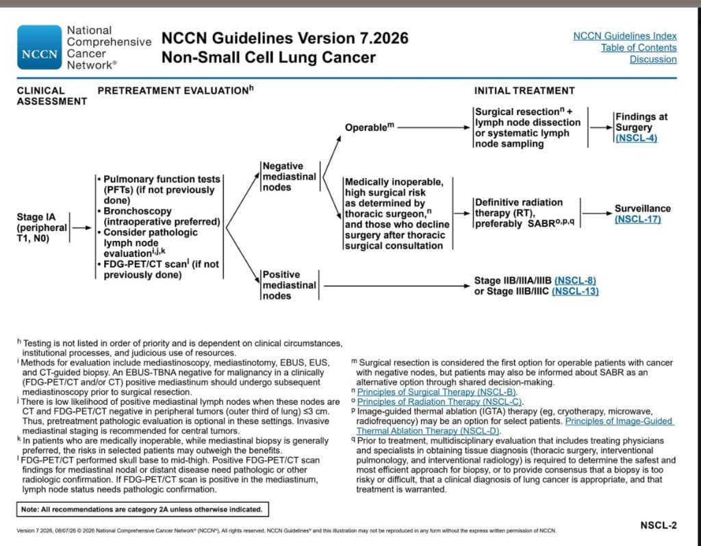
Who counts as inoperable. NCCN groups three situations together: medically inoperable, high surgical risk as determined by a thoracic surgeon, and patients who decline surgery after thoracic surgical consultation. Operability is a surgical judgement — do not label someone inoperable from PFTs alone without that consult. [31][35]
Pretreatment evaluation (same work-up whether or not surgery is planned): [35]
| Step | Why it matters |
|---|---|
| PFTs (if not already done) | Defines lung reserve and whether any resection would be tolerated |
| Bronchoscopy (intraoperative preferred if going to theatre) | Maps the airway and can obtain tissue |
| Consider pathologic mediastinal LN evaluation | EBUS, EUS, mediastinoscopy/mediastinotomy, or CT-guided biopsy. Optional for peripheral ≤ 3 cm tumours that are CT- and FDG-PET/CT-negative (occult N2 is uncommon). Invasive staging is recommended for central tumours |
| FDG-PET/CT (skull base to mid-thigh, if not already done) | Occult mediastinal or distant disease. PET-positive mediastinal nodes still need pathologic confirmation before denying local therapy |
Node-status branch:
- Positive mediastinal nodes → this is no longer Stage IA. Manage as Stage IIB/III (chemoradiation ± systemic therapy), not with SABR alone. [35]
- Negative mediastinal nodes → decide operability.
Initial treatment if inoperable / high-risk / declines surgery: definitive radiation therapy, preferably SABR (stereotactic ablative body radiotherapy). Then surveillance. [33][35]
- SABR/SBRT delivers a high, tightly conformal dose in few fractions (e.g. 54 Gy in 3 fractions). Local control is ~90% for T1–2 N0 tumours — the non-surgical curative option for Stage I. [33]
- Conventional fractionated RT is inferior to SABR when SABR is available.
- Image-guided thermal ablation (IGTA) — cryotherapy, microwave, or radiofrequency — is an option for selected patients (typically very peripheral, small lesions when SABR is unsuitable). [35]
- Operable patients should still hear about SABR as an alternative via shared decision-making; resection remains first-line. [35]
Exam Point — Inoperable Stage I
Three traps: (1) Positive mediastinal nodes are not Stage I — do not offer SABR as if the patient were N0. (2) Mediastinal biopsy is generally still preferred in medically inoperable patients, but the risk may outweigh the benefit in selected frail patients. (3) Tissue before treatment is the rule; if biopsy is too dangerous, an MDT (thoracic surgery, interventional pulmonology, radiation oncology, radiology) can agree a clinical diagnosis is enough to treat. [35]
3.2 Stage III: Locally Advanced NSCLC
Stage III is a heterogeneous group ranging from potentially resectable (some IIIA-N2) to unresectable (IIIB-IIIC). Management requires multidisciplinary team (MDT) discussion.
Concurrent chemoirradiation as treatment of choice [21]
- Why concurrent rather than sequential: Concurrent administration provides better local control because chemotherapy acts as a radiosensitiser — it makes tumour cells more susceptible to radiation-induced DNA damage. The survival benefit outweighs the increased toxicity.
- Chemotherapy regimen: usually cisplatin + etoposide or weekly carboplatin + paclitaxel [21]
- Radiotherapy: usually full-dose IMRT (60 Gy in 30 daily fractions) [21]
- "IMRT" = Intensity-Modulated Radiation Therapy — allows precise dose delivery to the tumour while sparing surrounding normal tissue (heart, oesophagus, spinal cord).
Consolidation after CRT without progression: osimertinib for eligible EGFR exon 19 deletion/L858R disease; durvalumab for other appropriate patients
- PACIFIC trial: Durvalumab (anti-PD-L1) for up to 12 months after completion of concurrent chemoRT in unresectable stage III NSCLC → significantly improved PFS and OS. This is now standard of care.
- Durvalumab = "dur-val-u-mab" — dur = durable, mab = monoclonal antibody.
- CheckMate 816 trial: Neoadjuvant nivolumab + platinum-doublet chemotherapy → surgery in resectable stage IB-IIIA → significant improvement in pathological complete response and event-free survival.
- This is an evolving paradigm — MDT discussion determines whether neoadjuvant → surgery or concurrent chemoRT is optimal for each patient.
3.3 Stage IV: Advanced / Metastatic NSCLC
This is where the molecular revolution has transformed outcomes. The approach depends entirely on the tumour's molecular profile.
Initial genetic testing to identify driver mutations as biomarkers [21]
- Indications: ALL adenocarcinoma and ALL never or minimal remote smokers [21]
- Current standard of care is to perform EGFR, ALK, and ROS1 analyses, but 10% patients have other potentially actionable mutations (e.g., other EGFR, CMET, BRAF V600, NTRK, HER2) [21]
- In East Asian adenocarcinoma, classic EGFR exon 19 deletion / L858R is the largest slice (~40%); the remaining actionable drivers are each uncommon but still matched to a drug — ALK ~4%, MET exon 14 ~3%, ROS1 ~3%, HER2 exon 20 insertion ~2%, BRAF V600E ~2%, KRAS G12C ~3%, RET ~2%, NTRK < 0.3%. About 1 in 5 have no actionable alteration.
Key targeted therapies:
| Target | Type of alteration | What it is | Drug Class | First-Line Agent | Mechanism | Key Side Effects |
|---|---|---|---|---|---|---|
| EGFR mutation (exon 19 del / L858R) | In-frame deletion (exon 19) or missense SNV (L858R) | Epidermal growth factor receptor — activating exon 19 deletion or exon 21 L858R point mutation → receptor tyrosine kinase stuck "on" | EGFR-TKI | Osimertinib (3rd gen; preferred 1st line — FLAURA trial) | Binds irreversibly to mutant EGFR tyrosine kinase → blocks downstream RAS-RAF-MEK-ERK and PI3K-AKT signalling → inhibits tumour proliferation | Diarrhoea, skin rash (acneiform), stomatitis, interstitial lung disease (rare but serious), QTc prolongation |
| EGFR exon 20 insertion | In-frame insertion (indel) | Same EGFR gene, but an in-frame insertion in exon 20 — still activating, yet the ATP-pocket shape makes classic EGFR-TKIs (including osimertinib) bind poorly | EGFR–MET bispecific antibody | Amivantamab ± chemotherapy | Bispecific antibody binds EGFR and MET → receptor internalisation and ADCC | Infusion-related reactions, rash, paronychia, hypoalbuminaemia, oedema |
| ALK rearrangement | Gene fusion (chromosomal rearrangement) | Anaplastic lymphoma kinase — chromosomal rearrangement (usually EML4-ALK fusion) creates a constitutively active fusion kinase | ALK inhibitor | Alectinib or lorlatinib (1st line — ALEX/CROWN trials) | Blocks ALK fusion protein kinase activity | Myalgia, oedema, visual disturbance (lorlatinib: CNS effects — mood, cognition) |
| MET exon 14 skipping / mutation | Splice-site mutation → exon skipping | Mesenchymal–epithelial transition factor (HGF receptor) — exon 14 skipping removes the degradation signal → MET protein accumulates and keeps signalling | MET inhibitor | Capmatinib or tepotinib | TKI blocks the accumulated MET kinase | Peripheral oedema, nausea, interstitial lung disease |
| ROS1 rearrangement | Gene fusion (chromosomal rearrangement) | ROS proto-oncogene 1 — chromosomal rearrangement creates a ROS1 fusion kinase (structurally similar to ALK) | ROS1 inhibitor | Crizotinib, entrectinib | Inhibits ROS1 kinase | GI upset, visual disturbance, oedema |
| HER2 exon 20 insertion | In-frame insertion (indel) | Human epidermal growth factor receptor 2 (ERBB2) — in-frame exon 20 insertion → constitutively active HER2 kinase | HER2 antibody–drug conjugate | Trastuzumab deruxtecan | Antibody delivers a topoisomerase-I payload to HER2-mutant cells | Interstitial lung disease / pneumonitis (must not miss), nausea, myelosuppression |
| BRAF V600E | Missense SNV | B-Raf serine/threonine kinase — valine → glutamate at codon 600 → MAPK pathway locked on | BRAF + MEK inhibitor | Dabrafenib + trametinib | Dual blockade of MAPK pathway | Pyrexia, skin toxicity, cardiomyopathy |
| KRAS G12C | Missense SNV | Kirsten rat sarcoma GTPase — glycine → cysteine at codon 12 locks KRAS in the GTP-"on" state (only G12C is currently druggable) | KRAS G12C inhibitor | Sotorasib, adagrasib | Covalently binds the mutant cysteine → locks KRAS inactive | Diarrhoea, hepatotoxicity |
| RET rearrangement | Gene fusion (chromosomal rearrangement) | REarranged during Transfection — chromosomal rearrangement creates a constitutively active RET fusion kinase | RET inhibitor | Selpercatinib or pralsetinib | Blocks RET fusion kinase | Hypertension, hepatotoxicity, QT prolongation, dry mouth |
| NTRK rearrangement | Gene fusion (chromosomal rearrangement) | Neurotrophic tyrosine receptor kinase (NTRK1/2/3) — fusion with a partner gene → constitutively active TRK kinase (tumour-agnostic target) | TRK inhibitor (tumour-agnostic) | Larotrectinib or entrectinib | Blocks NTRK fusion kinase; indicated across solid tumours harbouring NTRK fusions | Dizziness, weight gain, hepatotoxicity; entrectinib — cognitive / CNS effects |
Why "tyrosine kinase inhibitor"? "Tyrosine" = amino acid that gets phosphorylated; "kinase" = enzyme that adds phosphate groups; "inhibitor" = blocks. TKIs block the phosphorylation step that activates downstream proliferation pathways. When the tumour is driven by a single oncogene ("oncogene addiction"), blocking that pathway has dramatic effects.
Liquid biopsy: [21] Sample required: [21]
- On biopsy sample: traditional method, but risk of resistance heterogeneity
- On plasma/urine (liquid biopsy): detection of cell-free DNA in plasma and urine → minimally invasive (allows serial testing) and representative of dominant tumour molecular profile
Screening for programmed death ligand 1 (PD-L1) expression by immunohistochemistry [29]
Mechanism:
- Tumour cells upregulate PD-L1 on their surface → PD-L1 binds to PD-1 receptor on T-cells → sends an "off" signal to the T-cell → T-cell undergoes apoptosis or becomes anergic → tumour escapes immune surveillance.
- Monoclonal antibody against PD-1 and PD-L1 [29]
- Prevention of apoptosis of T-cells and enhance immune system [29]
- By blocking the PD-1/PD-L1 interaction, the T-cell remains active and can attack the tumour.
Key checkpoint inhibitors:
| Target | Drug | Indication |
|---|---|---|
| PD-1 blocking antibodies | Nivolumab, Pembrolizumab [29] | Advanced NSCLC (pembrolizumab most widely used 1st line) |
| PD-L1 blocking antibodies | Atezolizumab, Durvalumab [29] | Atezolizumab: advanced NSCLC, ES-SCLC. Durvalumab: consolidation after chemoRT in stage III |
PD-L1 expression guides treatment selection (for patients WITHOUT driver mutations):
| PD-L1 Score (TPS) | Preferred 1st-Line Treatment |
|---|---|
| ≥ 50% | Pembrolizumab monotherapy OR chemo + pembrolizumab |
| 1–49% | Chemo + pembrolizumab (combination preferred) |
| < 1% | Chemo + pembrolizumab or chemo + nivolumab + ipilimumab or chemo alone |
Side effects of immunotherapy: [29]
- Dermatological: Rash / Mucositis / Dry mouth
- Gastrointestinal: Diarrhoea / Immune-mediated colitis / Hepatotoxicity
- Endocrinological: Autoimmune thyroid disease / Adrenal insufficiency / Hypophysitis / Type 1 DM
Exam Point — Immune-Related Adverse Events
Be aware of flare of pre-existing autoimmune diseases with immunotherapy [29]. Checkpoint inhibitors release the brakes on the immune system → can trigger immune-related adverse events (irAEs) affecting any organ. The key ones to remember are immune-mediated pneumonitis (can be fatal), colitis, hepatitis, thyroiditis, hypophysitis, adrenalitis, and type 1 DM. Management involves holding the drug and giving high-dose corticosteroids.
For patients without actionable mutations and with low PD-L1, or in combination with immunotherapy:
- Platinum-based doublet: Standard backbone.
- Cisplatin + pemetrexed (non-squamous) or gemcitabine (squamous)
- Carboplatin + paclitaxel or nab-paclitaxel (often preferred due to better tolerability)
- Usually 4–6 cycles → then maintenance therapy (pemetrexed or immunotherapy).
| Agent | Mechanism | Key Toxicities |
|---|---|---|
| Cisplatin | Cross-links DNA → prevents replication | Nephrotoxicity (requires hydration), ototoxicity, neurotoxicity, severe emesis |
| Carboplatin | Same as cisplatin but less nephrotoxic | Myelosuppression (especially thrombocytopenia) |
| Pemetrexed | Multi-targeted antifolate — inhibits thymidylate synthase, DHFR, GARFT | Myelosuppression, mucositis. Requires folic acid + B12 supplementation |
| Paclitaxel | Stabilises microtubules → prevents mitotic spindle disassembly | Peripheral neuropathy, alopecia, myelosuppression |
| Gemcitabine | Nucleoside analogue — inhibits DNA synthesis | Myelosuppression, flu-like symptoms |
| Etoposide | Topoisomerase II inhibitor — prevents DNA unwinding | Myelosuppression, alopecia, secondary leukaemia |
3.4 SCLC Management
SCLC is managed very differently from NSCLC because of its exquisite chemosensitivity and radiosensitivity but rapid relapse and early metastasis.
For cT1-2N0M0 limited stage disease: [21]
- Primary surgery: lobectomy + mediastinal LN sampling/dissection
- Adjuvant chemotherapy: 4 cycles of cisplatin-based chemotherapy
- ± Adjuvant chemo/RT if LN involvement identified intra-operatively
This is the only scenario where surgery plays a role in SCLC — and it is uncommon because SCLC is rarely caught this early.
For unresectable limited stage (LS-SCLC) disease: [21]
- Concurrent platinum–etoposide + thoracic RT, followed by durvalumab in eligible patients without progression (ADRIATIC)
- Prophylactic cranial irradiation (PCI): offer after response to definitive chemo/RT if brain MRI shows no metastases and the patient is suitable [21][36]
Rationale for PCI: [21]
- Occult brain mets occur frequently in SCLC without neurological symptoms
- Brain mets often as sole site of relapse
- Effect in stage II–III LS-SCLC: prophylactic cranial irradiation → increased overall survival + decreased incidence of brain metastasis [21][36]
- PCI is less compelling after complete resection of pathologic stage I disease. Consider MRI surveillance instead in older/frail patients, those with poor performance status or baseline cognitive impairment, and those who prioritize avoiding neurotoxicity [36].
For extensive stage (ES-SCLC) disease: [21]
- Induction chemotherapy: mainstay, usually given for 4-6 cycles (± atezolizumab, PD-L1 mAb)
- Further thoracic EBRT if good response to initial chemotherapy
- Scheduled brain MRI surveillance is generally preferred over routine PCI. PCI reduces brain-metastasis risk but has not shown a clear survival benefit when MRI surveillance is available; reserve it for selected responders after shared decision-making [36].
The addition of atezolizumab (IMpower133 trial) or durvalumab (CASPIAN trial) to EP chemotherapy is now standard of care for ES-SCLC — this represented the first improvement in OS for ES-SCLC in decades.
After progression on or after platinum chemotherapy: tarlatamab-dlle (Imdelltra) is a DLL3-directed CD3 bispecific T-cell engager. FDA traditional approval (19 November 2025) followed DeLLphi-304, where median OS was 13.6 vs 8.3 months with standard chemotherapy (HR 0.60). Step-up dosing is required; boxed warnings cover cytokine-release syndrome and neurologic toxicity, including ICANS [37].
Role of radiotherapy in lung cancer management: [33]
| Setting | Indication | Details |
|---|---|---|
| Radical RT | Early-stage NSCLC in patients unfit for surgery | Stereotactic ablative body radiotherapy (SABR/SBRT): delivers high-dose focused radiation in few fractions (e.g., 54 Gy in 3 fractions). Local control rates ~90% for T1-2 N0 tumours. Preferred NCCN treatment for medically inoperable Stage IA (see 3.1.5). [33][35] |
| Concurrent chemoRT | Unresectable NSCLC; LS-SCLC | NSCLC commonly 60 Gy/30 fractions. LS-SCLC commonly 45 Gy/30 twice-daily fractions or an accepted once-daily course; see the SCLC section |
| Adjuvant RT | Consider for positive margins or selected high local-risk situations | Not routine solely for completely resected pN2 disease; balance local control and cardiopulmonary harm |
| Palliative RT | Brain metastases (whole-brain RT or stereotactic radiosurgery), bone mets (pain), SVCO, haemoptysis, spinal cord compression | Symptom relief. Whole-brain RT: 20 Gy / 5 fractions or 30 Gy / 10 fractions. SRS (stereotactic radiosurgery) for limited brain mets. |
| PCI | Stage II–III LS-SCLC with response and negative brain MRI [21][36] | Reduces incidence of brain mets; historical evidence supports improved OS. Individualize against MRI surveillance and neurocognitive risk. In ES-SCLC, MRI surveillance is usually preferred. |
Pleural mesothelioma: consider it in an asbestos-exposed patient with pleural thickening, effusion and chest-wall pain. Obtain adequate pleural tissue for subtype assessment (epithelioid, biphasic, sarcomatoid); negative fluid cytology does not exclude it. For unresectable disease, first-line options include nivolumab–ipilimumab or pembrolizumab with platinum–pemetrexed, selected by histology, fitness and contraindications. FDA pembrolizumab indication, September 2024.
Do not present pleurectomy/decortication or extrapleural pneumonectomy as routine curative treatment for all operable mesothelioma. MARS 2 found worse survival and more serious adverse events with extended pleurectomy/decortication plus chemotherapy versus chemotherapy alone; surgery requires expert selection and discussion of uncertain benefit. MARS 2, PMID 38740044.
Thymoma/thymic carcinoma: an anterior mediastinal mass with myasthenia gravis or pure red-cell aplasia should prompt thymic evaluation. CT defines extent; AFP/β-hCG help assess germ-cell alternatives. All thymic epithelial tumours have malignant potential, including encapsulated thymoma. Resectable disease usually proceeds to complete surgery; biopsy and induction/systemic treatment are considered for advanced/unresectable disease. Postoperative RT depends on stage, histology and margins, and chemotherapy is not automatic after every thymoma resection. NCI thymic tumour PDQ.
These topics come from the faculty Other thoracic malignancies lecture (pp. 3–8); they use separate staging and treatment pathways from bronchogenic carcinoma.
Palliative care, including pain control is an integral part of management for all stages, not just end-stage disease. [23]
Supportive treatment modalities: [21]
| Problem | Intervention |
|---|---|
| Pain | Analgesics for pain relief — WHO pain ladder: paracetamol → weak opioids → strong opioids ± adjuvants (gabapentin for neuropathic pain). Palliative RT for bone pain. |
| Cough | Cough suppression — codeine, morphine linctus, dexamethasone if lymphangitis |
| Malignant pleural effusion | Pleurodesis for malignant pleural effusion [21][27] — Chemical pleurodesis (talc via chest drain) as 1st line; surgical pleurodesis (VATS) if fit; indwelling pleural catheter (IPC) if trapped lung or short life expectancy |
| Airway obstruction | Bronchoscopic laser Tx or stenting to relieve airway obstruction [21] |
| SVCO | Endovascular stenting (rapid relief) + RT/chemo for underlying tumour [34] |
| Brain metastases | Dexamethasone (reduce oedema) + whole-brain RT or stereotactic radiosurgery |
| Spinal cord compression | Emergency dexamethasone + urgent surgical decompression or RT |
| Bone metastases | Bisphosphonates (zoledronic acid) or denosumab (RANKL inhibitor) to reduce skeletal events + palliative RT |
| Electrolyte imbalance | Management of complications, e.g., electrolyte imbalance [21] — hyponatraemia (SIADH → fluid restriction ± tolvaptan), hypercalcaemia (IV fluids + bisphosphonates) |
| Psychosocial | Other support: end-of-life care, psychosocial support [21] |
Malignant Pleural Effusion — Management Detail
Occur in 50% of all metastatic malignancy (especially NSCLC) [27]
| Option | Indication | Details |
|---|---|---|
| Repeated thoracocentesis | Small volume, slow recurrence, poor prognosis | Simple drainage every few weeks |
| Chemical pleurodesis (1st line) [27] | Recurrent symptomatic MPE, lung re-expandable | Agents: Talc (5g in 100mL NS), minocycline (300mg in 100mL NS) [27]. Instilled via chest drain after lung re-expansion. |
| Surgical pleurodesis [27] | Good performance status, failed chemical pleurodesis | VATS mechanical abrasion, pleurectomy |
| Long-term ambulatory indwelling pleural catheter (IPC) [27] | Short life expectancy / trapped lung — lung cannot re-expand fully, so pleurodesis will fail | Patient drains at home |
| Pleuroperitoneal shunt [27] | Short life expectancy / trapped lung | Shunts fluid from pleural to peritoneal cavity |
6. Special Situations
Treatment of malignancy-related SVCO: [34]
- Principle: Treatment is tailored to specific neoplasm and therefore tissue diagnosis is essential prior to empirical treatment which can potentially jeopardise histological evaluation [34]
- Current management guidelines emphasise: [34]
- Accurate histological diagnosis of underlying aetiology before starting therapy
- Upfront use of endovenous stents in severely symptomatic patients to provide more rapid relief than can be achieved using radiotherapy
Approach based on clinical status:
| Scenario | Management |
|---|---|
| Clinically stable | Clinical examination and investigations targeted to establish tissue diagnosis by minimally invasive methods. Deferring empirical treatment until a full diagnostic work-up has been completed does not pose a hazard for most patients provided that evaluation is efficient and patient is clinically stable. [34] |
| Urgent life-threatening (stridor, cerebral oedema) | Stabilise ABC → urgent oncological consultation → urgent endovascular stenting (provides most rapid relief without affecting subsequent tissue diagnosis) [34] |
Medical treatment: [34]
- Selected patients with limited metastatic burden (1-3 metastases — "oligometastatic") may benefit from local ablative treatment of all sites (surgery, SABR, or radiofrequency ablation) + systemic therapy.
- This is an evolving area — MDT discussion essential.
Prognosis of different types of lung cancer: [29]
SCLC: [29]
- Worst prognosis
- Few patients survive more than a year (without treatment)
- With treatment: LS-SCLC median OS ~20-25 months; ES-SCLC median OS ~12-15 months with chemo-IO.
NSCLC: [29]
- Better prognosis for patients with early surgical excision
- 30-40% of patients with completely excised early-stage cancer may survive for > 5 years
- Stage IV with driver mutation on matched TKI: median OS now > 3 years for EGFR-mutant disease.
High Yield Summary — Management of CA Lung
- Early NSCLC (I-II): Surgery (lobectomy) is the gold standard → ± adjuvant chemo (stage II+) → ± adjuvant osimertinib if EGFR-mutant. Medically inoperable / high-risk / decline surgery Stage I → definitive RT, preferably SABR.
- Unresectable NSCLC (III): CRT → osimertinib for eligible EGFR 19del/L858R disease, or durvalumab for other eligible patients (PACIFIC regimen). Selected patients may have neoadjuvant chemo-IO → surgery.
- Advanced NSCLC (IV): Molecular testing is mandatory → driver mutation → matched TKI; no mutation → immunotherapy ± chemotherapy based on PD-L1 expression.
- SCLC: Chemo-sensitive but relapses fast → LS: chemoRT → durvalumab if eligible without progression; assess PCI after response if MRI-negative and suitable; ES: chemo + atezolizumab/durvalumab ± thoracic RT, with MRI brain surveillance generally preferred to routine PCI. Tarlatamab is an option after platinum progression [36][37].
- Surgical fitness assessed by spirometry + DLCO → calculate ppo values → CPET if borderline. Low ppo values indicate increased risk; they are not isolated absolute exclusion criteria.
- Key post-op complications: air leak, haemorrhage, bronchopleural fistula.
- Palliative care is integral at ALL stages — pleurodesis for MPE, stenting for SVCO/airway obstruction, RT for bone/brain mets, pain management.
- irAEs from checkpoint inhibitors can affect any organ — pneumonitis, colitis, hepatitis, endocrinopathies. Manage with steroids.
- SVCO: tissue diagnosis first unless life-threatening → endovenous stent provides most rapid relief.
Active Recall - Management of CA Lung
References
[2] Senior notes: Maksim Medicine Notes.pdf (Clinical oncology — Lung cancer, p.51) [14] Lecture slides: Clinical manifestation of lung cancer (1).pdf (p.21 — summary) [21] Senior notes: Ryan Ho Respiratory.pdf (p.143-150 — staging, surgical options, adjuvant therapy, advanced NSCLC, SCLC, palliative care) [23] Lecture slides: Diagnostic work up and investigations for lung cancer (1).pdf (p.26 — management framework) [27] Senior notes: Maksim Medicine Notes.pdf (p.292-294 — Malignant pleural effusion, pleurodesis) [29] Senior notes: MBBS Final MB (Medicine) (Felix PY Lai).pdf (p.254 — Immunotherapy, PD-L1, prognosis) [31] Lecture slides: CMB40 - Lung cancer surgery - Perioperative aspects - Thoracic surgery (Dr Lucius KF Lee) rev2 (1).pdf (p.26 — summary) [33] Lecture slides: CMB42 - Role of radiotherapy in lung cancer management (Professor Victor Lee) rev2.pdf [34] Senior notes: MBBS Final MB (Medicine) (Felix PY Lai).pdf (p.1436-1439 — SVCO management) [35] NCCN Clinical Practice Guidelines in Oncology: Non-Small Cell Lung Cancer, Version 7.2026 (NSCL-2 — Stage IA peripheral T1 N0; medically inoperable pathway) [36] Käsmann L, et al. Update 2026: Management of Small Cell Lung Cancer. Lung. 2026. (PCI indications and MRI-surveillance approach in limited- and extensive-stage SCLC) [37] U.S. Food and Drug Administration. FDA grants traditional approval to tarlatamab-dlle for extensive-stage small cell lung cancer. 19 November 2025.
Complications of CA Lung
Complications of lung cancer arise from three distinct sources: the disease itself (local progression, metastasis, paraneoplastic), the treatment (surgery, chemotherapy, radiotherapy, immunotherapy, targeted therapy), and the interaction between the two. Understanding each complication from first principles — why does this happen? — transforms a list into genuine clinical reasoning.
1. Complications of the Disease
These complications arise because the tumour grows within the thorax and invades or compresses surrounding structures. The specific complication depends on which structure is involved — this is fundamentally an anatomy question.
| Complication | Structure Involved | Pathophysiology | Clinical Features | Management |
|---|---|---|---|---|
| Post-obstructive pneumonia / lung abscess | Bronchus (endobronchial tumour) | Tumour partially or completely obstructs airway → mucus retention distal to obstruction → bacterial superinfection → consolidation or frank abscess formation [35] | Fever, productive cough (may be foul-smelling if abscess with anaerobes), non-resolving pneumonia in the same lobe despite antibiotics | Antibiotics (cover anaerobes if abscess), bronchoscopic debulking or stenting to relieve obstruction, treat underlying cancer |
| Lobar/lung collapse (atelectasis) | Bronchus (complete obstruction) | Complete endobronchial obstruction → absorption of distal trapped air → collapse of distal lung segment/lobe | Dyspnoea, reduced chest expansion, tracheal deviation towards lesion, dullness, absent breath sounds | Bronchoscopic recanalisation (laser, cryotherapy, stent), treat underlying cancer |
| Pleural effusion | Pleura (visceral and/or parietal) | Tumour invades pleura → increased capillary permeability + lymphatic obstruction → exudative effusion. Malignant pleural effusion occurs in 50% of all metastatic malignancy, especially NSCLC. [27] | Progressive dyspnoea, pleuritic chest pain, stony dull percussion, reduced breath sounds | Thoracocentesis, chemical pleurodesis (talc), surgical pleurodesis (VATS), indwelling pleural catheter (IPC) if trapped lung [27] |
| Pericardial effusion / cardiac tamponade | Pericardium | Direct invasion or lymphatic spread to pericardium → fluid accumulation. If rapid onset → pericardium cannot stretch → cardiac chambers compressed → tamponade (obstructive shock) [13] | Beck's triad (hypotension, distended JVP, muffled heart sounds), pulsus paradoxus, tachycardia, electrical alternans on ECG | Urgent pericardiocentesis (echo-guided), pericardial window (surgical), treat underlying cancer |
| Recurrent laryngeal nerve palsy | RLN (left > right) | Left RLN has a long intrathoracic course looping under the aortic arch → vulnerable to compression by tumour or enlarged aortopulmonary window lymph nodes → vocal cord paralysis [2][3] | Hoarseness of voice, breathy voice, aspiration risk (incomplete glottic closure), bovine cough (weak, non-explosive) | Speech therapy, injection laryngoplasty (medialization of paralysed cord), treat underlying cancer |
| Phrenic nerve palsy | Phrenic nerve (C3-5) | Tumour or lymph nodes compress/invade phrenic nerve → hemidiaphragm paralysis → reduced tidal volume [2] | Dyspnoea (especially supine), elevated hemidiaphragm on CXR, paradoxical diaphragm movement on fluoroscopy (sniff test) | Treat underlying cancer; diaphragm plication rarely indicated |
| Superior vena cava obstruction | SVC | External compression or direct invasion of the SVC by tumour (especially right-sided tumours, SCLC, or mediastinal lymph nodes) → impaired venous drainage of head, neck, and upper extremities [2][13][34] | Facial/upper limb oedema, dilated neck and chest wall veins, facial plethora, Pemberton's sign. May progress to stridor (laryngeal oedema) or cerebral oedema (confusion, coma) | Endovenous stent (rapid relief) [34], RT/chemotherapy for the tumour, dexamethasone + furosemide |
| Pancoast syndrome | Apical structures: brachial plexus (C8-T1), stellate ganglion, first/second ribs, vertebral bodies | Superior sulcus tumour invading multiple adjacent structures simultaneously [2][3] | Horner's syndrome (miosis, ptosis, anhidrosis) + brachial plexopathy (shoulder/arm pain, small hand muscle wasting) + rib/vertebral body destruction | Concurrent chemoRT ± surgery in selected cases |
| Massive haemoptysis | Pulmonary artery or bronchial artery | Tumour erodes into a large vessel → catastrophic bleeding. Kills by asphyxia (drowning in blood), not exsanguination [10] | Sudden large-volume haemoptysis ( > 100-200 mL/day), respiratory distress, haemodynamic instability | Secure airway FIRST: intubation (double-lumen ETT ideal), lie on side of bleeding lung, urgent bronchoscopy ± bronchial artery embolisation, surgery if localised and patient fit [10] |
| Airway obstruction / stridor | Trachea or main bronchus | Tumour narrows the central airway > 50% → turbulent flow through narrowed lumen → stridor (inspiratory with extrathoracic, biphasic with intrathoracic obstruction). This is an oncological emergency. [13] | Stridor, dyspnoea, respiratory distress | Urgent bronchoscopic intervention (laser debulking, cryotherapy, endobronchial stenting), emergency RT, dexamethasone |
| Lymphangitis carcinomatosis | Pulmonary lymphatics | Tumour spreads through pulmonary lymphatics → interstitial oedema → thickened interlobular septa → impaired gas exchange | Progressive dyspnoea (out of proportion to CXR findings), dry cough, hypoxia. CXR: reticulonodular pattern, Kerley B lines. HRCT: thickened interlobular septa | Treat underlying cancer, supplemental O2, corticosteroids (limited benefit) |
| Oesophageal compression / invasion | Oesophagus | Extrinsic compression by tumour or mediastinal nodes, or direct invasion [2] | Dysphagia (usually to solids first), weight loss | Oesophageal stenting, palliative RT |
| Chest wall invasion | Ribs, intercostal muscles, intercostal nerves | Direct tumour extension through parietal pleura into chest wall | Severe, constant, localised chest wall pain (worse at night), palpable mass, rib destruction on imaging | Palliative RT for pain, en bloc resection if operable |
Oncological Emergencies from CA Lung
The four oncological emergencies most commonly caused by CA lung that require immediate action are:
- SVCO — especially if stridor or cerebral oedema → urgent endovenous stent [13][34]
- Spinal cord compression — back pain + limb weakness + sensory level → urgent MRI + dexamethasone + surgical decompression or RT [13]
- Cardiac tamponade — Beck's triad → urgent pericardiocentesis [13]
- Massive haemoptysis — secure airway first [10]
Failure to recognise and act on these immediately can result in death within hours.
These arise because lung cancer spreads haematogenously and lymphatically to distant organs. The complication depends on the metastatic site and the burden of disease.
| Metastatic Site | Complications | Pathophysiology | Management |
|---|---|---|---|
| Brain | Headache, seizures, focal neurological deficits, raised ICP, cognitive decline, herniation | Mass effect of metastatic deposits + surrounding vasogenic oedema → raised intracranial pressure. Focal deficits depend on the location (frontal → personality change; motor cortex → contralateral weakness; cerebellum → ataxia). [2] Complicates ~25% of extracranial malignancies; lung is the most common primary for brain mets [36] | Dexamethasone (reduce oedema), whole-brain RT, stereotactic radiosurgery (SRS) for limited mets ( ≤ 3), anti-epileptic drugs if seizures, surgical resection for solitary accessible met |
| Bone | Bone pain, pathological fractures, hypercalcaemia, spinal cord compression | Osteolytic metastases (via osteoclast activation by tumour-derived factors including PTHrP, RANKL, IL-6) weaken bone → fractures through diseased bone with minimal or no trauma ("pathological fracture"). Vertebral collapse → epidural compression of spinal cord → neurological emergency [2][13] | Bisphosphonates (zoledronic acid) or denosumab (RANKL inhibitor), palliative RT, surgical fixation for pathological fractures, urgent decompressive surgery/RT for cord compression |
| Liver | Hepatomegaly, RUQ pain, deranged LFTs, jaundice, hepatic failure (rare unless massive) | Tumour deposits replace hepatocytes + compress biliary canaliculi [2] | Systemic therapy, palliative care |
| Adrenal | Usually asymptomatic; rarely adrenal insufficiency | Bilateral adrenal destruction (both adrenals need to be > 90% destroyed before clinical insufficiency occurs — explains why it is rare). [2] | Hydrocortisone replacement if adrenal insufficiency confirmed |
| Contralateral lung | Additional respiratory impairment | Haematogenous or lymphatic spread → further compromised gas exchange | Systemic therapy |
These are remote effects of cancer NOT caused by direct invasion or metastasis, but by tumour-secreted substances. They are complications because they cause end-organ damage independent of the tumour mass itself. (Detailed mechanisms were covered in Clinical Features; here we focus on the complications they produce.)
| Syndrome | Most Common Subtype | Complication | Why It Matters |
|---|---|---|---|
| SIADH | SCLC | Severe hyponatraemia → confusion, seizures, coma, cerebral oedema, pontine myelinolysis if corrected too rapidly [15] | Most common paraneoplastic cause of hyponatraemia. Can be the presenting feature. Fluid restriction ± tolvaptan. |
| Ectopic ACTH | SCLC | Severe hypokalaemia → cardiac arrhythmias, muscle weakness. Hyperglycaemia. Immunosuppression (opportunistic infections). | Unlike classic Cushing's, onset is too rapid for typical Cushingoid habitus. Potassium replacement, ketoconazole/metyrapone to block cortisol synthesis. |
| Hypercalcaemia (PTHrP) | SCC | Severe hypercalcaemia → dehydration (nephrogenic DI), renal failure, confusion, cardiac arrhythmias (short QT, prolonged PR), coma [8] | Oncological emergency if Ca > 3.5 mmol/L. IV 0.9% saline (rehydrate) + IV bisphosphonate (zoledronic acid) + treat underlying cancer. |
| Lambert-Eaton (LEMS) | SCLC | Respiratory failure (diaphragmatic weakness), falls (proximal weakness) | Can be mistaken for myasthenia gravis. Treat cancer + 3,4-DAP (enhances ACh release). |
| Hypercoagulability (Trousseau syndrome) | Adenocarcinoma | DVT, PE, migratory thrombophlebitis, DIC [16] | In our locality, the most common condition associated with VTE is underlying malignancy [16]. Anticoagulation with LMWH preferred over warfarin in cancer-associated VTE. |
| HPOA | NSCLC | Painful swollen wrists/ankles, periostitis | Treat underlying cancer → symptoms resolve |
2. Complications of Treatment
Describe common and key postoperative complications and its management: [31] Air leak, hemorrhage, and bronchopleural fistula [31]
| Complication | Incidence | Pathophysiology | Clinical Features | Management |
|---|---|---|---|---|
| Air leak [31] | Common (5-15%) | Disruption of visceral pleura at staple line or lung parenchymal surface → air escapes from alveoli into pleural space. Prolonged air leak (PAL) = persistent > 5-7 days | Continuous bubbling in underwater seal drainage system, subcutaneous emphysema | Conservative (water seal, avoid suction), autologous blood patch pleurodesis, re-operation if persistent > 7 days |
| Haemorrhage [31] | Uncommon (~1-3%) | Bleeding from intercostal vessels, bronchial arteries, pulmonary artery/vein stump, or chest wall | Excessive chest drain output ( > 200 mL/hr), haemodynamic instability, dropping Hb | Transfusion, re-exploration (thoracotomy/VATS) if ongoing significant bleeding |
| Bronchopleural fistula (BPF) [31] | Rare but devastating (1-4% post-pneumonectomy) | Dehiscence of the bronchial stump → communication between bronchial tree and pleural space. Risk factors: long bronchial stump, ischaemia, infection, post-operative RT, right pneumonectomy (shorter bronchial stump, less tissue coverage) | Early ( < 7 days): sudden large air leak, tension pneumothorax, expectoration of serosanguinous fluid when lying on the operated side. Late ( > 7 days): empyema, fever, persistent air leak | Emergency chest drain (prevents tension PTX), antibiotics, bronchoscopic assessment, surgical repair or completion pneumonectomy. Mortality 20-70%. |
| Atelectasis / post-op pneumonia | Very common (15-20%) | Pain inhibits coughing → mucus retention → segmental/lobar collapse → bacterial superinfection. Risk factors: thoracotomy, one-lung ventilation, smoker status, pre-existing COPD [37] | Fever, productive cough, tachypnoea, hypoxia, CXR shows collapse/consolidation | Chest physiotherapy, incentive spirometry, adequate analgesia (to enable coughing), bronchoscopy for suctioning if refractory, antibiotics |
| Cardiac arrhythmia (esp. AF) | 10-20% post-lobectomy, up to 40% post-pneumonectomy | Pericardial irritation, vagal nerve handling, fluid shifts, sympathetic activation, intra-operative manipulation of heart | Palpitations, hypotension, irregular pulse | Rate control (beta-blocker, CCB, or amiodarone), rhythm control if haemodynamically unstable, anticoagulation to prevent stroke |
| Post-pneumonectomy pulmonary oedema | 2-5% post-pneumonectomy | Entire cardiac output is forced through one lung → increased capillary hydrostatic pressure + capillary endothelial damage from over-distension + possible fluid overload peri-operatively | Acute dyspnoea, hypoxia, diffuse alveolar shadowing on CXR in the remaining lung, typically day 2-3 post-op | Fluid restriction, diuretics, ventilatory support. High mortality (~50%). Prevention: restrict IV fluids peri-operatively. |
| Empyema | 2-5% | Infection in the pleural space, especially if BPF creates ongoing contamination | Fever, pleuritic pain, purulent chest drain output | Drainage (chest tube or VATS decortication), prolonged IV antibiotics |
| Wound infection / intercostal neuralgia | Variable | Post-thoracotomy pain syndrome (chronic) — intercostal nerve damage during rib retraction/spreading | Chronic pain along the intercostal distribution ( > 2 months post-op) | Gabapentin/pregabalin, intercostal nerve block, physiotherapy |
| Right heart failure | Rare | Loss of vascular bed (especially post-pneumonectomy) → increased pulmonary vascular resistance → right ventricular afterload increases beyond capacity | Elevated JVP, peripheral oedema, hepatomegaly, low cardiac output | Supportive, pulmonary vasodilators, avoid fluid overload |
Surgical Complication Prevention
Principles of postoperative care after lung resection and concept of early recovery after surgery: [31]
- Multimodal analgesia — thoracic epidural or paravertebral block + paracetamol + gabapentinoids ± NSAIDs (avoid NSAIDs if pleurodesis planned). Good analgesia → enables coughing and early mobilisation → reduces atelectasis and pneumonia.
- Early removal of chest drain — when no air leak, drainage < 200-400 mL/day, lung re-expanded on CXR. Reduces infection risk and facilitates mobilisation.
- Chest physiotherapy — deep breathing exercises, incentive spirometry, early ambulation.
- Smoking cessation — ideally ≥ 4 weeks pre-operatively to reduce post-op pulmonary complications.
| Complication | Responsible Agent(s) | Pathophysiology | Clinical Features | Management |
|---|---|---|---|---|
| Myelosuppression (neutropenia, anaemia, thrombocytopenia) | Nearly all agents (carboplatin → thrombocytopenia; etoposide → all lines) | Cytotoxic drugs kill rapidly dividing bone marrow progenitor cells → pancytopenia. Nadir typically day 7-14. | Neutropenic fever ( ≥ 38.3°C + ANC < 0.5 × 10⁹/L), fatigue, bleeding | G-CSF (filgrastim) prophylaxis for high-risk regimens; empirical broad-spectrum antibiotics for neutropenic fever; transfusion PRN |
| Nephrotoxicity | Cisplatin | Direct tubular epithelial cell damage (cisplatin accumulates in proximal tubular cells → mitochondrial dysfunction → apoptosis) | Rising creatinine, AKI, electrolyte wasting (Mg²⁺, K⁺) | Prevention: aggressive IV hydration pre/post cisplatin. If nephrotoxicity occurs → switch to carboplatin. |
| Ototoxicity | Cisplatin | Damage to cochlear outer hair cells → irreversible sensorineural hearing loss (high-frequency first) | Bilateral high-frequency hearing loss, tinnitus | Irreversible; audiometry monitoring; consider switching agent |
| Peripheral neuropathy | Cisplatin, paclitaxel | Damage to dorsal root ganglia (cisplatin) or microtubule disruption in axons (paclitaxel) → axonal degeneration | "Stocking-and-glove" paraesthesia/numbness, reduced vibration/proprioception | Dose reduction or cessation; duloxetine may help symptoms |
| Nausea and vomiting | Cisplatin (highly emetogenic), carboplatin (moderately) | Stimulation of chemoreceptor trigger zone (area postrema) in medulla + direct GI mucosal irritation → vagal afferents → vomiting centre | Acute ( < 24h), delayed (day 2-5), anticipatory | Triple antiemetic regimen: 5-HT₃ antagonist (ondansetron) + NK1 antagonist (aprepitant) + dexamethasone |
| Pulmonary toxicity | Bleomycin (if used; rare in lung cancer regimens), checkpoint inhibitors | Bleomycin: free radical-mediated damage to alveolar epithelium → pulmonary fibrosis. CPI: immune-mediated pneumonitis. | Dry cough, progressive dyspnoea, hypoxia, bilateral GGO/reticular changes on CT | Stop offending drug. Bleomycin fibrosis: irreversible. CPI pneumonitis: high-dose steroids. |
| Secondary malignancy | Alkylating agents (cyclophosphamide), etoposide | DNA damage to normal stem cells → secondary MDS/AML (therapy-related myeloid neoplasm). Etoposide-related AML: typically MLL rearrangement, peaks 2-3 years. Alkylating: peaks 5-7 years. [38] | Cytopenias, constitutional symptoms | Treat as secondary AML |
| Alopecia | Paclitaxel, etoposide | Damage to rapidly dividing hair follicle matrix cells → hair shaft breaks at or below scalp level | Diffuse hair loss 2-4 weeks after starting chemotherapy | Reversible on drug cessation; scalp cooling may reduce severity |
| Timing | Complication | Pathophysiology | Clinical Features | Management |
|---|---|---|---|---|
| Acute ( < 6 weeks) | Radiation oesophagitis | Radiation damages rapidly dividing oesophageal squamous epithelium → mucosal inflammation + ulceration | Odynophagia, dysphagia, retrosternal pain | Liquid/soft diet, viscous lidocaine, PPI, sucralfate |
| Radiation pneumonitis | Radiation damages type I and II pneumocytes + endothelial cells → inflammatory exudate fills alveoli (typically 1-3 months post-RT) | Cough, dyspnoea, low-grade fever, GGO/consolidation on CT within the radiation field | High-dose prednisolone (taper over weeks), supplemental O₂ | |
| Fatigue | Multifactorial: cytokine release, anaemia, nutritional depletion | Profound tiredness during and weeks after RT | Supportive | |
| Skin reaction | Radiation dermatitis — damage to basal layer of epidermis | Erythema, desquamation (dry → moist in severe cases) | Moisturiser, topical steroid, wound care | |
| Late ( > 6 months) | Radiation fibrosis | Chronic inflammation → fibroblast activation → irreversible collagen deposition in lung parenchyma | Progressive dyspnoea, restrictive pattern on PFT, fibrotic changes on CT corresponding to radiation field | Irreversible; supportive care, supplemental O₂ |
| Brachial plexopathy | Fibrosis around brachial plexus (especially after Pancoast tumour RT) | Arm pain, weakness, paraesthesia — may mimic tumour recurrence | Distinguished from recurrence by MRI ± PET (fibrosis = non-FDG avid) | |
| Radiation myelopathy | Spinal cord damage if cord dose exceeds tolerance (~45-50 Gy) | Progressive lower limb weakness, sensory loss, Brown-Séquard syndrome | Prevention (strict dose constraints); irreversible once established | |
| Cardiac toxicity | Pericarditis, accelerated coronary artery disease, cardiomyopathy | Chest pain, heart failure, constrictive pericarditis (years later) | Cardiology involvement; pericardiocentesis if effusion | |
| Second malignancy [38] | Radiation-induced DNA damage in normal cells | New cancer within or near the radiation field years later (breast, oesophageal, thyroid, sarcoma) | Screening, treat as new primary |
Side effects of immunotherapy: [29]
Checkpoint inhibitors release the immune "brakes" — this can lead to autoimmune-like damage to any organ (immune-related adverse events, irAEs). They differ fundamentally from chemotherapy side effects because they are immune-mediated, not directly cytotoxic.
| Organ System | irAE | Incidence | Pathophysiology | Management |
|---|---|---|---|---|
| Skin | Rash, mucositis, dry mouth [29] | Most common (~30%) | T-cell-mediated attack on skin keratinocytes | Topical steroids, hold drug if severe, systemic steroids if grade ≥ 3 |
| GI | Diarrhoea, immune-mediated colitis, hepatotoxicity [29] | 10-20% | T-cell infiltration of colonic/hepatic epithelium | Colitis: systemic steroids → infliximab if refractory. Hepatitis: steroids → mycophenolate if refractory. |
| Endocrine | Autoimmune thyroid disease, adrenal insufficiency, hypophysitis, Type 1 DM [29] | 5-10% | T-cell destruction of endocrine glands. Thyroiditis often presents as transient thyrotoxicosis (destructive phase) → hypothyroidism. | Hormone replacement (levothyroxine, hydrocortisone, insulin). Unlike other irAEs, endocrinopathies are usually permanent — the gland is destroyed. |
| Lung | Immune-mediated pneumonitis | 3-5% (higher with combination CPI) | T-cell-mediated alveolitis → GGO and organising pneumonia pattern on CT | Hold drug, high-dose IV methylprednisolone. Can be fatal if not recognised early. |
| Neurological | Myasthenia-like syndrome, Guillain-Barré, encephalitis | Rare ( < 1%) | Autoimmune neuronal/NMJ damage | Steroids, IVIG, plasmapheresis |
| Cardiac | Myocarditis | Rare ( < 1%) but high mortality (~50%) | T-cell infiltration of myocardium | Hold drug immediately, high-dose steroids, cardiac monitoring in ICU |
| Renal | Immune-mediated nephritis | 1-2% | Tubulointerstitial nephritis | Steroids |
Exam Pitfall – irAEs
Be aware of flare of pre-existing autoimmune diseases with immunotherapy [29]. Patients with pre-existing autoimmune conditions (e.g., SLE, inflammatory bowel disease, rheumatoid arthritis) are at higher risk of severe irAEs and are often excluded from clinical trials. If an irAE occurs:
- Grade 1 (mild): Continue drug, monitor closely.
- Grade 2 (moderate): Hold drug, oral prednisolone 0.5-1 mg/kg.
- Grade 3-4 (severe/life-threatening): Permanently discontinue drug, IV methylprednisolone 1-2 mg/kg, ± additional immunosuppression (infliximab, mycophenolate).
Endocrine irAEs are the exception — they are managed with hormone replacement rather than immunosuppression, as the glandular destruction is usually irreversible by the time it is detected.
| TKI Class | Key Side Effects | Pathophysiology |
|---|---|---|
| EGFR-TKIs (osimertinib, gefitinib, erlotinib) | Acneiform rash (paradoxically correlates with response), diarrhoea, stomatitis, interstitial lung disease/pneumonitis (1-3%, can be fatal), QTc prolongation (osimertinib), hepatotoxicity | EGFR is expressed in normal skin/GI epithelium → blocking it causes epithelial damage |
| ALK inhibitors (alectinib, lorlatinib) | Myalgia, oedema, visual disturbance (alectinib), CNS effects with lorlatinib (mood changes, cognitive impairment, hallucinations — because lorlatinib crosses BBB) | ALK is expressed in developing nervous system → CNS effects |
| KRAS G12C inhibitors (sotorasib) | Diarrhoea, hepatotoxicity | Off-target effects |
| Category | Examples | Key Management Principles |
|---|---|---|
| Local tumour effects | Airway obstruction, post-obstructive pneumonia, pleural effusion, SVCO, RLN palsy, phrenic nerve palsy, massive haemoptysis, tamponade | Treat the tumour (chemo/RT/surgery) + manage the emergency (stent, drain, secure airway) |
| Metastatic | Brain mets (seizures, raised ICP), bone mets (fractures, cord compression, hypercalcaemia), liver mets (LFT derangement) | Dexamethasone for brain/cord, bisphosphonates for bone, palliative RT, systemic therapy |
| Paraneoplastic | SIADH, ectopic ACTH, hypercalcaemia (PTHrP), LEMS, VTE | Treat the underlying cancer + correct metabolic derangement |
| Surgical | Air leak, haemorrhage, BPF, pneumonia, arrhythmia, post-pneumonectomy pulmonary oedema | Prevention (analgesia, physio, early drain removal) + early recognition and intervention |
| Chemotherapy | Myelosuppression, nephrotoxicity, neuropathy, emesis, secondary malignancy | Prophylaxis (G-CSF, antiemetics, hydration), dose modification, supportive care |
| Radiotherapy | Oesophagitis, pneumonitis, fibrosis, cardiac toxicity, secondary malignancy | Steroids for pneumonitis, dose constraints, supportive care |
| Immunotherapy | irAEs: pneumonitis, colitis, hepatitis, endocrinopathies, myocarditis | Hold drug, systemic steroids, hormone replacement for endocrinopathies |
| Targeted therapy | Skin rash, diarrhoea, ILD, hepatotoxicity, CNS effects | Dose reduction, hold/stop drug, supportive care |
High Yield Summary — Complications of CA Lung
- Local complications are anatomy-driven: know which structure is invaded → predict the complication (RLN → hoarseness, phrenic → hemidiaphragm, SVC → SVCO, brachial plexus → Pancoast).
- Four oncological emergencies: SVCO (stent), spinal cord compression (MRI + dexamethasone + decompression), cardiac tamponade (pericardiocentesis), massive haemoptysis (secure airway first — kills by asphyxia).
- Malignant pleural effusion occurs in ~50% of metastatic NSCLC — manage with pleurodesis or IPC.
- Paraneoplastic syndromes cause organ damage independently of tumour mass: SIADH (hyponatraemia), PTHrP (hypercalcaemia), LEMS (weakness), Trousseau (VTE).
- Post-surgical complications: air leak, haemorrhage, BPF (most feared), atelectasis/pneumonia, AF. Prevention = multimodal analgesia + chest physio + early drain removal.
- irAEs can affect ANY organ — pneumonitis, colitis, hepatitis, endocrinopathies, myocarditis. Manage with steroids except endocrine irAEs (hormone replacement). Endocrine irAEs are usually permanent.
- Cisplatin complications: nephrotoxicity (prevent with hydration), ototoxicity (irreversible), neuropathy, severe emesis.
- Radiation pneumonitis (1-3 months post-RT) is steroid-responsive; radiation fibrosis (late) is irreversible.
Active Recall - Complications of CA Lung
References
[2] Senior notes: Maksim Medicine Notes.pdf (Clinical oncology — Lung cancer, p.51) [3] GC Lecture slides: GC 077. Pleural effusion in a chronic smoker.pdf [8] Senior notes: Ryan Ho Chemical Path.pdf (p.23 — Hypercalcaemia and malignancy) [10] Senior notes: Ryan Ho Fundamentals.pdf (p.226 — Approach to haemoptysis, massive haemoptysis management) [13] Lecture slides: CMB09 - Oncologic Emergency (Professor Aya El Helali)rev2.pdf [15] Senior notes: Block A - Electrolyte and Acid-Base Disorders.pdf (p.21 — SIADH) [16] Senior notes: Block A - Leg swelling and chest pain deep vein thrombosis; pulmonary embolism; Thrombophilia.pdf (p.16 — malignancy-associated VTE) [21] Senior notes: Ryan Ho Respiratory.pdf (p.126 — asbestos complications; p.147-150 — surgical Mx, complications, palliative care) [27] Senior notes: Maksim Medicine Notes.pdf (p.292-294 — Malignant pleural effusion, pleurodesis) [29] Senior notes: MBBS Final MB (Medicine) (Felix PY Lai).pdf (p.254 — Immunotherapy side effects) [31] Lecture slides: CMB40 - Lung cancer surgery - Perioperative aspects - Thoracic surgery (Dr Lucius KF Lee) rev2 (1).pdf (p.2, 26) [34] Senior notes: MBBS Final MB (Medicine) (Felix PY Lai).pdf (p.1436-1439 — SVCO management) [35] Senior notes: Ryan Ho Respiratory.pdf (p.65 — unresolving pneumonia, complications) [36] Senior notes: Ryan Ho Neurology.pdf (p.164 — brain metastasis) [37] Senior notes: MBBS Final MB (Surgery) (Felix PY Lai).pdf (p.387 — prevention of respiratory complications) [38] Senior notes: Ryan Ho Haemtology.pdf (p.96 — treatment-related complications, second malignancies)
High Yield Summary
Staging update: use TNM 9 (2025): N2a = one mediastinal/subcarinal station, N2b = multiple stations; M1c1 = multiple metastases in one organ system, M1c2 = multiple systems. T1N2a is IIB. Keep older trial eligibility labelled by edition; see the Ix table.
- CA Lung = most common cancer cause of death worldwide and in Hong Kong.
- Adenocarcinoma is the most common subtype globally and in HK (peripheral, weak smoking association, test for EGFR/ALK in Asians).
- SCC is central, strongest smoking association; SCLC is central, aggressive, early metastasis, paraneoplastic.
- Clinical features are driven by anatomy: endobronchial symptoms (cough, haemoptysis, obstruction), local invasion (RLN → hoarseness, phrenic → hemidiaphragm, SVC → SVCO, brachial plexus → Pancoast), metastasis (bone, brain, liver, adrenal), paraneoplastic syndromes (SIADH/SCLC, PTHrP/SCC, LEMS/SCLC).
- Non-resolving or recurrent same-location pneumonia in a smoker = think CA lung.
- Massive haemoptysis kills by asphyxia, not exsanguination → secure airway first.
- In HK, ~50% adenocarcinoma harbour EGFR mutations → tyrosine kinase inhibitor therapy.
- LDCT screening reduces lung cancer mortality in high-risk smokers.
High Yield Summary – Differential Diagnosis of CA Lung
- The DDx depends on the clinical presentation — lung mass, haemoptysis, non-resolving pneumonia, pleural effusion, or incidental nodule.
- In HK, the "Big Three" for haemoptysis are CA lung, TB, and bronchiectasis.
- Solitary pulmonary nodule: malignancy probability depends on the population and nodule features — use risk stratification (age, smoking, size, margin, calcification, PET avidity, interval growth).
- A negative biopsy does NOT exclude malignancy — if suspicion remains, consider repeat biopsy or surgical excision with frozen section.
- TB and CA lung can co-exist — always consider malignancy in a patient with "treated TB" who develops new symptoms.
- Non-resolving or same-location recurrent pneumonia in a smoker → always exclude endobronchial CA lung.
- Features favouring malignancy over benign: spiculated margin, lack of calcification, interval growth, age ≥ 50, smoker, raised SUV, associated atelectasis/adenopathy.
High Yield Summary — Investigations for CA Lung
- CXR raises suspicion; CT diagnoses and stages; PET-CT detects occult metastases; MRI brain screens for brain mets.
- Tissue is mandatory before treatment — method depends on tumour location (central → bronchoscopy; peripheral → CT-guided biopsy; nodes → EBUS-TBNA; accessible mets → biopsy there).
- Molecular testing (EGFR, ALK, ROS1, PD-L1) is essential for all advanced non-squamous NSCLC — determines eligibility for targeted therapy/immunotherapy.
- CT nodal staging is limited by size criteria — cannot distinguish reactive from metastatic; EBUS-TBNA provides tissue confirmation.
- PET-CT is poor for brain (high background activity) — use MRI brain instead.
- Functional assessment (FEV1, DLCO, VO2max, ECOG) determines whether a patient can tolerate surgery or radical RT.
- Negative biopsy ≠ no cancer — if suspicion remains, pursue wedge resection with intra-operative frozen section.
- Malignant pleural effusion is exudative, often blood-stained, with low glucose and pH; cytology ~60% sensitive.
High Yield Summary — Management of CA Lung
- Early NSCLC (I-II): Surgery (lobectomy) is the gold standard → ± adjuvant chemo (stage II+) → ± adjuvant osimertinib if EGFR-mutant. Medically inoperable / high-risk / decline surgery Stage I → definitive RT, preferably SABR.
- Unresectable NSCLC (III): CRT → osimertinib for eligible EGFR 19del/L858R disease, or durvalumab for other eligible patients (PACIFIC regimen). Selected patients may have neoadjuvant chemo-IO → surgery.
- Advanced NSCLC (IV): Molecular testing is mandatory → driver mutation → matched TKI; no mutation → immunotherapy ± chemotherapy based on PD-L1 expression.
- SCLC: Chemo-sensitive but relapses fast → LS: chemoRT → durvalumab if eligible without progression; assess PCI after response if MRI-negative and suitable; ES: chemo + atezolizumab/durvalumab ± thoracic RT, with MRI brain surveillance generally preferred to routine PCI. Tarlatamab is an option after platinum progression.
- Surgical fitness assessed by spirometry + DLCO → calculate ppo values → CPET if borderline. Low ppo values indicate increased risk; no isolated cutoff decides operability.
- Key post-op complications: air leak, haemorrhage, bronchopleural fistula.
- Palliative care is integral at ALL stages — pleurodesis for MPE, stenting for SVCO/airway obstruction, RT for bone/brain mets, pain management.
- irAEs from checkpoint inhibitors can affect any organ — pneumonitis, colitis, hepatitis, endocrinopathies. Manage with steroids.
- SVCO: tissue diagnosis first unless life-threatening → endovenous stent provides most rapid relief.
High Yield Summary — Complications of CA Lung
- Local complications are anatomy-driven: know which structure is invaded → predict the complication (RLN → hoarseness, phrenic → hemidiaphragm, SVC → SVCO, brachial plexus → Pancoast).
- Four oncological emergencies: SVCO (stent), spinal cord compression (MRI + dexamethasone + decompression), cardiac tamponade (pericardiocentesis), massive haemoptysis (secure airway first — kills by asphyxia).
- Malignant pleural effusion occurs in ~50% of metastatic NSCLC — manage with pleurodesis or IPC.
- Paraneoplastic syndromes cause organ damage independently of tumour mass: SIADH (hyponatraemia), PTHrP (hypercalcaemia), LEMS (weakness), Trousseau (VTE).
- Post-surgical complications: air leak, haemorrhage, BPF (most feared), atelectasis/pneumonia, AF. Prevention = multimodal analgesia + chest physio + early drain removal.
- irAEs can affect ANY organ — pneumonitis, colitis, hepatitis, endocrinopathies, myocarditis. Manage with steroids except endocrine irAEs (hormone replacement). Endocrine irAEs are usually permanent.
- Cisplatin complications: nephrotoxicity (prevent with hydration), ototoxicity (irreversible), neuropathy, severe emesis.
- Radiation pneumonitis (1-3 months post-RT) is steroid-responsive; radiation fibrosis (late) is irreversible.